
Scimitar syndrome (SS) is a rare congenital anomaly characterized by anomalous drainage of the right pulmonary veins into the inferior vena cava (IVC), with an estimated incidence of 1–3 per 100,000 live births [1]. It may be associated with right lung hypoplasia, pulmonary sequestration, dextroposition of the heart, or atrial septal defect. The clinical presentation varies widely – from asymptomatic cases to severe pulmonary hypertension [2] and heart failure – and may manifest in infancy [3] or adulthood [4].
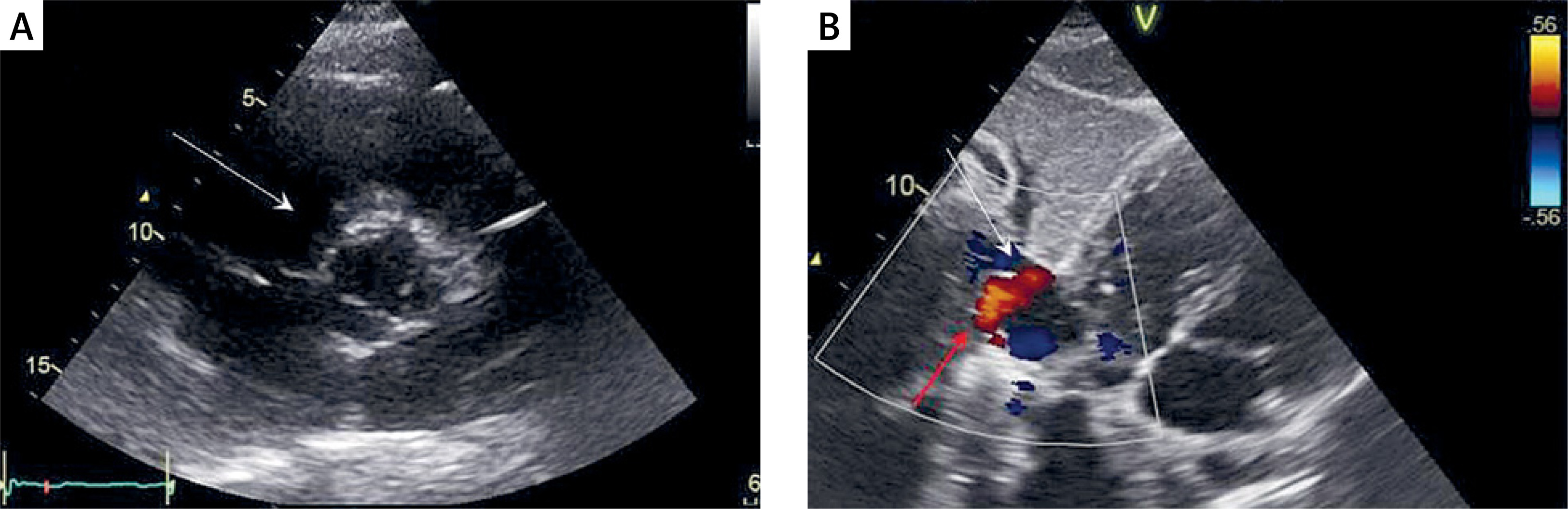
A 52-year-old male patient was referred for evaluation of ascending aorta (AAo) dilatation and partial anomalous pulmonary venous return. He was active but reported a recent decline in exercise tolerance and episodic palpitations. Transthoracic echocardiography (TTE) revealed an ascending aortic diameter of 52 mm with a tricuspid aortic valve, mild tricuspid regurgitation, right ventricular systolic pressure < 35 mm Hg, and suspected anomalous pulmonary venous drainage to the IVC (Figure 1). Cardiac computed tomography (cCT) confirmed AAo dilatation to 56 mm and anomalous right pulmonary venous connection with a membranous structure at the site of junction. Electrocardiography showed first-degree atrioventricular block and right bundle branch block. Cardiac magnetic resonance (CMR) additionally provided indirect evidence of a substantial shunt volume. Specifically, it demonstrated a pulmonary vein (22 mm in diameter) draining into the IVC, with anomalous venous return from the majority of the right lung (entire upper and lower lobes) to the IVC. Quantitative CMR data revealed a right ventricular stroke volume of 170 ml and a left ventricular stroke volume of 105 ml. The estimated Qp/Qs ratio was approximately 1.6, consistent with a significant left-to-right shunt.
Figure 1
Transthoracic echocardiography. A – Tricuspid aortic valve – white arrow (parasternal short-axis view at the level of the aortic valve in mid-systole). B – Anomalous pulmonary venous drainage (red arrow showing drainage direction) to the inferior vena cava – white arrow (subcostal view)
According to the heart-team decision, the patient met the criteria for surgical intervention due to a hemodynamically significant left-to-right shunt with evidence of right ventricular (RV) volume overload, demonstrated by RV dilatation in TTE and cardiac magnetic resonance. Therefore, surgical correction was performed: tunneling of the anomalous pulmonary venous return to the left atrium (LA) and replacement of the ascending aorta with a vascular graft. Postoperative TTE demonstrated unobstructed pulmonary venous drainage and normal flow through the tunnel, with a graft diameter of 35 mm. The postoperative course was uneventful. Two years later, the patient presented with atrial arrhythmia; after excluding thrombus in the LA, successful electrical cardioversion was performed (Supplementary Figures S1–S3).
This case demonstrates the exceptional coexistence of SS and an ascending aortic aneurysm in a patient with a tricuspid aortic valve. To our knowledge, this constellation has not been described previously. However, coexistence of SS with a bicuspid aortic valve (with aortic aneurysm) has been reported before [5]. The absence of a bicuspid aortic valve or connective tissue disorder further emphasizes the rarity of this association, though any causal relationship remains speculative.
Preoperative cCT and CMR were crucial in delineating the complex venous anatomy and quantifying the aortic enlargement, providing indispensable information for surgical planning. Three-dimensional assessment of the relationship between the right pulmonary veins, IVC, and LA enabled safe tunneling and minimized intraoperative uncertainty.
The combined repair required addressing two anatomically distinct regions – the anomalous venous connection and AAo – within one operative session. Careful coordination was necessary to limit bypass and cross-clamp times. The successful outcome confirms the feasibility of this approach in experienced centers.
Clinically, this report highlights that SS may remain mildly symptomatic for decades, with incidental detection in adulthood. Nevertheless, surgical correction is recommended in symptomatic patients or in those with right heart volume overload [6].
In conclusion, this unique case expands the clinical spectrum of SS, underscores the pivotal role of cCT in preoperative evaluation, and highlights potential technical challenges of combined repair. Individualized, multidisciplinary management is essential for favorable outcomes in such rare presentations.










