Spontaneous coronary artery dissection (SCAD) is a rare condition, presenting similarly to myocardial infarction and most commonly occurring in women under 50 years of age. It is responsible for 1–4% of all cases of acute coronary syndromes (ACS). However, among women of reproductive age it accounts for up to 30% of ACS cases and remains a significant diagnostic challenge [1–3].
The exact mechanism underlying SCAD remains unclear. Several factors, such as emotional stress, sex hormones, dyslipidemia, and hypertension, have been found to be associated with SCAD [1, 3, 4].
A 59-year-old woman with a history of tobacco use and a family history of cardiovascular disease was admitted to the emergency department with suspected ACS due to chest pain. On admission, electrocardiography (ECG) showed a QS pattern, ST-segment elevation in leads V1-V2 and T-wave inversion in leads V1-V3 (Figure 1 A). Pretreatment included 300 mg of aspirin, 5000 IU of unfractionated heparin, and fluids. Echocardiography revealed impaired contractility of the anterior wall.
Figure 1
A – 12-lead electrocardiography (ECG), during admission, B – coronary angiography of left coronary artery (LCA) (arrow: dissection of LAD), C – coronary angiography of right coronary artery (RCA), D – persistent coronary artery dissection in coronary computed tomography angiography, 11 months after dissection (arrow: persistent dissection of LAD)
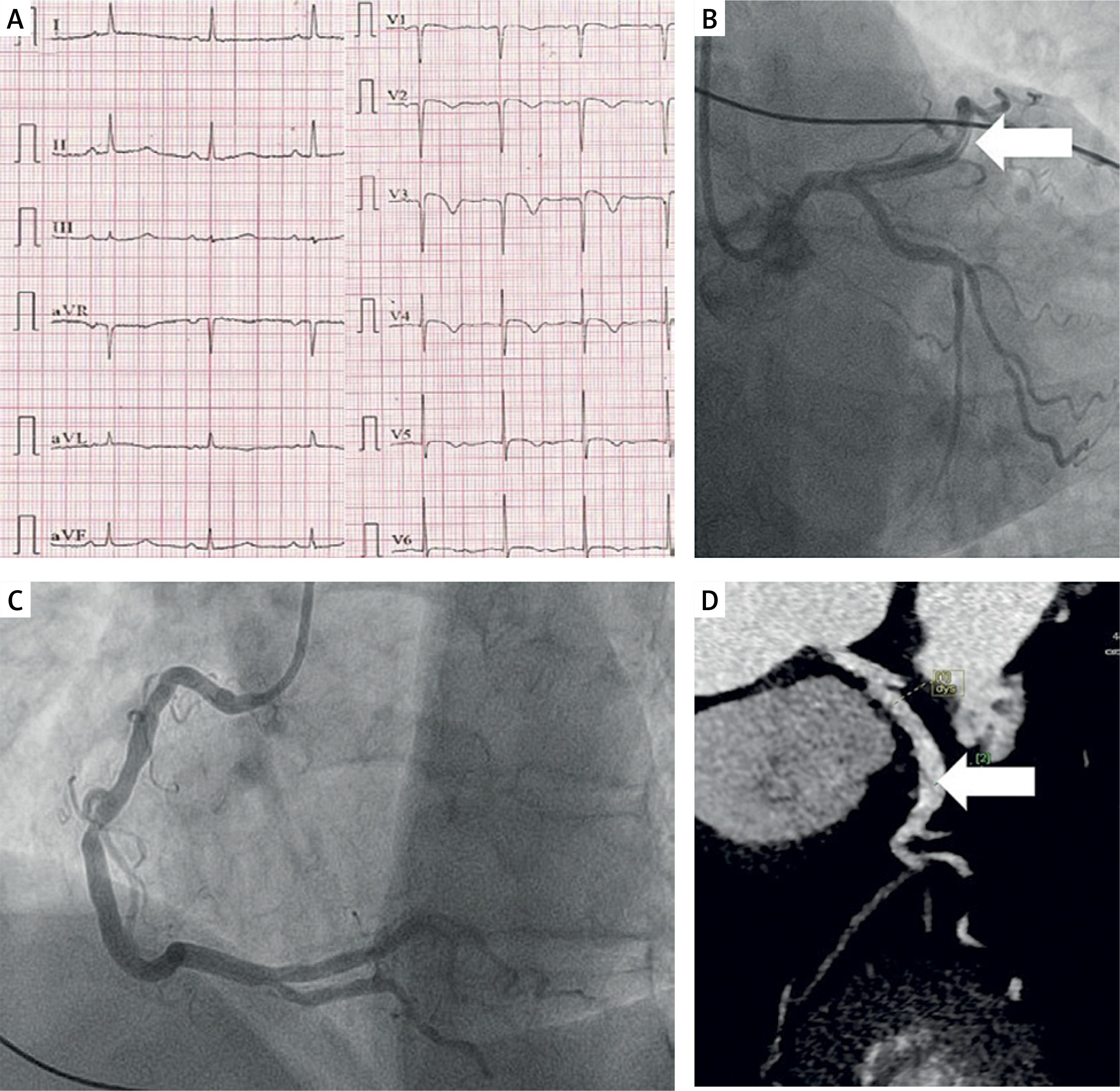
Emergency coronary angiography revealed type 1 SCAD. The left anterior descending artery (LAD) was patent with TIMI 3 flow and no contrast retention. The remaining coronary arteries were angiographically normal (Figures 1 B, C). The patient was free of resting chest pain at the time of examination and was therefore managed conservatively. During hospitalization, she was diagnosed with arterial hypertension and mixed hyperlipidemia. A second antiplatelet agent was not administered.
Further treatment was complicated by pneumonia, but after empiric antibiotic therapy, the patient was discharged home in good condition after 1 week of hospitalization. On discharge, echocardiography showed persistent akinesis of the apical segment of the interventricular septum and a left ventricular ejection fraction (LVEF) of 50%.
During the 3-month-follow-up, the patient underwent cardiac rehabilitation without experiencing any cardiovascular events. Eleven months after hospitalization, computed tomography angiography revealed a persistent long-segment dissection of the LAD (Figure 1 D). Despite this finding, the patient remained asymptomatic and reported no cardiovascular complaints. The LVEF was 55%. Her level of physical activity remained limited, without engagement in significant physical exertion, and she was not receiving dual antiplatelet therapy during follow-up.
This case highlights the importance of a comprehensive diagnostic approach, which also includes SCAD in the differential diagnosis, even in patients with traditional cardiovascular risk factors. As in numerous other instances, the patient was treated conservatively, with a favorable outcome, illustrating the potential effectiveness of this strategy.