Uncrossable lesions may occur in up to 10% of chronic total occlusions (CTOs) [1]. Rotational atherectomy (RA) is a well-established procedure, especially in cases where passage of a balloon catheter or its inflation is not possible due to severe calcifications [2]. Additionally, treatment options for balloon-uncrossable lesions include the use of excimer laser coronary atherectomy (ELCA). The ELCA catheter can cross the lesion through its photochemical, photothermal, and photomechanical action, resulting in softening of hard plaque, allowing passage of subsequent devices through the CTOs [3].
An 81-year-old male patient with a history of coronary artery disease, heart failure with midrange ejection fraction (HFmrEF), prior percutaneous coronary intervention (PCI) of the marginal branch, hypertension, diabetes mellitus type 2, dyslipidemia, and prior dual chamber (DDD) pacemaker implantation was admitted to reattempt PCI of the CTO of the left anterior descending artery (LAD) (Figure 1 A). The patient remained symptomatic, with Canadian Cardiovascular Society (CCS) class III angina, and had documented ischemia of the anterior wall and interventricular septum on single-photon emission computed tomography (SPECT). An initial PCI attempt of the LAD resulted in successful wire crossing, but neither a microcatheter (MC) nor the smallest available balloon was able to cross the lesion. Six months later, another PCI attempt was performed. This time the MC was able to pass further, and a Rotawire reached the distal segments of the LAD. However, despite the use of a 7F Guidezilla guide extension catheter for additional support, the lesion remained uncrossable due to severe calcification (Figure 1 B).
Figure 1
A – Chronic total occlusion of the left anterior descending artery (LAD) was confirmed by coronary angiography. B – Rotational atherectomy performed using two 1.25 mm burrs and one 1.5 mm burr at speeds of up to 220,000 revolutions per minute (RPM) was ineffective. C – ELCA with a 0.9 mm catheter (80 mJ/mm2, 60 Hz). D – Balloon predilation was performed at pressures up to 20 atm. E – Optimal results were confirmed by angiography. F – Final IVUS image of the LAD after revascularization, showing a final MSA of 6.14 mm2
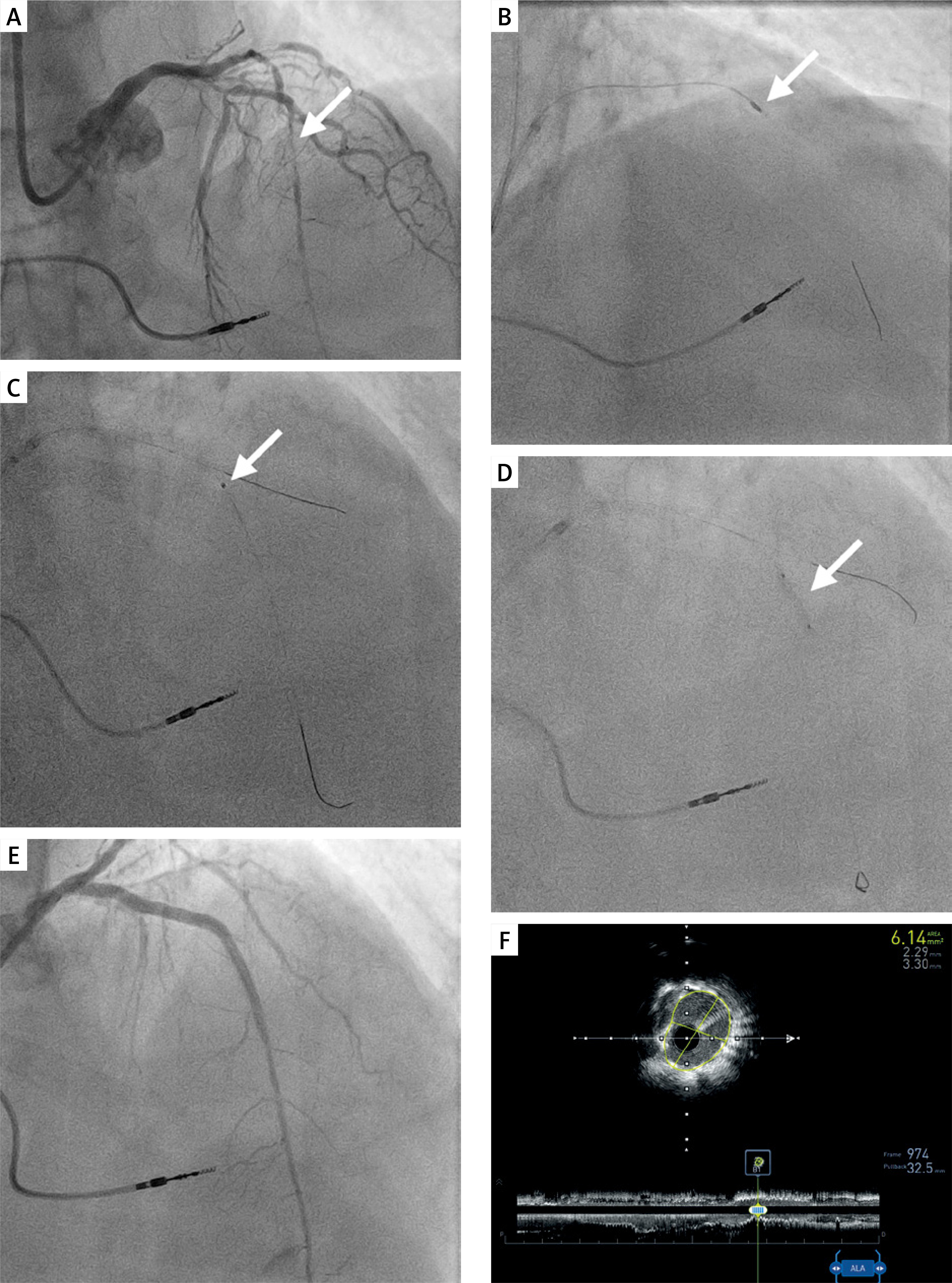
Given the availability of ELCA in our cardiac catheterization laboratory, considering its plaque-modifying properties, the patient was admitted for a final PCI attempt. Right radial access was used. Control angiography showed a CTO of the LAD. Wire crossing was effective, but the MC could not advance. ELCA was performed using a 0.9 mm catheter with seven passages of 80 mJ/mm2 fluence and 60 Hz frequency with a concomitant saline flush (Figure 1 C). The catheter could cross the lesion, allowing balloon predilation up to 20 atm (Figure 1 D). Balloon angioplasty was performed with subsequent 3.0 × 26 mm drug-eluting stent (DES) implantation in the 6th/7th segment, followed by 3.5 mm proximal optimization technique (POT) post-dilation at up to 20 atm. In the 6th segment, a 3.5 × 33 mm DES was implanted, overlapping with the previous stent, followed by post-dilation at up to 18 atm. Final angiography and intravascular ultrasound (IVUS) confirmed optimal results, with TIMI 3 flow and a minimal stent area (MSA) of 6.14 mm2 in the mid-LAD and 10 mm2 in the proximal LAD (Figures 1 E, F). The procedure was completed without complications, and the patient was discharged home in good condition. To conclude, ELCA is a valuable tool for the treatment of complex balloon-uncrossable CTO lesions, particularly after unsuccessful RA. Nevertheless, potentially serious complications, such as the no-reflow phenomenon, should be considered [4]. Further studies are warranted to evaluate its efficacy and safety compared with RA in the treatment of CTOs.