
Spontaneous coronary artery dissection (SCAD) is a non-atherosclerotic, non-traumatic and non-iatrogenic disease, characterized by intramural hematoma formation, which results in acute coronary syndrome, predominantly affecting young and middle-aged women [1]. Although SCAD is not an inherited disease, it occasionally occurs in patients with genetically based connective tissue diseases, like Ehlers-Danlos, Marfan and Loeys-Dietz syndromes as well as autosomal dominant polycystic kidney disease [1, 2].
Bicuspid aortic valve (BAV) is the most frequent congenital heart disease among adults [3]. The occurrence of BAV is genetically determined, exhibiting an autosomal dominant inheritance pattern with incomplete penetrance and variable expressivity [4]. Furthermore, BAV is known to co-occur in patients with genetic syndromes like Marfan syndrome [5].
A 56-year-old female patient was referred due to a non-ST elevation myocardial infarction (NSTEMI). She reported recurrent, short-lasting chest pain that had initiated 15 h prior to admission. The admission electrocardiogram (ECG) showed no significant acute ischemic changes, however cardiac biomarkers, including high-sensitivity troponin and CK-MB, were significantly elevated, confirming the diagnosis of NSTEMI.
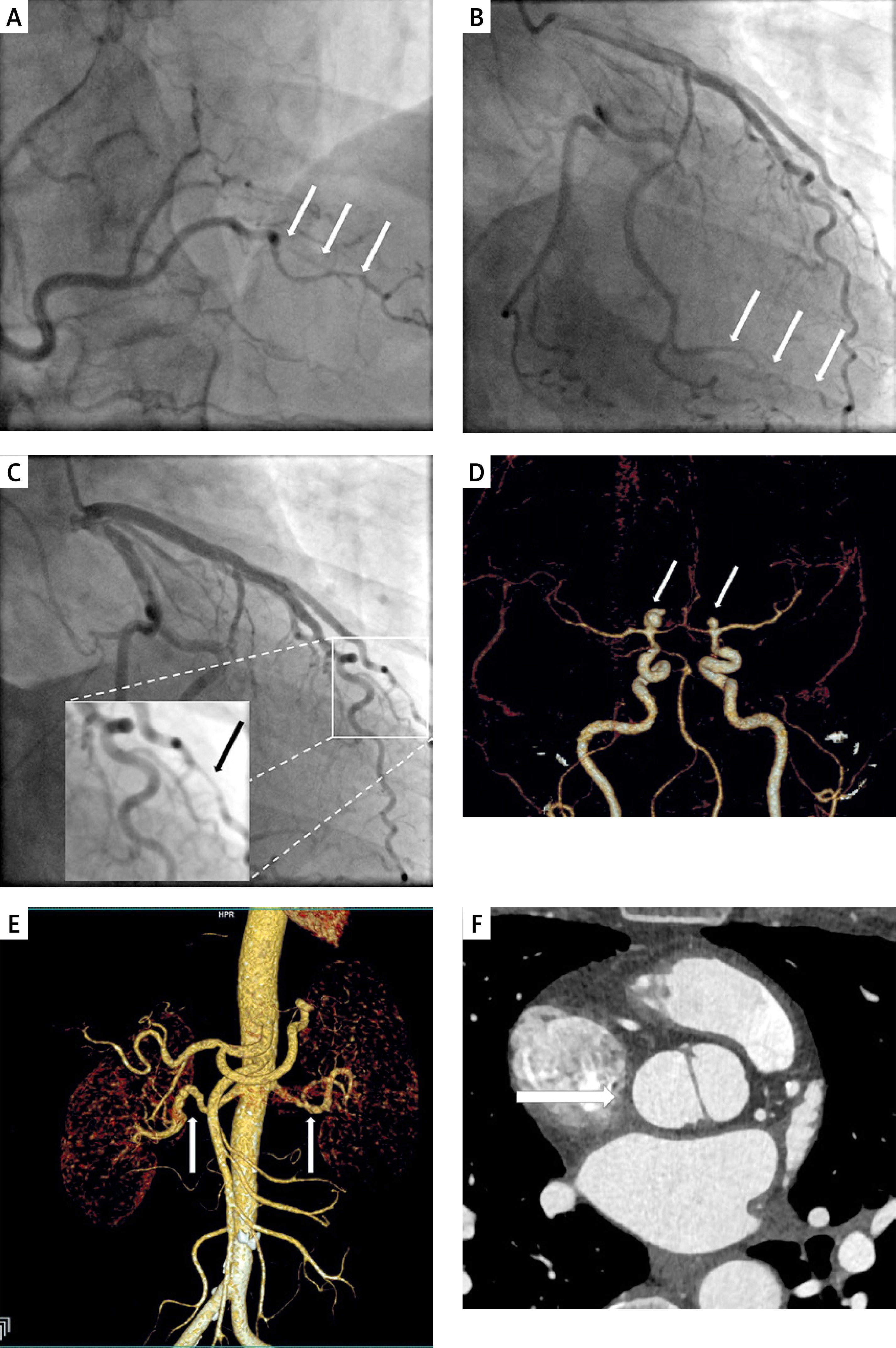
Urgent coronary angiography demonstrated multivessel SCAD involving the posterior descending branch of the right coronary artery, the marginal branch of the left circumflex artery and the second diagonal branch with TIMI 3 flow in all affected vessels (Figures 1 A–C). Given the patient’s stable clinical condition, distal location of dissections and preserved flow, conservative management was initiated including aspirin and bisoprolol.
Figure 1
SCAD in the posterior descending branch of the right coronary artery (A), the marginal branch of the left circumflex artery (B), and the second diagonal branch (C) on coronary angiography; high-risk intracranial aneurysms in both middle cerebral arteries (D); bilateral FMD in renal arteries (E); bicuspid aortic valve on CT angiography (F)
During hospitalization, transthoracic echocardiography revealed preserved systolic function of the left ventricle with ejection fraction of approximately 60%. The aortic valve was found to be bicuspid without evidence of stenosis or regurgitation, and the ascending aorta was not dilated. The patient remained clinically stable and was discharged home after 5 days.
In accordance with current expert consensus on SCAD, screening for extracoronary vascular abnormalities (EVA) was performed after 2 months [1]. Head-to-pelvis computed tomography angiography revealed two high-risk aneurysms: one right middle cerebral artery 9 × 5 × 7 mm polycyclic aneurysm with focal outpouchings and one 3.5 × 4.5 × 5 mm aneurysm located in the left middle cerebral artery (Figure 1 D). Additionally, mild multifocal fibromuscular dysplasia lesions were identified in both renal arteries and the right iliac artery, without hemodynamically significant stenoses (Figure 1 E). The presence of BAV was also confirmed on CT angiography (Figure 1 F). The intracranial aneurysms were subsequently treated successfully with endovascular two-stage procedure. The first stage consisted of right-sided, stent-assisted coiling using a Leo stent. During the second stage, coil embolization was performed on the left side. Dual antiplatelet therapy was recommended for 1 year since the first procedure. The 12-month follow-up was uneventful.
To date, only few case reports of SCAD in male individuals with BAV have been published [6, 7]. Our case is probably the first one presenting a female with BAV affected by SCAD and so many extracoronary vascular complications. These cases suggest that shared features of vascular fragility and aortopathy may contribute to the coexistence of both conditions, warranting comprehensive vascular assessment in such patients. In the presented female patient, despite the presence of multivessel coronary dissections, left ventricular impairment was remarkably mild. Crucially, the correct identification of SCAD prompted extensive vascular screening leading to detection of high-risk intracranial aneurysms. This enabled timely endovascular intervention and prevented potentially life-threatening cerebral complications.
In a young or middle-aged woman with BAV presenting with acute coronary syndrome, SCAD should always be considered in the differential diagnosis and screening for vascular abnormalities remains mandatory.










