
Patients with severe aortic stenosis (AS) and heavily calcified aortic valves, particularly those with bicuspid morphology, represent a challenging subgroup for transcatheter aortic valve implantation (TAVI) due to an increased risk of procedural complications such as paravalvular leak or post-TAVI conduction disturbances [1].
A 68-year-old male patient with multimorbidity, including hypertension, dyslipidemia, type 2 diabetes mellitus, morbid obesity, and heart failure with preserved ejection fraction, was admitted to the Cardiology Department with suspected acute coronary syndrome. Coronary angiography showed no significant coronary artery disease, and echocardiography revealed severe AS.
After Heart Team consultation, the patient was deemed unsuitable for surgical aortic valve replacement (SAVR) and referred for TAVI in a tertiary cardiology care unit due to third-degree obesity (BMI 39.2 kg/m2), persistent contractures of the upper and lower limbs, impaired mobility requiring two canes, and consequently a very high perioperative risk.
Pre-TAVI coronary computed tomography angiography (CCTA) demonstrated a massively calcified aortic valve, likely functionally bicuspid due to a calcified pseudo-raphe and slit-like opening (Figure 1 A), with no significant coronary stenoses (Figure 1 B). It also revealed severe tortuosity of the iliac arteries and the aorta (Figure 1 C).
Figure 1
A – Computed tomography angiography (CCTA) (arrow: heavily calcified, functionally bicuspid aortic valve (BAV)), B – CCTA of the coronary arteries before transcatheter aortic valve implantation (TAVI), C – CCTA of the iliac arteries and aorta before TAVI (top arrow: aortic tortuosity; bottom arrow: iliac arteries tortuosity), D – Final result after TAVI
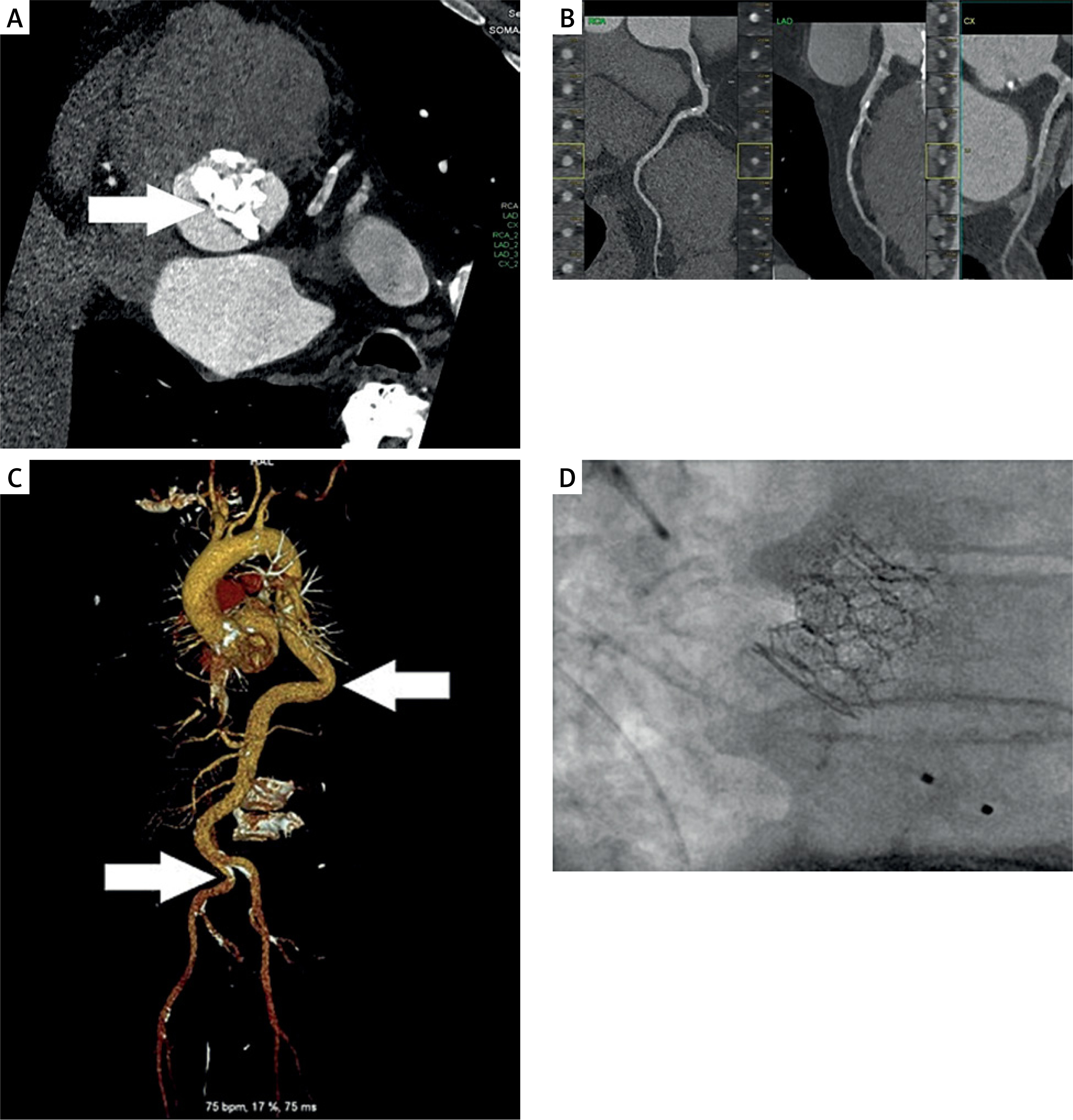
TAVI was performed via the right common femoral artery. After placement of ProStyle vascular closure devices (Abbott Vascular Inc., United States) and predilation with a Mammoth 25/40 mm balloon (Meril Life Sciences, India), a Myval Octacor XL 32 bioprosthetic valve (Meril Life Sciences, India) was implanted. An optimal procedural result was achieved (Figure 1 D). The vascular access sites were successfully closed using ProStyle and AngioSeal devices.
Further hospitalization was complicated by a transient third-degree atrioventricular block and pneumonia, requiring a temporary electrode for percutaneous stimulation. Ten days after the procedure, following a follow-up 24-hour Holter ECG showing only sinus bradycardia, the patient was successfully discharged home.
Beyond extensive valve calcification, severe obesity and metabolic comorbidities are key contributors to increased perioperative risk, precluding surgical treatment and highlighting TAVI as a less invasive alternative [2]. Careful preprocedural CCTA enables accurate valve sizing and access planning and, given its high negative predictive value for significant coronary artery disease, may also help identify AS as the primary cause of symptoms and stratify the risk of major adverse cardiovascular events [3, 4].
Despite postprocedural complications, the overall clinical course was favorable, underscoring the importance of a multidisciplinary Heart Team approach, a meticulous procedural strategy, and close post-TAVI monitoring in high-risk patients. Moreover, this case emphasizes the importance of detailed CCTA analysis prior to TAVI, especially in complex cases, as the combination of a heavily calcified raphe and the use of a large balloon-expandable valve represents a recognized substrate for annular injury and requires a cautious procedural strategy, as demonstrated in this case.










