A 73-year-old man with a history of inferior ST-elevation myocardial infarction treated with percutaneous coronary intervention (PCI) of the circumflex artery (drug-eluting stent [DES] and bare-metal stent [BMS] implantation in 2010), hypertension, and advanced chronic kidney disease (CKD stage G5A1, eGFR 14–15 ml/min/1.73 m²) with congenital absence of the left kidney was admitted due to progressive angina (CCS III), consistent with chronic coronary syndrome.
Laboratory testing demonstrated severe renal impairment (creatinine 4.5 mg/dl) and mildly elevated troponin levels without a dynamic pattern, interpreted in the context of advanced CKD rather than acute coronary syndrome. Electrocardiography showed sinus rhythm with subtle ST-T abnormalities in leads I, II, and V3–V6. Transthoracic echocardiography demonstrated preserved left ventricular ejection fraction (60%) with regional wall motion abnormalities.
Coronary angiography revealed diffuse atherosclerosis with a significant calcified stenosis involving the distal left main (LM) and proximal left anterior descending artery (LAD) (up to 80–90%), as well as additional non-critical lesions in other vessels (Figure 1 A). The patient was discussed by the Heart Team and deemed eligible for coronary artery bypass grafting (CABG); however, he refused surgical treatment. Given the advanced CKD and high risk of contrast-induced nephropathy, a decision was made to proceed with ultra-low contrast PCI under intravascular ultrasound (IVUS) guidance [1]. Ultra-low contrast PCI refers to a strategy that minimizes contrast use, typically targeting a contrast volume-to-eGFR ratio of around 1.
Figure 1
Ultra-low contrast IVUS-guided PCI of the left main (LM) and left anterior descending artery (LAD). A – Coronary angiography showing a calcified, severe stenosis involving the distal LM (arrow) and proximal LAD (arrowhead). B – Pre-intervention IVUS demonstrating severe concentric calcification involving the distal LM/proximal LAD segment with a calcium arc approaching 360°. The distal LM MLA measured 8.3 mm2 (reference diameter 4.9 mm), while the proximal LAD MLA measured 1.5 mm2 (reference diameter 3.6 mm), followed by post-stenotic ectatic enlargement to approximately 4.5 mm distally. C – Lesion preparation with intravascular lithotripsy (IVL) balloon in the LM/LAD segment (arrow). D – Final angiographic result showing optimal stent expansion and TIMI 3 flow
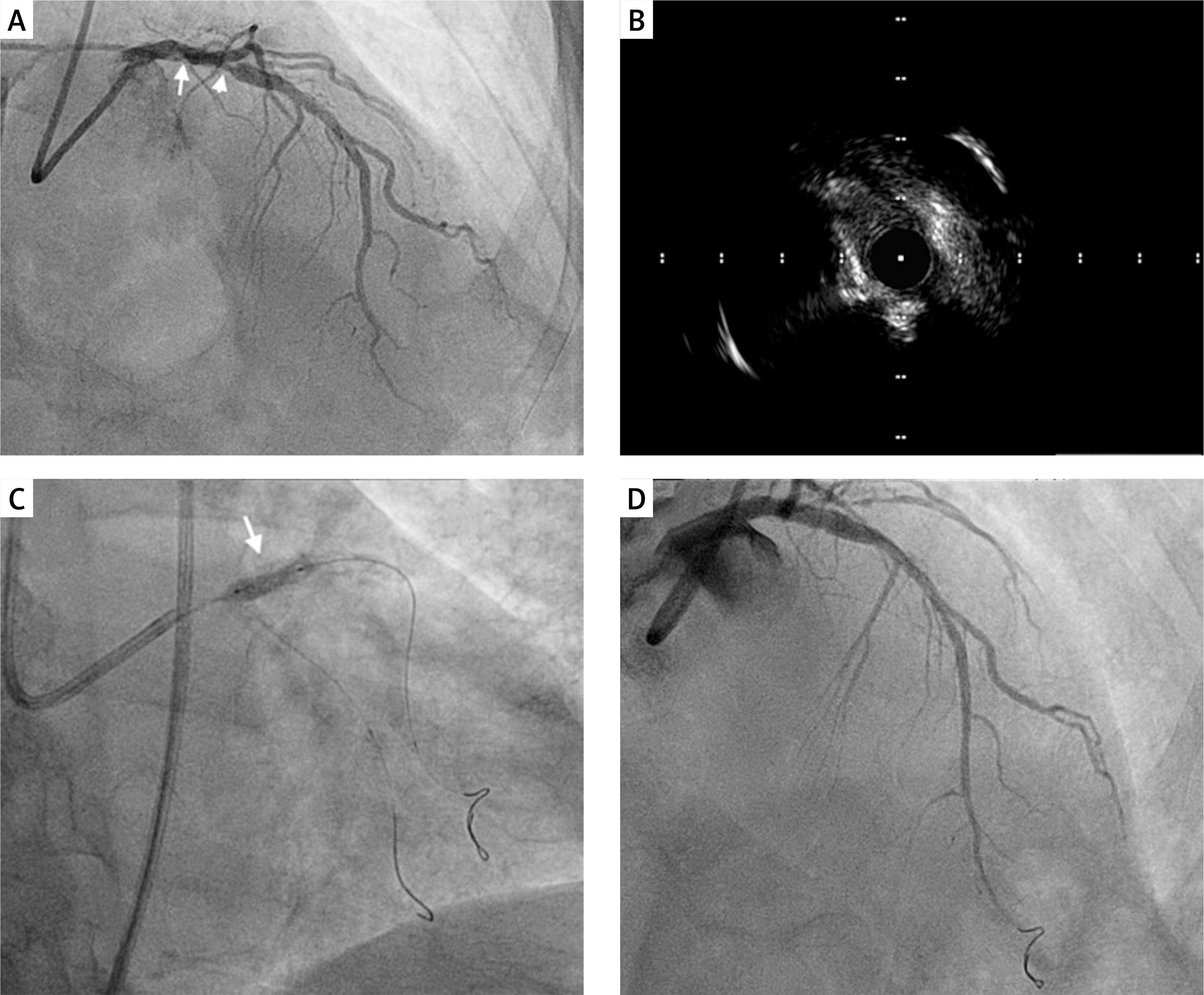
The procedure was performed via right femoral access (7 Fr, EBU 4.0 guiding catheter). This strategy was chosen primarily to preserve the radial arteries for potential future arteriovenous fistula creation in the setting of advanced CKD. A Pilot 50 guidewire was selected as the primary wire according to operator preference, providing adequate support and deliverability for IVUS-guided treatment of the severely calcified LM/LAD lesion. IVUS imaging was used for lesion assessment, vessel sizing, and optimization of stent implantation, thereby minimizing contrast use (Figure 1 B). Due to severe calcification, lesion preparation included intravascular lithotripsy (Shockwave 4.0 × 12 mm) followed by non-compliant (NC) balloon predilatation (Figure 1 C).
A drug-eluting stent (Promus ELITE 4.0 × 24 mm) was implanted from the LM into the LAD, fully covering the left main. Proximal optimization technique (POT) was performed with a 5.0 mm NC balloon. Due to distal edge dissection, an additional overlapping stent (Promus ELITE 4.0 × 8 mm) was implanted distally. Postdilatation was performed with a 4.5 mm NC balloon. IVUS confirmed optimal stent expansion and apposition without residual dissection. Final angiography showed TIMI 3 flow (Figure 1 D). The circumflex artery was managed conservatively without dedicated intervention, as IVUS pullback from the LAD into the LM demonstrated no evidence of circumflex ostial compromise. Importantly, baseline diagnostic coronary angiography had been performed using 15 ml of iodinated contrast. The PCI itself was performed using 20 ml of iodinated contrast, corresponding to a contrast volume-to-eGFR ratio of approximately 1.3. Renal function remained clinically stable after PCI (creatinine 4.26 → 4.52 mg/dl during hospitalization; eGFR 15 → 14 ml/min/1.73 m²), without requirement for renal replacement therapy. At 1-month follow-up, renal function improved (creatinine 3.49 mg/dl; eGFR 18 ml/min/1.73 m²). No renal replacement therapy was required during hospitalization or follow-up.
During hospitalization, hemoglobin decreased from 11.6 g/dl to 8.0 g/dl. Ultrasound excluded pseudoaneurysm and retroperitoneal bleeding, and gastrointestinal assessment did not demonstrate active bleeding. One unit of packed red blood cells was transfused, after which hemoglobin stabilized. Given the high ischemic risk after complex LM PCI, dual antiplatelet therapy was maintained with proton pump inhibitor protection and planned outpatient completion of the gastrointestinal work-up.
Several practical technical measures may facilitate ultra-low contrast PCI in advanced CKD. First, before starting PCI, the diagnostic angiogram should be reviewed frame-by-frame and displayed alongside live fluoroscopy, using identical projections during the intervention. In calcified lesions, fluoroscopic calcium often provides a useful “natural roadmap” for device positioning. Second, IVUS pullback should be performed early to define lesion length, reference vessel diameters, and proximal/distal landing zones before device selection. When selecting landing zones, operators should target “normal-to-normal” segments or, in diffuse disease, segments with plaque burden < 50% [2].
Third, stent positioning can be facilitated by using fixed anatomical landmarks rather than repeated angiography. In left main PCI, useful landmarks include the LM ostium, carina position, guide catheter tip, calcified plaque distribution, and side-branch take-off. Fourth, complex ultra-low contrast interventions benefit from a 7 Fr system, which improves guide support and allows simultaneous use of IVUS and adjunctive devices. In this case, severe calcification identified on IVUS prompted upfront calcium modification with intravascular lithotripsy before stent implantation [3].
Finally, contrast use should be reserved for uncertainty or safety checkpoints rather than being part of the routine workflow. If there is doubt regarding wire position, vessel injury, or the final procedural result, a very small contrast injection may be used selectively rather than repeatedly. Final optimization should rely primarily on IVUS confirmation of expansion, apposition, and exclusion of edge dissection [4].
This case demonstrates that ultra-low contrast PCI of the left main can be safely performed even in the presence of severe calcification, using an IVUS-guided strategy combined with intravascular lithotripsy. In such scenarios, intravascular imaging plays a central role in enabling precise lesion assessment and stent optimization while minimizing contrast exposure.