
Transcatheter aortic valve implantation (TAVI) has become a recognized method of treatment of aortic valve stenosis for high-risk patients. The list of potential complications covers acute or delayed prosthesis migration and embolization. The latter occurs in approximately 1% of patients [1]. The report presents a new and unusual technique for valve snaring during the procedure.
An 89-year-old woman was admitted to our department for the TAVI procedure. During the local Heart Team meeting, the patient was disqualified from a surgical approach to aortic valve replacement due to high perioperative risk. Based on computed tomography (CT) scan and measurements (perimeter 64.9 mm, mean size of annulus 20.5 mm), Medtronic Evolut Pro valve was chosen (26 mm). Surgical access was performed and balloon predilatation was made (balloon 20 mm in diameter). The valve was prepared and inserted in a typical way. Intentionally, the valve was implanted in a high position because of the small LVOT diameter (19 mm), but during the valve’s positioning, the operator changed the X-ray projection and missed the previous settings. The valve opened above the annulus, which was observed after carefully reviewing the X-ray loops later (Figure 1 A). Subsequently, echocardiographic assessment revealed a significant aortic insufficiency and perivalvular leakage. It resulted in sustained ventricular tachycardia, and cardioversion was needed to reverse the normal cardiac rhythm. The operator decided to re-dilate (balloon 24 mm) the migrated valve. Planning to implant a new valve, we tried to snare the first one but failed when using a regular coronary snare device. On fluoroscopy, the distance between the valve and the aortic wall was large, and therefore we decided to use another technique.
Figure 1
The figure shows 4 important steps of the procedure. A – the angiographic position of the first valve above the patient’s annulus. B – (white circle) a Judkin’s right guiding catheter and coronary guidewire crossing the outflow stent. C – an inflated coronary balloon pulling up the valve. D – the final position of both valves
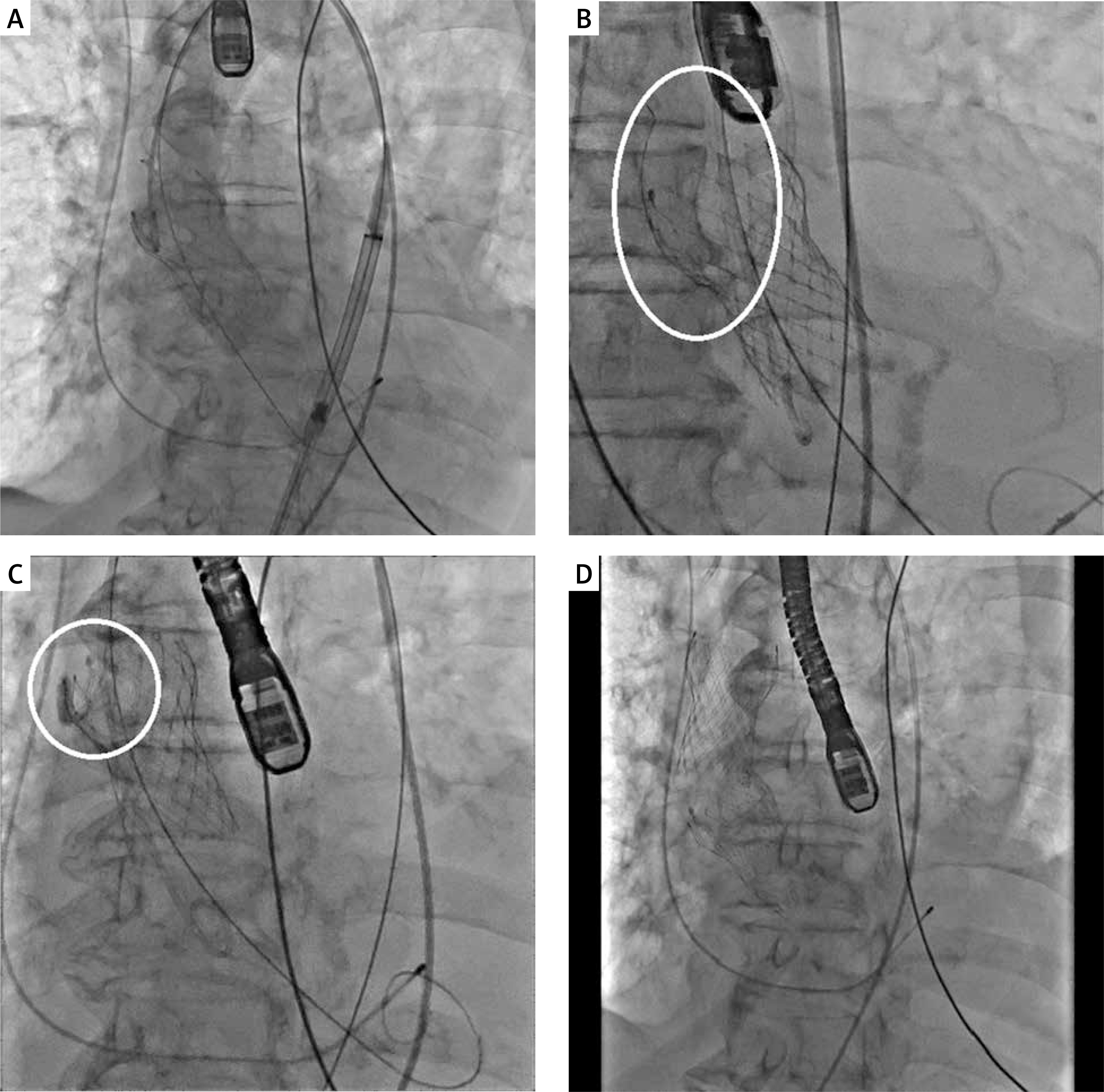
By right radial approach, the operator introduced a coronary guiding catheter (Judkins JR 4.0) and placed it into the valve stent cells (Figure 1 B). Then, the coronary guidewire was carried out of the cells, and the non-compliant 4.0 mm balloon was inflated (Figure 1 C). The valve was pulled using slow traction all the way to the brachiocephalic trunk and held there until the end of the procedure, i.e. the implantation of a new valve (Figure 1 D).
The periprocedural outcome and the further course of hospitalization were uncomplicated. The patient was discharged home without signs of aortic valve stenosis or insufficiency.
The described technique of valve snaring is a new idea that was developed ad hoc because of the difficulties of the usual procedure. It could be an emergency means of management of a similar issue. In this case, valve migration was caused by the wrong position of the valve caused by a change in the X-ray tube position.


