A 61-year-old man with a history of left-sided pneumonectomy due to lung cancer, chronic obstructive pulmonary disease, nicotine dependence, and alcohol use disorder (complicated by gastric varices) was admitted to a tertiary care center with symptoms of acute ischemic stroke. Symptom onset occurred approximately 2 h prior to admission and was preceded by a generalized seizure. Subsequently, the patient developed right-sided hemiparesis and dysphasia.
On admission, the patient was hemodynamically and respiratory stable. Neurological examination revealed mixed dysarthria, right-sided hemiparesis (Medical Research Council grade 2/5), sensory deficits, hemispatial neglect, and homonymous hemianopia. The National Institutes of Health Stroke Scale (NIHSS) score was 17. Initial computed tomography angiography (CTA) demonstrated occlusion of the P1 segment of the left posterior cerebral artery (PCA), critical stenosis of the proximal left vertebral artery (VA), and an anatomical variant in which the vertebral artery terminated in the posterior inferior cerebellar artery (PICA-VA) (Figures 1 A, B).
Figure 1
Baseline computed tomography angiography (CTA) showing an axial view of extracranial portions of carotid and vertebral arteries with stenosis of the left VA (white triangle) (A). 3D reconstruction revealed occlusion of the P1 segment of the left posterior cerebral (white arrow) artery and absence of the distal part of the right vertebral artery, confirming the anatomical variant (PICA-VA) (B), which was further demonstrated by angiography (white star) (C). Angiography performed via the left subclavian artery showed filling of the left vertebral artery via branches of the thyrocervical trunk (white arrow) and a visible stump of the left VA (white triangle) (D). After passing through the stenosis, PCA occlusion was confirmed (E – white arrow). Final digital subtraction angiography (DSA) showed complete recanalization (F)
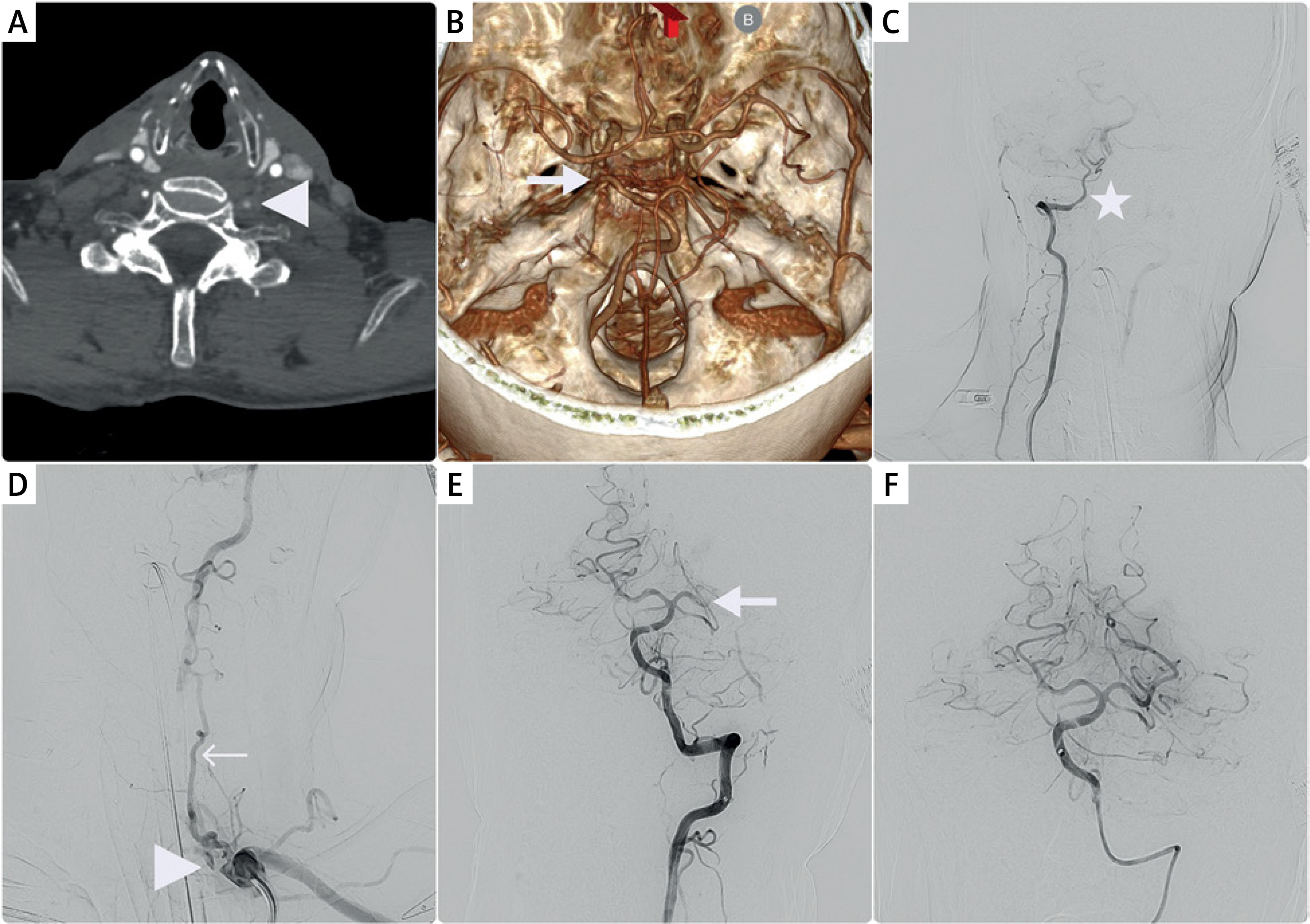
The patient was scheduled for endovascular treatment. The procedure was performed under general anesthesia via a femoral approach. Initial angiography confirmed the anatomical variant of the right vertebral artery (Figure 1 C), filling of the left vertebral artery via branches of the thyrocervical trunk, and a visible stump of the left VA (Figure 1 D). A 5Fr aspiration catheter was navigated through the stenotic segment of the left VA, and mechanical thrombectomy of the PCA was performed using a stent retriever. Complete recanalization (TICI 3) was achieved after a single pass. Given the patient’s history of gastric varices and the presence of well-developed collateral circulation, stenting of the vertebral artery was not performed.
In the following days, gradual neurological improvement was observed. Follow-up CT performed 24 h after the intervention did not show evidence of intracranial hemorrhage. At discharge, the patient was conscious, with mild dysarthria and slight right-sided weakness (MRC 4/5), along with persistent sensory deficits and neglect. The NIHSS score was 6, and the modified Rankin Scale (mRS) score was 3. The patient was transferred in good general condition to the Neurology Department for further diagnostic evaluation and rehabilitation. Follow-up Doppler ultrasonography of the extracranial arteries demonstrated good blood flow. At 3-month follow-up, further clinical improvement was noted, with an NIHSS score of 4 and an mRS score of 2.
Among anatomical variations of the vertebral arteries, termination of the vertebral artery in the posterior inferior cerebellar artery (PICA-VA) is estimated to occur in approximately 2% of the population [1]. Studies demonstrated that this variant is associated with an increased risk of vertebrobasilar stroke; however, the underlying mechanism remains a matter of debate [2]. It has been hypothesized that PICA-VA may reflect hypoplasia or underdevelopment of the vertebrobasilar system leading to unfavorable hemodynamic conditions in the posterior circulation and consequently a higher susceptibility to vessel occlusion [3]. Successful bailout access to the VA via collateral circulation during endovascular procedures was reported; however, no clear recommendations are available [4]. Our case highlights the feasibility and effectiveness of mechanical thrombectomy in posterior circulation stroke in the presence of PICA-VA and concomitant contralateral stenosis of the vertebral artery. Careful individualized decision-making allowed avoidance of stenting in a high bleeding-risk patient while achieving a favorable clinical outcome.