
Ostium secundum atrial septal defect (secundum ASD) is a congenital heart defect resulting from tissue deficiency in the fossa ovalis, allowing shunting between the systemic and pulmonary circulations. Large ASDs causing right ventricular volume overload are closed percutaneously when the anatomy is favorable, while small, hemodynamically insignificant defects are rarely treated, mainly for secondary prevention of cryptogenic stroke [1]. Although generally safe, the procedure may rarely lead to device migration, usually early and into the pulmonary circulation [2]. The presented case illustrates a rarely observed systemic migration occurring later than the periprocedural period.
A 44-year-old man was admitted with acute ischemic stroke and was successfully treated with intravenous thrombolysis, resulting in near-complete neurological recovery. Standard stroke work-up revealed no evidence of thrombophilia, atrial arrhythmia on 24-hour Holter monitoring, or carotid artery pathology on duplex ultrasound. Transesophageal echocardiography (TEE) demonstrated a 7 × 9 mm secundum ASD with a left-to-right shunt and no significant hemodynamic impact (Figures 1 A1-2). Although small, the defect was considered a likely cause of paradoxical embolization, and the patient was referred for percutaneous closure, which was performed 2 months later under fluoroscopic and TEE guidance. Due to the absence of an aortic rim (Figure 1 A3), defect sizing was based on pre-procedural TEE measurements and the TEE color Doppler stop-flow technique, as fluoroscopic balloon sizing risked oversizing, and a 12-mm Amplatzer ASD occluder was selected (Figure 1 A4-5).
Figure 1
A – First ASD closure procedure: 1) Three-dimensional TEE planimetry of the ASD (7 × 9 mm; white star); 2) left-to-right shunt through the ASD on color Doppler; 3) absence of the aortic rim on TEE (white arrow); 4) fluoroscopic balloon sizing showing unilateral minimal indentation of the defect (black arrow); 5) TEE color Doppler stop-flow sizing; 6) stability assessment with the tug test during fluoroscopy; 7) TEE superior–inferior view after occluder deployment demonstrating complete rim coverage (red arrows); 8) TEE anterior–posterior view confirming posterior rim coverage (red dashed arrow) without aortic overcompression by the occluder (red X). B – Percutaneous removal of embolized occluder: 1) TTE at 6 months post-procedure showing absence of the occluder in the heart; 2) fluoroscopy showing migration of the occluder into the abdominal cavity; 3) CT with 3D reconstruction demonstrating device displacement to the aortic bifurcation (navy circle); 4) fluoroscopy showing occluder captured with a vascular snare; 5) occluder after removal. C – Re-implantation of the new occluder: 1) TEE color Doppler stop-flow sizing; 2) occluder position on TEE and its effect on the aorta (Ao) before release; 3) fluoroscopy after device deployment; 4) TEE showing post-implantation relationship between the occluder and the aorta (Ao); 5) TEE with bubble test at 3 months confirming correct device position and absence of interatrial shunt; 6) TEE at 6 months showing a favorable result; 7) stable occluder position on 12-month 3D TEE follow-up; 8) color Doppler TEE at 12 months showing no aortic erosion
TEE – transesophageal echocardiography, TTE – transthoracic echocardiography, 3D – three-dimensional.
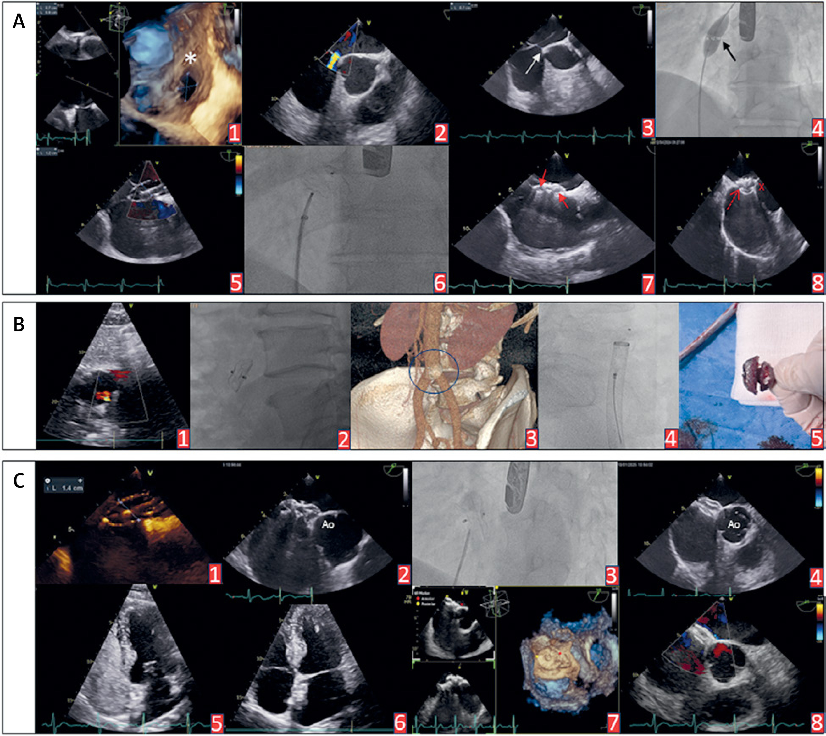
The device was deployed without difficulty, its stability was verified using the “tug test” (Figure 1 A6). TEE confirmed complete rim coverage and the absence of residual shunt on color Doppler (Figures 1 A7-8). The following day, transthoracic echocardiography (TTE) confirmed the correct device position. The patient was discharged on dual antiplatelet therapy with aspirin and clopidogrel.
At the scheduled 6-month follow-up visit, TTE unexpectedly showed absence of the device at the implantation site (Figure 1 B1). Fluoroscopy revealed the occluder within the abdominal cavity (Figure 1 B2), and computed tomography confirmed its migration to the abdominal aortic bifurcation (Figure 1 B3). Given the risks of distal embolization, limb ischemia, vessel injury, or hemorrhage, urgent percutaneous retrieval was undertaken. The right femoral artery was surgically exposed, and a 20F vascular sheath was inserted. Using a 15-mm Multi-Snare device (PFM Medical), the occluder was grasped under fluoroscopic guidance (Figure 1 B4). Partial endothelial overgrowth limited device mobility during capture, but the occluder was ultimately removed successfully via the femoral artery (Figure 1 B5).
Three months later, repeat percutaneous closure was performed using an occluder 2 mm larger than the TEE balloon-derived defect size (Figure 1 C1), namely a 16-mm Amplatzer ASD occluder, deployed after careful TEE assessment of its interaction with the aortic wall (Figure 1 C2-4), with durable success confirmed on serial TTE and 12-month TEE follow-up (Figure 1 C5-8).
Device migration remains a rare but significant complication of ASD closure. More than 80% of embolized devices dislocate into the pulmonary arteries, usually due to incorrect sizing, unfavorable morphology, or technical challenges during implantation. Migration into the systemic circulation is exceedingly unusual and is typically associated with immediate procedural complications during the closure of large, irregular defects [3].
This case demonstrates that even small defects, closed in accordance with operative standards and initially stable, do not preclude late device migration. Percutaneous closure is feasible in the absence of an aortic rim, but sizing is challenging. The TEE color Doppler stop-flow method is safer than fluoroscopic balloon sizing, avoiding oversizing and potential aortic erosion, and devices up to 2 mm larger than the balloon-derived diameter may be acceptable when other rims are compliant [4].
This experience prompted revision of our local surveillance protocol to include TTE at 1 week and at 1, 3, and 6 months, and TEE at 12 months, enabling early detection and percutaneous retrieval before endothelialization. Despite the overall safety of ASD closure, accurate defect sizing is the key procedural step, and systematic imaging follow-up is essential for early detection of device dislocation.










