
Introduction
Basaloid squamous cell carcinoma usually arises from various anatomic sites, including the laryngeal part of the throat, salivary glands, basics of the tongue, anus, esophagus, prostate, thymus, bladder and lungs [1]. It is characterized by an ulcerated, infiltrating growth pattern and poor clinical outcome. It rarely affects the female genital tract. Only six cases of squamous basaloid cervical cancer have been described yet.
Sarcomas are malignant tumors of tissues, derived from mesoderm, involve the transformation of the mesenchymal stem cell. Uterine sarcomas are rare tumors of the female genital tract. Their incidence is estimated at about 1-3% of all tumors in female reproductive organs, of which only 0.2% is undifferentiated sarcoma. They are characterized by poor prognosis, high rates of relapse and distant metastases. The most important risk factors for undifferentiated sarcoma is tamoxifen therapy, excessive stimulation with estrogen, pelvic irradiation and polycystic ovary syndrome (PCOS). Sarcomas may be manifested by abnormal uterine bleeding, pelvic pain, enlargement of the abdominal circumference or may be asymptomatic [2].
Case report
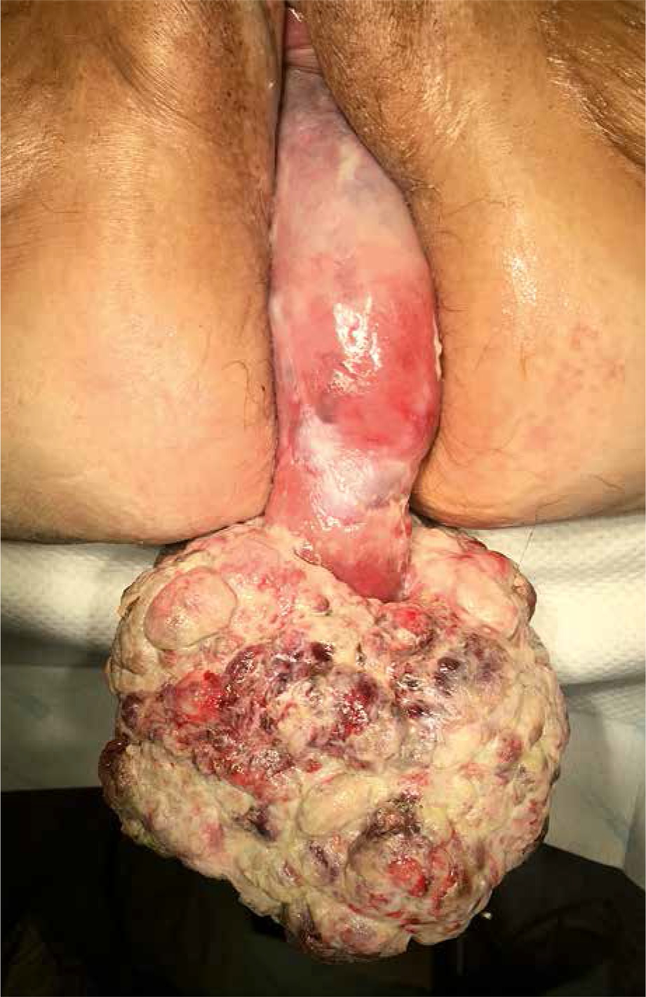
We present a rare and interesting case of a basaloid squamous cell carcinoma of the uterine cervix in a 71-year-old female with prolapse of the uterus with co-existing atypical undifferentiated sarcoma. The patient was admitted to the Department of Gynecology due to a tumor from the district hospital on 27.07.2018. For the last 4 years, the patient have had abnormal uterine bleedings and cervical cancer was suspected.
The patient had her first menstruation at the age of 10, next she had regular but painful menstruations, approximately every 30 days. In the interview she delivered naturally twice, and had last menstruation until about 55 years old. Woman referred urinary incontinence. Her medical history included diabetes mellitus (DM) since she was 14 years old, which had been controlled by metformin, hypertension, cholelithiasis, dementia, immobilized for six months, intestinal disease, lead toxicity (was exposed to high concentrations of lead). About 30 years ago, the patient underwent appendectomy, complicated by abnormal wound healing for a year.
She received spironolactone, pantoprazole, cycklonamine, folic acid, iron preparations, enoxyparin, metformin, metamizol, perfalgan, and fluid therapy. She did not report any allergies, smoking or alcohol; in addition, her sister died of breast cancer at age 57, father – cancer of unknown origin. The son fell ill with colon cancer and asthma.
Physical examination showed ulcerated uterine cervical tumor measuring 20 × 19 cm, weighing about 2 kg, excising the uterus outside (Fig. 1). Ultrasound examination showed no pathological changes in pelvis. The concentrations of tumor markers were normal (CA125 13.36 U/ml, CA 19.9-3.93 U/ml). The rest of the laboratory tests was normal except for findings of anemia (Hb 9.0 g/dl), and elevated inflammatory parameters (leukocytosis – 12, 7 × 10³/ul, elevated CRP concentration 109.2 mg/l) were found.
The cervical tumor was surgically removed under general anesthesia, the transvaginal hysterectomy was performed with the right salpingo-oophorectomy and the anterior and posterior vaginal plasty. Pathologic findings were consistent with an undifferentiated sarcoma and basaloid squamous cell carcinoma.
No further treatment was administered. Patient was discharged. Follow-up after 8 months of discharge, no clinical evidence of recurrence were detected. The performed pelvic CT scan did not reveal any pathological structures.
Discussion
Basaloid squamous cell carcinoma is uncommon tumor and has received little recognition, there are only a few cases in the medical literature. This cancer is included in the World Health Organization's classification of cervical neoplasms. The morphologic characteristics include nests of small basaloid cells, hyperchromatic cells and tendency to palisade at the periphery of the tumor islands. The stroma features desmoplasia.
Oh et al. indicate the possibility of developing basaloid squamous cell carcinoma after radio- and chemotherapy [3]. It is interesting to be able to influence the exposure to high concentrations of lead for the emergence of a basaloid and undifferentiated sarcoma. Lead interacts with selenium, abolishing its anti-carcinogenic effect. On chronic exposure it also accelerates tumor growth rates [4]. There is increased activity of superoxide dismutase in the serum exposed to lead due to adoptive mechanism against the increased amount of reactive oxygen species, which are involved in carcinogenesis [5].
Correct diagnosis is important due to the high malignancy of this cancer and poor prognosis. Immunocytochemistry may be helpful in identifying. Six cases of basaloid cervical cancer available in PubMed database were analyzed. The first was a case of adenoid cystic carcinoma occurring in a young woman in 1999, associated with invasive basaloid squamous cell carcinoma, and HPV-related lesions (condyloma acuminata, VIN, CIN) [6]. Valeri et al. described a 54-year-old patient with basaloid squamous cell carcinoma recurrent 15 years after hysterectomy with metastasis in the left iliac region [7]. Kwon et al. presented a 70-year-old woman with squamous cell carcinoma, in whom radical hysterectomy with negative twelve-month observation was performed [8].
Tsai et al. described the case of a 35-year-old female admitted to the Department due to profuse bleeding from the genital tract (Hb 3.6 g/dl), which was 12 months earlier the normal result of cytological examination, the patient was diagnosed with an ulcerative exophyte of 5 cm in diameter Ber-EPE4 immunohistochemistry, a patient treated with chemo-radiotherapy [1]. Majhi et al. described a 67-year-old woman with basaloid cancer with bleeding from the genital tract. with a cervical tumor of pron. 3.5 cm × 2.5 cm × 2.5 cm. Hysterectomy with adnexa was performed, followed by radiotherapy [9]. Giron et al. described the case of a 54-year-old woman with basaloid cancer, with abnormal bleeding from the genital tract lasting four months. The change included the front lip of the cervix at the external estuary, it was ulcerative and necrotic, infiltrating tissues up to 3.5 mm [10].
Conclusions
Histopathological diagnosis of basaloid squamous cell carcinoma of the uterine cervix is not difficult, but due to the rare occurrence and lack of good knowledge of its biology it is difficult to define the prognosis and optimal treatment. It is important to accumulate data of these rare tumors to determine whether their behavior differs from that of conventional cervical carcinomas.


