A 79-year-old woman was referred to our department due to severe symptomatic aortic valve stenosis requiring cardiac surgery. Her medical background consisted of chronic obstructive pulmonary disease, obesity (body mass index (BMI): 43.4 kg/m2), hypertension, dyslipidemia, and poor mobilization due to orthopedic problems. She received neither anticoagulant nor corticosteroid therapy. Cardiac ultrasound findings were as follows: severe aortic valve stenosis with aortic valve area (AVA): 0.7 cm2, peak gradient 104 mm Hg and maximum velocity 4 m/s with mild mitral valve regurgitation, mild tricuspid valve regurgitation and preserved ejection fraction of the left ventricle (ejection fraction (EF): 60%). The measurements of the ascending aorta and aortic root were found to be within normal parameters, with no evidence of aortic dilatation, aneurysm or dissection identified. There was no documented history of connective tissue disorders in this patient. No pathological findings were detected from coronary catheterization.
Transcatheter aortic valve replacement was not feasible due to an extended waiting period exceeding 1 year; therefore, conventional open surgery was selected as the preferred approach. The conventional full sternotomy approach was chosen as a partial sternotomy or right thoracotomy approach was not deemed eligible for this case due to the body characteristics and the need for short duration surgery.
A rapid deployment bioprosthesis (Edwards Intuity aortic valve) was chosen to reduce cross-clamp, cardiopulmonary bypass (CPB), and procedure times, aiming to improve short-term outcomes for this elderly high-risk patient. After CPB initiation and aortotomy, the native tricuspid aortic valve was excised, and excessive atheromatous lesions were carefully removed from the annulus. It should be noted that aortic wall thickness was within normal limits.
Valve sizing was conducted with great precision to prevent oversizing of the prosthetic device. A 21 mm rapid deployment balloon-expandable surgical bioprosthesis was placed in the aortic annulus, and the aortotomy was closed.
The patient was successfully weaned from cardiopulmonary bypass, with an ischemia time of 47 minutes and a total CPB duration of 62 minutes. The cannulas were subsequently removed without complications. Before sternal closure, a significant hemorrhage was observed from the aortic annulus as the systolic arterial pressure was elevated to 160 mm Hg.
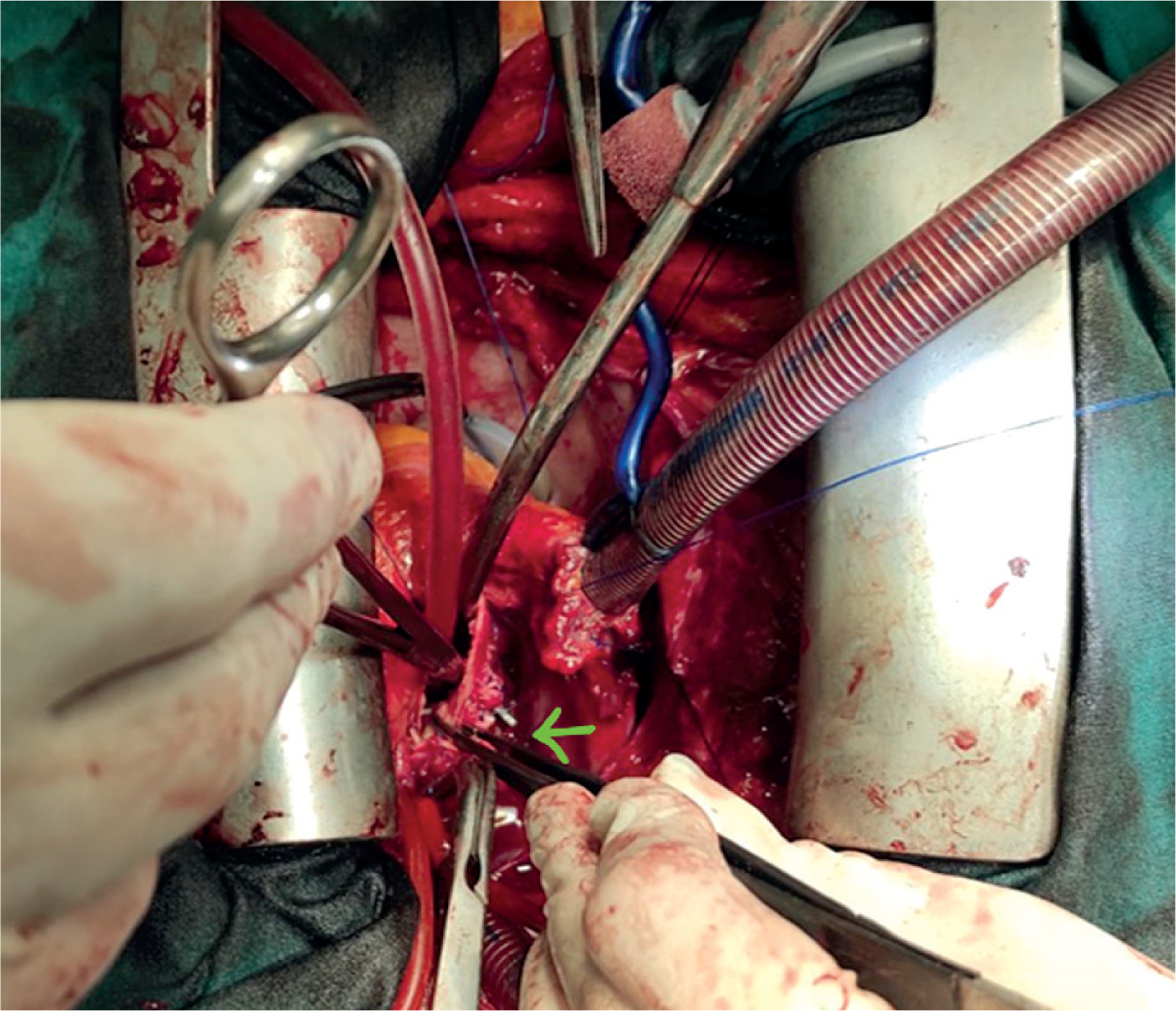
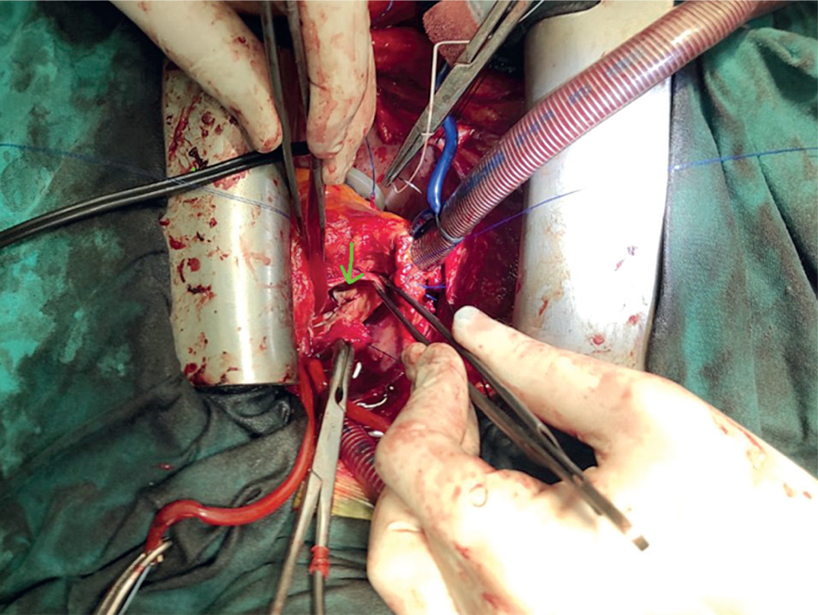
After an initial unsuccessful effort of direct suturing, CPB was initiated urgently as the aortic rupture increased over time and the patient became hemodynamically unstable. After the aortic cross clamp was placed, the bioprosthesis was removed and the rupture point was detected at the level of the posterior aortic annulus (Figures 1 and 2).
Figure 1
Intraoperative image. The aortic annular rupture is at the tip of the forceps and is indicated with a green arrow
Subsequently, a stented surgical bioprosthesis of size 21 mm was placed in order to reinforce the aortic annulus at the point of the rupture while isolated 4-0 prolene pledgeted sutures were placed internally. The operation was completed without further bleeding or other complications (ischemia time 82 minutes and total CPB 104 minutes).
The patient was extubated on the same day. She remained in the intensive care unit (ICU) for 36 hours and was discharged on postoperative day eight. In the follow-up examination 18 months after surgery, the bioprosthesis functioned properly and no pathological findings were detected in the aortic root or aortic annulus.
In this article, an extremely rare and life-threatening intraoperative complication, rupture of the aortic annulus, is presented. It is reported that after transcatheter aortic valve replacement the incidence of aortic annular rupture is approximately 0.4–2.3% and the 30-day mortality rate of this complication is 49–67% [1].
Several risk factors for annular rupture during transcatheter aortic valve implantation have been reported, including female sex, balloon valvuloplasty prior to valve implantation, use of balloon-expandable aortic valves, oversized bioprosthesis, heavily calcified aortic annulus and aortic leaflets, and technical problems [2, 3].
Rapid deployment and sutureless surgical aortic valves use transcatheter technology to reduce cross-clamp and bypass times. These valves are ideal for minimally invasive procedures and are often chosen for elderly or high-risk patients. The SURD-IR study (Aortic Valve Replacement International Registry) indicates that they are a safe alternative to conventional valves, with low complication rates and satisfactory mid-term results [4].
In studies of rapid deployment and sutureless aortic valves, intraoperative aortic annular rupture has not been reported as a complication [4–6]. Only 1 case report documents a delayed rupture following sutureless bioprosthetic valve replacement [7].
The association between rapid deployment aortic valves and aortic rupture is a nuanced topic that involves several factors including prosthetic valve design, surgical technique and experience, preexisting aortic pathology and the presence of comorbidities. Elderly, female and frail patients with severe aortic valve calcification or connective tissue disorders are at higher risk for aortic complications. Extremely thin-walled aorta, aortic dilatation, aneurysm, and dissection are factors that constitute contraindications for the implantation of sutureless and rapid deployment aortic valve bioprostheses. Special surgical training is required for the safe implantation of these valves. During the procedure, aggressive manipulations of the aortic root and annulus should be avoided.
Preoperative imaging and planning involve detailed assessment of the aortic valve, aortic annulus, aortic root, and proximal aorta using computed tomography or echocardiography.
Preoperative assessment of the bioprosthesis size and accurate intraoperative sizing are crucial in order to avoid aortic annular rupture or paravalvular leakage. Valve oversizing must be avoided because it exacerbates radial stress on the aortic wall and may lead to aortic rupture.
After reviewing the relevant parameters of the present case, we hypothesize that a possible factor leading to aortic annular rupture was the severe aortic valve calcification and the extensive removal of the atheromatic burden that led to thickening of the aortic annulus. A heavily calcified aortic wall and aortic annulus usually lacks the elasticity of normal aortic tissue, making it more vulnerable to aortic rupture. Conversely, retaining a substantial amount of calcified native aortic tissue can greatly decrease the size of the implanted prosthesis and raise the likelihood of paravalvular leakage.
In conclusion, while rapid deployment and sutureless aortic valves offer significant advantages in terms of hemodynamic performance and procedure time, aortic rupture remains a rare and potentially fatal complication. The risk appears multifactorial, emphasizing the need for meticulous patient selection, precise sizing and technical proficiency to avoid serious complications [8].
We hypothesize that extensive removal of atheromatosis from the aortic annulus caused its thinning and subsequent rupture.
Accurate reporting and analysis of these events are important for improving techniques and materials. There is a need for multicenter registries, standardized training for rapid deployment valves, and imaging-based risk stratification. Additional long-term studies are necessary to further clarify this association.