Introduction
Polycystic ovary syndrome (PCOS) is one of the most prevalent reproductive and endocrinological disorders affecting women of reproductive age worldwide [1]. The three primary clinical signs and symptoms of PCOS include elevated androgen levels, menstrual irregularities, and polycystic ovarian morphology [2]. The global prevalence rate of PCOS in women of reproductive age is between 6% and 13% as per the World Health Organization reports (2025); while in India it ranges between 7.2–19.6% [3]. The wide differences in the prevalence rates may be attributed to the different criteria followed for diagnosis, ethnicity, and the populations surveyed. Polycystic ovary syndrome is reported to be the primary cause of anovulatory infertility, and the patients have a heightened risk of developing endometrial cancer [4]. Polycystic ovary syndrome patients also have a significant risk of neonatal complications such as pre-eclampsia, early pregnancy loss, premature birth, gestational diabetes, etc. [5]. In addition, associated cardio-metabolic conditions such as hypertension, hyperlipidaemia, insulin resistance, type 2 diabetes mellitus, and cardiovascular disease have also been documented [6]. The aetiopathophysiology of PCOS is complex and multifaceted, with genetic, epigenetic, lifestyle, nutrition, ethnicity, and environmental factors being cited as contributory factors. However, the quantum of each factor contributing to the overall disease pathophysiology is not clear.
There are at least three major classifications for the diagnosis of PCOS, namely, the NICHD criteria (1990) [7], the Rotterdam criteria [8] and the Androgen Excess and PCOS Society [9] criteria. Polycystic ovary syndrome has been reported to follow different modes of inheritance, which include autosomal dominant mode with incomplete penetrance, X-linked and co-dominant inheritance with complete penetrance [10]. Genetic studies such as genome-wide association studies, linkage studies, twin studies, studies on first-degree relatives and male relatives of PCOS patients have reported more than a dozen candidate genes/loci for PCOS across different ethnicities by independent research groups [10]. These candidate genes have been broadly grouped into six different pathways that comprise of the gonadotropin action and regulation pathway [luteinizing hormone/choriogonadotropin receptor gene (LHCGR), anti-Mullerian hormone gene (AMH), follicle stimulating hormone receptor gene (FSHR), AMH type II receptor (AMHR2) gene], ovarian and adrenal steroidogenesis pathway [cytochrome P450 family 11 subfamily A member 1 gene (CYP11A1), cytochrome P450 family 17 subfamily A member 1 gene (CYP17A1), cytochrome P450 family 19 subfamily A member 1 gene (CYP19A1), etc.], insulin action and secretion pathway [insulin gene (INS), insulin receptor gene (INSR), calpain 10 gene (CAPN10)], steroid hormone effects pathway [androgen receptor gene (AR), sex hormone binding globulin gene (SHBG)], energy homeostasis pathway (leptin and receptor genes, adiponectin, C1Q and collagen domain containing gene (ADIPOQ), and peroxisome proliferator-activated receptor γ gene (PPARG) and chronic inflammation pathway tumor necrosis factor α (TNF-α) gene [11].
The AMH gene is reported to be an important candidate gene in the pathophysiology of PCOS [12]. The AMH gene (ENSG00000104899) is located on chromosome 19p13.3, and has five exons with an approximate size of 2.8 kbp. The AMH gene codes for three transcripts, out of which only one transcript (ENST00000221496.5) is functional, and encodes for the anti-Mullerian hormone (AMH) protein (https://asia.ensembl.org/index.html). The AMH is a homo-dimeric protein composed of 560 amino acids, with an approximate molecular weight of 140 kDa. The AMH is also known as Mullerian-inhibiting factor, and is responsible for sexual differentiation during foetal development [13].
Role of AMH in polycystic ovary syndrome pathogenesis
The primary pathophysiological condition for the initiation of PCOS is follicular arrest, which is caused by the AMH interfering with follicular growth and recruitment. Elevated AMH levels reduces the sensitivity of ovarian follicles to follicle stimulating hormone, a crucial hormone that regulates follicular growth and maturity. This prevents a single follicle from becoming dominant and eventually ovulating, resulting in the typical polycystic appearance of the ovaries and ovulatory dysfunction [14]. Studies by independent researchers have reported a two- to three-fold increase in serum AMH levels in PCOS patients, when compared to healthy women of reproductive age [15]. An increase in AMH levels leads to an elevated Luteinizing hormone secretion and neuroendocrine dysregulation thereby contributing to impaired ovulation and follicular development. The AMH suppresses the production of the enzyme aromatase in granulosa cells through a pathway involving the transforming growth factor β (TGF-β) and small mothers against decapentaplegic D proteins. When aromatase is suppressed, less estrogen is produced and there is an increase in the androgen levels [16]. Few studies have also reported that AMH plays a role in metabolic co-morbidities associated with PCOS such as lipid metabolism, inflammatory signalling, and insulin sensitivity [17].
While there are multiple reports of pathogenic mutations in the AMH gene from different ethnicities, there are no reports from Assam, India. Therefore, in the current study, we aimed to screen for the novel, as well as the reported sequence variations in the coding and the flanking regions of AMH in 100 PCOS patients from Assam. This would give an insight into the genetic predisposition of the AMH in this cohort of patients.
Material and methods
Recruitment of study participants
The current study is a prospective, case-control, tri-centric, hospital-based study. After obtaining signed informed consent, 100 PCOS patients with a confirmed diagnosis of PCOS (sporadic/familial) presenting to the outpatient departments of the Gogoi Nursing Home, Tezpur, Dorika Hospital, Tezpur, Assam and Matrikas IVF Centre, Guwahati, Assam were enrolled into the study. The diagnosis was based on a thorough evaluation by senior gynaecologists following the Rotterdam criteria. Women in the age group of 18–45 years, native to Assam, and with a clear diagnosis of PCOS were included in the study. All the relevant clinical signs and symptoms were recorded and tabulated. Patients with other hormonal disorders such as thyroid disorders, hyperprolactinemia, adrenal hyperplasia, and Cushing’s syndrome, pregnant women, and unwilling participants were excluded from the study. Similarly, 50 willing participants with no history of PCOS, in the same reproductive age group, with other gynaecological complications, presenting to the collaborating institutes were recruited as controls.
Blood sample collection, genomic DNA extraction, and gene amplification
2–5 ml of blood sample was collected from the study participants through a simple venipuncture, in an EDTA-coated vacutainer, by the trained staff of our collaborating institutes. Using the standard phenol-chloroform extraction method with minor modifications or using commercially available kits, genomic DNA was isolated from the frozen blood samples. The isolated genomic DNA was then subjected to quantitative and qualitative assessments, using a NanoDrop spectrophotometer (Nanodrop, 2000 Spectrophotometer, Thermo Scientific, India). The absorbance was measured at 260 nm and 280 nm; followed by 0.8% agarose gel electrophoresis using ethidium bromide staining and visualisation; using the UV-Visible gel documentation system (Bio-Rad: Chemidoc XRS+, USA.)
The AMH gene sequence, cDNA, and the protein sequences were retrieved from the Ensembl database (https://asia.ensembl.org/index.html) in .fasta and .rtf formats with exon-intron mark up. Primer3 online tool (http://frodo.wi.mit.edu/primer3/) was used for designing the primers, for the amplification of the coding and the flanking regions, by supplying the AMH in .fasta sequences, and altering the default parameters. Few important parameters that were set included: GC% of the primers (30–50%), the amplicons size (500–700 bp), the difference between the Tm of the forward and reverse primer (≤ 2°C), etc. From the list of the output generated, the optimal pairs of the primers were selected (Table 1); and the desalted lyophilised oligos, at a concentration of 25 nM, were procured commercially from GCC Biotech (I) Pvt. Ltd, Kolkata, India. The primers were reconstituted with sterile deionised water to 100 mM concentration (stock), and further diluted to a 2.5 pm/µl (working stock). The primer pairs were standardised for optimal polymerase chain reaction (PCR) amplification by varying the different PCR conditions of Mg+2 ion concentration (1–2 mM), primer concentration (7.5–10 pM), and annealing temperature (54–60°C for 30–45 s), using a 96-well gradient thermocycler (Applied Biosystems™: Veriti™ 96-Well Thermal Cycler), for a 40 µl reaction volume. The final reaction mixture comprised of dNTPs (0.2 mM), PCR buffer (1X), forward and reverse primers (5–10 pm), MgCl2 (1–2 mM), Taq DNA Polymerase (1 Unit) (Genei Laboratories Pvt. Ltd., Bengaluru, India), and template DNA (50–100 ng). The cycling conditions for the PCR included initial denaturation at 94°C for 5 min, and 35 cycles of denaturation at 94°C for 30 s, annealing between 54–60°C for 30 s to 45 s, extension at 72°C, for 60 s. The final extension was performed at 72°C for 8 min followed by termination at 4°C for 5 min.
Table 1
List of primers designed for screening the AMH gene with their names, sequences, size along with their polymerase chain reaction optimization conditions and product size
The polymerase chain reaction amplicons were then subjected to 1.5% agarose gel electrophoresis with ethidium bromide and subsequently visualized using UV-visible gel documentation system for the desired size and amplicon intensity. The optimised PCR conditions for each pair of primers were then applied, followed by the PCR reaction using the DNA from the patients. Further, PCR amplicons were purified using a QIAquick PCR Purification Kit (Qiagen India Pvt. Ltd., India), following the manufacturer’s protocol, and resuspended in 50 µl of deionized sterile water. The purified PCR amplicons were commercially outsourced to Biokart India Pvt. Ltd., Bengaluru, India for unidirectional automated Sanger’s sequencing, using either forward or reverse primers. The electropherograms so obtained were analysed with Chromas 2.6.6 (Technelysium Pty Ltd., Australia) and aligned with the wild-type gene sequences that were retrieved earlier from the Ensembl database, using the pairwise sequence alignment tool (https://www.ebi.ac.uk/jdispatcher/psa). The novel sequence variants were re-confirmed by PCR amplification and resequencing. Similarly, control screening was done in 50 unrelated ethnically matched control samples by unidirectional automated Sanger’s sequencing.
In silico analysis
Clustal Omega (https://www.ebi.ac.uk/jdispatcher/msa/clustalo) tools were used to perform the multiple sequence alignment (MSA) for predicting the conservation of the AMH protein sequence across different species. The sequence variant was analysed for its possible impact on the structure and function of the protein, using online prediction tools namely-SIFT (http://sift.jcvi.org/), PolyPhen2 (http://genetics.bwh.harvard.edu/pph2/), Align-GVGD (http://agvgd.hci.utah.edu/agvgd_input.php), MutationTaster (https://www.mutationtaster.org/) and I-Mutant2.0 (https://folding.biofold.org/i-mutant/imutant2.0.html).
A SIFT score between to 0 and 0.05 is considered to be ‘affecting protein function’ and deleterious, and a score greater than or equal to 0.05 is considered to be ‘tolerated’ [18]. PolyPhen-2 scores categorizes the sequence variation into ‘benign, possibly damaging and probably damaging’, with a score ranging from 0.0 (benign) to 1.0 (damaging) [19]. Align-GVGD scores range from ‘Classes C0’, indicating the substitution is least likely to interfere with function, to Class C65, indicating it is most likely to be pathogenic [20]. MutationTaster result is a qualitative prediction, classifying the variant as disease causing (pathogenic) or polymorphism (benign) [21]. I-Mutant2.0 predicts whether the mutation is predicted to significantly decrease or increase the protein’s stability, which can either be negative (destabilizing mutation) or positive (stabilizing mutation) [22].
Results
A total of 100 PCOS patients and 50 controls were recruited for the study. The mean and median age of the PCOS patients at the time of presentation were calculated to be 27 years. We had observed that our cohort of PCOS patients comprised of phenotype A, C and D with a frequency of 14%, 7% and 79% respectively, and there were no patients with phenotype B. A positive family history was observed in only 7 cases, and the rest were sporadic in nature. At the time of enrolment, 77 participants were married, and 23 participants were unmarried.
We identified a novel sequence variation c.698T>A in a homozygous condition (Figure 1), in exon 4 of the AMH gene, resulting in a p.Leu233Gln amino acid substitution in a 31-year-old, married patient. The patient had a history of a spontaneous abortion, and no positive family history of PCOS. The patient had a body mass index of 28.7, and a waist-to-hip ratio of 0.91, at the time of presentation. The patient presented with oligomenorrhea (menstrual cycles > 35 days), with mild primary dysmenorrhea, light menstrual flow, and occasional bloating. Transvaginal ultrasonography was performed using Samsung Ultrasound System HS50 at 5–12 Hz frequency (Figure 2).
Figure 1
A representative electropherogram depicting the pathogenic sequence variation observed, c.698T>A, in homozygous condition
This specific alteration at the DNA level results in a corresponding change in the AMH protein, where the amino acid leucine (L) is replaced by glutamine (Q) at position 233.
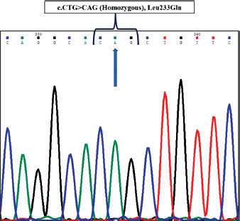
Figure 2
The 2D transvaginal ultrasound image depicting a cross-section of the ovaries of the patient, demonstrating features consistent with polycystic ovarian morphology
The key findings include ovarian enlargement (volume > 10 ml) and numerous small dark, circular antral follicles. A, B) represent the images of right and left ovary respectively. D1, D2, and D3 represent the length, height, and width of the ovary in millimetre (mm).
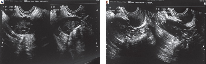
The findings revealed the presence of bilateral polycystic ovarian morphology, with multiple follicles arranged peripherally in both the ovaries. Both the ovaries had an increased volume of > 8 cc (right ovary: 15 cc with approximately 24 follicles; left ovary: 8.1 cc with 21 follicles); with 3–5 mm follicle size at the time of presentation. The biochemical parameters of the random blood glucose level, prolactin levels, and thyroid hormone levels were all found to be within normal limits. The serum AMH level, assessed using the Biomerieux Mini-Vidas ®Anti-Mullerian Hormone assay, was found to be elevated at 5.27 ng/ml. The patient also presented with acanthosis nigricans. Based on all the observed clinical signs and symptoms the patient was classified under phenotype D as per the Rotterdam criteria.
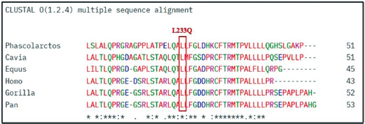
This novel sequence variation was not found in fifty control samples. Multiple sequence alignment confirmed that the p.Leu233Gln residue is highly conserved across species (Figure 3). SIFT analysis predicted the sequence variation to be ‘deleterious’ with a score of 0. PolyPhen2 predicted the variant to be ‘probably damaging’, with a confidence score of 0.992 (specificity of 0.97 and sensitivity of 0.7). Align-GVGD classified the variant as ‘class C65’, indicating a greater likelihood of damaging to protein function. MutationTaster predicted the variant to be ‘probably deleterious’, with a functional impact score of 113, suggesting possible disruption of important protein features. I-Mutant2.0 analysis predicted a decrease in protein stability, suggesting a potential destabilizing effect on the protein structure, at pH 7.0 and 25°C. Furthermore, this variant was absent in both ExAC and 1000 Genome databases, indicating it to be rare or novel substitution.
Figure 3
Multiple sequence alignment of the AMH protein depicting the conservation of the Leucine residue at position 233 (indicated in the box) across species
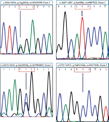
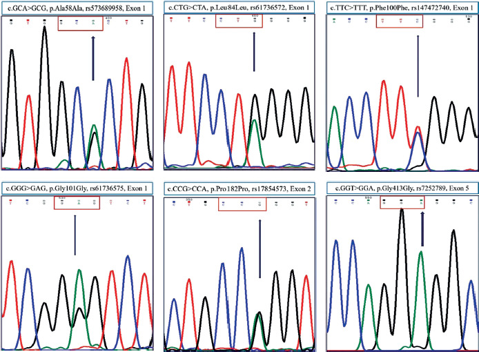
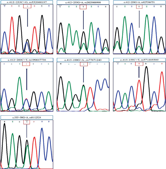
Besides these, a total of ten exonic sequence variations, and seven intronic sequence variations were also observed. Of the 10 exonic sequence variations, the four non-synonymous variants included: c.76A>G, p.Arg26Gly (rs142456399), c.146G>T, p.Ser49Ile (rs10407022), c.1154C>G, p.Ala385Gly (rs1447004865), c.1544T>C, p.Val415Ala (rs10417628) (Figure 4) and the six synonymous variants included: c.174A>G, p.Ala58Ala (rs573689958), c.252G>A, p.Leu84Leu (rs61736572), c.300C>T, p.Phe100Phe (rs147472740), c.303G>A, p.Gly101Gly (rs61736575), c.546G>A, p.Pro182Pro (rs17854573), c.1239T>A, p.Gly413Gly (rs7252789) (Figure 5). The seven intronic variants included: c.412+233C>G (rs532060197), c.412+255G>A (rs2025000998), c.412+258G>A (rs927386751), c.412+260C>T (rs190647736), c.413-100G>A (rs77671243), c.413-155C>T (rs571605589), c.555+50G>A (rs8112524) (Figure 6).
Figure 4
The representative electropherograms for the non-synonymous sequence variations identified within the exonic regions of the AMH gene
Each sequence variation with its corresponding cDNA position, resultant protein change, and single nucleotide polymorphism database reference ID is provided.
Discussion
Polycystic ovary syndrome is a multifactorial disorder affecting women of reproductive age. It is characterized by irregular menstrual cycle, increased androgen levels, and the presence of polycystic ovaries, which can lead to infertility and/or various metabolic co-morbidities [2]. The aetiology of PCOS is multifactorial, with a strong, well-established genetic component. The AMH has been reported to be one of the important candidate genes associated with PCOS, considering its role in normal ovarian functions [12].
The role of AMH in polycystic ovary syndrome pathophysiology
The AMH is a member of the transforming growth factor-β superfamily and is a key regulator of ovarian folliculogenesis [13]. The AMH is generated and secreted by ovarian granulosa cells in females, from around 36 weeks till menopause [14]. It plays an important role in transitioning of primordial follicles from a resting state to active development. Once recruited for development, the preantral and small antral follicles start secreting AMH, until the follicles reach a diameter of 8 mm in size, after which there is a decrease in its levels [12]. In females, the AMH level concentration reaches a maximum value around the age of 25, from which point it corresponds to ovarian reserve and systematically declines to values near zero at menopause [23, 24].
Studies by Durlinger et al. have demonstrated that the female AMH null mice [AMH (+/–), AMH (–/–)], and ovaries from 2-day-old C57Bl/6J mice, recruited primordial follicles more rapidly and experienced earlier follicular depletion when compared to wild-type ones. These reports suggested that the AMH suppressed the initiation of primordial follicular growth, and avoided premature follicular exhaustion [25]. Some of the target organs that express the AMH receptors include ovarian granulosa cells, testicular sertoli and Leydig cells, and the Mullerian duct [14].
The gene locus for AMH type II receptor (AMHR2) (ENSG00000135409) is 12q13.13 and it has 11 exons. AMHR2 codes for AMHR2 protein, a receptor in the TGFβ receptor family, with 573 amino acids. AMHR2 has an extracellular domain, which has high affinity for AMH and an intracellular domain with weak autophosphorylation activity [26]. The core pathophysiology of PCOS involves the AMH-AMHR2 interaction that impairs the ovarian as well as the central nervous system. In the hypothalamus and the pituitary glands, a high level of AMH passes through the blood-brain barrier and binds to AMHR2, where it hyperactivates GnRH neurons resulting in a neurological stimulation wherein the LH pulse frequency increases and further stimulates the production of androgens, leading to chronic anovulation and hyperandrogenism [27, 28]. Few independent research groups have also reported the association of AMHR2 sequence variations with PCOS [29–31].
Godin et al. [32] reported that an intramuscular, single administration of an adeno-associated viral vector delivering an AMH transgene in the prepubertal female domestic cats induced sterility once they reach adulthood. Animal studies by Racine et al. [33] reported that treatment of anovulatory Goto-Kakizaki rats using a blocking antibody against AMH, could reverse the major reproductive dysfunctions as observed in PCOS.
AMH gene variants across different populations
Polycystic ovary syndrome is genetically heterogenous and the pathogenic mutations in the AMH gene have been reported in different populations. Kevenaar et al. [34] reported the association of Ile49Ser mutation with PCOS phenotype in Dutch population and found that the mutant protein had a diminished bioactivity when compared to the wild type through in vitro studies. Zheng et al. [35] reported the association of AMH gene polymorphisms with insulin resistance in Chinese population with PCOS. Gorsic et al. [30, 31] initially reported fourteen pathogenic mutations in the AMH gene in PCOS patients from the European population and the same group subsequently reported an additional 20 variants in the AMH and AMHR2 genes. These reported variants largely had loss-of-function and were predicted to disrupt protein synthesis, stability, or signali. Moolhuijsen et al. [36] reported rs10406324 (−210 A>G) polymorphism in the AMH gene promoter region that was significantly associated with serum AMH levels in PCOS patients of the Northern Europe ancestry. To date, there have been no reports that suggest an association between the severity or grading of the PCOS with the mutations observed in the AMH gene.
Insights into the clinical, biochemical and in silico predictions for the pathogenic variant (p.Leu233Gln)
In the current study, we identified a novel homozygous pathogenic sequence variation, c.698T>A in exon 4, leading to p.Leu233Gln substitution, in the AMH gene of a PCOS patient from Assam, India. This variant was not present in 50 ethnically matched controls and in large genomic databases such as ExAC and 1000 Genomes.
In the present study, the patient with the p.Leu233Gln presented with ‘phenotype D’ that is characterized by the presence of ovulatory dysfunction and polycystic ovarian morphology and without any symptoms of hyperandrogenism. The patient also had and an elevated serum AMH level. This mutation might have resulted in the selective disruption of AMH signalling pathway related to follicle maturation without significantly impacting the pathways that leads to elevated AMH protein levels.
The AMH is a homodimeric glycoprotein with two identical subunits that are joined by sulfide bridges. It exists in a noncovalent complex formed by a large N-terminal pro-domain (≈ 115 kDa) and a small C-terminal mature domain (≈ 25 kDa). The 560-amino acid proAMH precursor is cleaved at an R-X-X-R motif by convertases to form the mature signalling protein. The C-terminal domain is responsible for receptor binding and biological activity. The N-terminal pro-domain exhibits an atypical two-domain structure (dimerizing and GF-binding domains) that binds to the mature domain, and while not having intrinsic activity, it is required for proper binding and action [37].
Howard et al. [38] reported that the leucine residue at position 233 is located in the pro-domain of the AMH protein, thus this mutation is located in the TPD component of the prodomain and does not directly interact with the growth factor. Comprehensive in silico analysis using multiple bioinformatic predictive tools (SIFT, PolyPhen2, Align-GVGD, MutationTaster and I-Mutant2.0) have classified this substitution as deleterious, likely damaging to protein function and decreasing its stability. The leucine residue at position 233 is highly conserved across multiple species, signifying its possible role in normal functioning of AMH protein.
Conclusions
The identification of this pathogenic variant (p.Leu233Gln) expands the known mutational spectrum of the AMH gene associated with PCOS. The absence of mutations in the AMH gene in rest of the PCOS patients analysed, suggests possible presence of other sequence variations/mutations, in the regulatory elements or the non-coding regions of AMH; further analysis is required to elucidate the same in other candidate genes, or the genetic elements. Our study signifies the importance of investigating rare genetic regional variants in PCOS from Assam, a northeastern state of India, and their potential impact on disease pathogenesis. It also emphasizes the need for future research with a larger sample size for validation. Further in vivo and in vitro studies are warranted to understand the role of the observed mutation in the etiopathology of PCOS.