Solitary fibrous tumor (SFT) is an uncommon mesenchymal tumor, first described as a pleural tumor [1]. It is now known that SFT can occur in virtually any location of the body, but the pleura continues to be one of the most frequent primary sites [2]. SFT is a rare tumor, constituting less than 2% of all soft tissue tumors. These tumors may be benign or malignant and are frequently difficult to diagnose because of a non-specific clinical picture and diverse histopathological features [3].
We report the case of a 64-year-old man with an SFT of the pleura, diagnosed by core needle biopsy (CNB) and confirmed by immunohistochemical examination. Particular attention is given to clinical and radiological diagnosis, management, and the relevance of histopathological and immunohistochemical assessment.
A 64-year-old man with a history of type 2 diabetes, arterial hypertension, and sleep apnea underwent follow-up computed tomography (CT) of the chest in 2022 following a COVID-19 infection. Imaging demonstrated a solid 11 mm isotropic nodule in segment 6/9 of the right lung. The lesion was not associated with mediastinal lymphadenopathy. The patient remained asymptomatic (Figure 1).
Figure 1
A – Axial chest computed tomography scan showing a well-circumscribed, solid 11 mm lesion in the posterior segment of the right lower lobe (arrow). B – Computed tomography-guided core needle biopsy of the lesion performed for histopathological verification
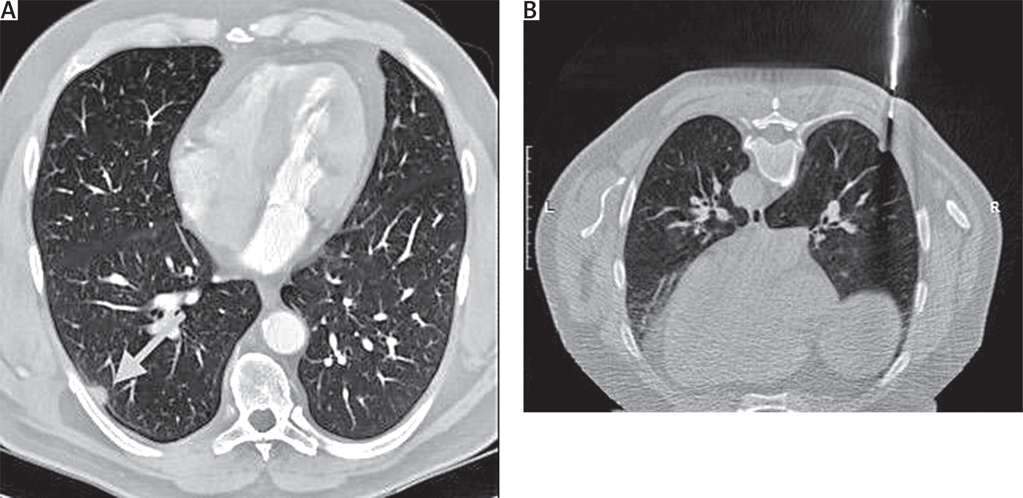
Six months later, a follow-up CT revealed fibrotic strands above the nodule and a ground-glass opacity area below it. A CT-guided core needle biopsy in 2023 revealed macrophages and lymphocytes without atypia. The patient was monitored radiologically during follow-up. Further diagnostic investigation was conducted because of an enlargement of the mass to 15 × 8 mm. In 2024, no pathological radiotracer uptake was observed in positron emission tomography (PET-CT) imaging. A repeat core needle biopsy later established the diagnosis of SFT.
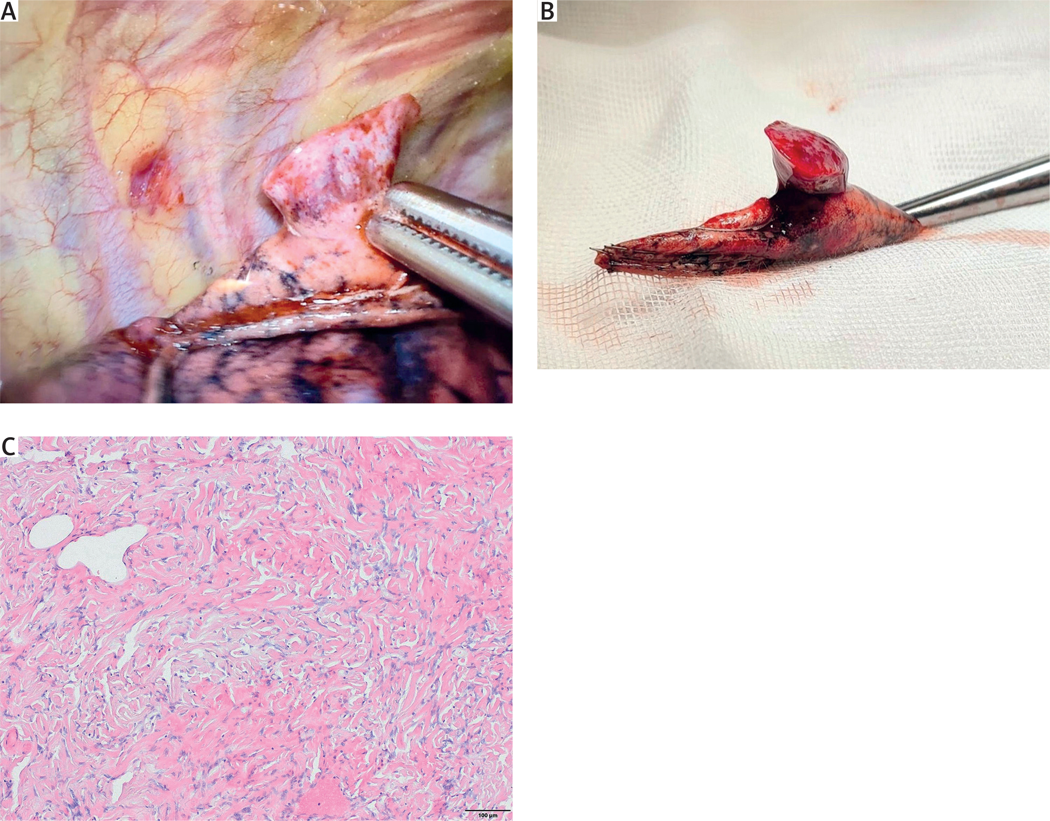
The patient was a candidate for surgical therapy. Right-sided video-assisted thoracoscopic surgery (VATS) was performed. Intraoperatively, a tumor was identified in the right lower lobe on the pedicle of the visceral pleura. The tumor was resected without difficulty using a linear three-line stapler at the base of the pedicle. The operation proceeded without complications, and postoperative outcomes were favorable. The histopathological examination confirmed a solitary fibrous tumor (STAT6+, CD34+, with mitotic index < 1/2 mm2, without necrosis). The lesion was completely excised with a clear margin (Figure 2).
Figure 2
A – Intraoperative thoracoscopic view of a solitary fibrous tumor arising from the visceral pleura of the right lower lobe. B – The completely resected specimen obtained via videoassisted thoracoscopic surgery. C – Histopathological image of described solitary fibrous tumor showing spindle cells arranged in a collagen-rich stroma. Hematoxylin and eosin (H&E) staining, ×10 magnification
Solitary fibrous tumors are rare mesenchymal lesions that occur most frequently in the pleura, although extrapleural locations have been documented, including rare and potentially life-threatening cardiac involvement [2, 4]. In the present case, the tumor arose from the pleura of the right lung, aligning with typical primary sites. Diagnosis is usually delayed or incidental because of the indolent growth and variable presentation [3]. The lesion in this case exhibited features suggestive of benign behavior, including small size and asymptomatic course – characteristics typical of many SFTs discovered incidentally on imaging performed for unrelated reasons [5]. Subsequent lesion growth and radiological findings such as fibrotic strands and ground-glass opacification prompted further investigation.
Core needle biopsy played a pivotal role in the diagnostic process. Although the first CNB was inconclusive, continued radiological follow-up was appropriate. The diagnosis of SFT was established by repeat biopsy following lesion progression. This emphasizes the need for continued radiological surveillance in suspected SFT when initial findings are nondiagnostic [6]. Immunohistochemical (IHC) markers including STAT6 and CD34 were essential for the final diagnosis. STAT6, in particular, is a highly specific marker that distinguishes SFT from other pleural and mediastinal mesenchymal tumors [7]. The low metabolic activity on PET-CT was consistent with low-grade biological behavior. The tumor was successfully treated with minimally invasive VATS, an established approach for selected small (< 5 cm) benign pleural tumors [8]. Histopathological results – low mitotic index and absence of necrosis – supported a favorable prognosis.
Even after radical excision, long-term follow-up is recommended because of the potential for late recurrence or malignant transformation [5].
This report demonstrates the diagnostic and therapeutic challenges of SFTs. It stresses the need for repeated histological and immunohistochemical evaluation and shows the usefulness of less invasive procedures such as VATS in the treatment of small benign pleural SFTs. Long-term follow-up is advisable after total resection of the SFT because of the potential for late recurrence.