Self-harm is a complex behavioral and public health issue that often requires a multidisciplinary therapeutic approach. It encompasses a wide range of behaviors, including cutting, burning, piercing, and self-inflicted blunt trauma, with skin-cutting being the most frequently reported form [1]. While many individuals engage in self-harm as a coping mechanism for emotional distress, some cases involve more severe injuries associated with substantial morbidity and, in rare cases, mortality. A particularly rare and dangerous form of self-injury involves the intentional insertion of foreign objects into the body, which can result in significant internal damage. The heart’s continuous motion and complex anatomy make such injuries particularly challenging to diagnose and manage, often requiring advanced imaging techniques and specialized medical intervention.
In this study, we present the case of a 61-year-old male patient who deliberately inserted two needles into his chest – one located precordially and the other near the interventricular septum – as an act of self-harm. Despite the potential severity of this condition, the patient remained hemodynamically stable and repeatedly refused surgical intervention. This case highlights not only the medical complexities associated with penetrating cardiac injuries but also the psychological factors that may contribute to such behaviors.
A 61-year-old male prisoner was admitted to the Clinical Emergency Department (CED) in December 2024 due to the presence of foreign bodies in the cardiac region. His medical history included epilepsy, though he was non-adherent to antiepileptic therapy. He denied any allergies. On admission, the patient was in hemodynamically stable condition. His vital signs were as follows: heart rate 82 bpm, blood pressure 110/70 mm Hg, and oxygen saturation 97%. On physical examination, he was alert, oriented, and responsive, scoring 15 points on the Glasgow Coma Scale (GCS). His heart rate was regular, with clear heart sounds and no pathological murmurs. Lung auscultation revealed a symmetrical vesicular murmur. The abdomen was soft and non-tender, with normal peristalsis. No peripheral edema was present, and the tongue was moist. A self-inflicted wound was observed in the precordial area.
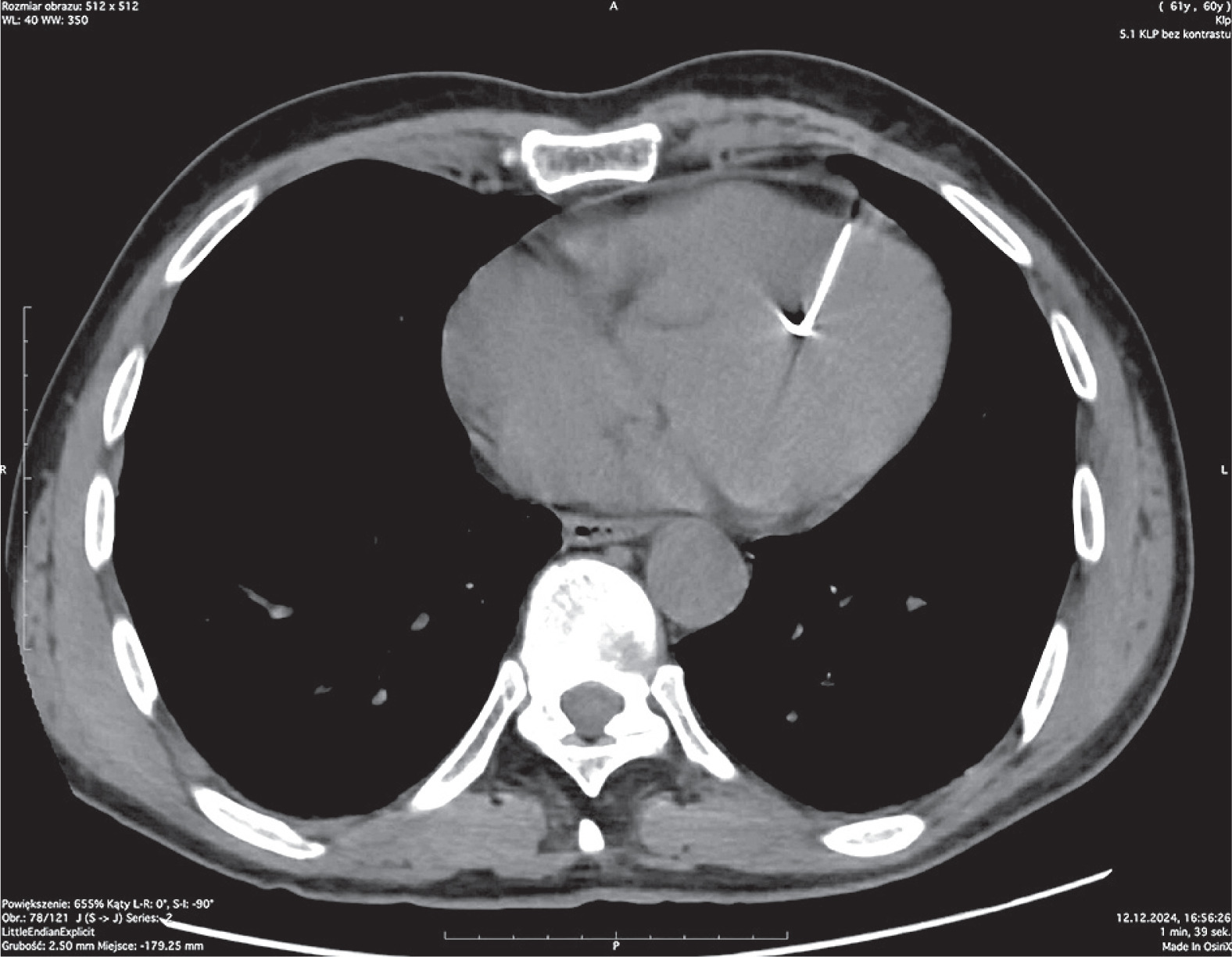
A chest X-ray confirmed the presence of two foreign bodies in the cardiac region. Subsequent chest computed tomography (CT) revealed hyperdense, linear foreign bodies up to 36 mm in length, located in the anterosuperior wall of the left ventricle and the interventricular septum. Another foreign body measuring 33 × 3 mm was identified in the left fourth intercostal space, embedded in the chest wall and pericardial fat. Additional findings included mild cardiomegaly, a small pericardial effusion, and a borderline-width pulmonary trunk. No signs of an acute life-threatening condition were observed.
A cardiothoracic surgery consultation was requested. After reviewing the imaging studies and comparing them with a previous CT scan from 2023, the surgical team found no progression of changes and no indications for urgent cardiac intervention. The patient remained hemodynamically stable, without signs of cardiac tamponade or other immediate complications. Due to his stable condition, the absence of acute risk, and – most importantly – his refusal of surgical treatment, a conservative management approach was chosen. The patient was informed of the risks associated with retaining foreign bodies, including potential migration of the needles within the heart. Despite this, he categorically declined surgical intervention. The patient remained under observation at the CED for another day and was then discharged in good general condition. A review of his medical records revealed that he had been hospitalized multiple times for the same issue since 2006. According to the patient, the foreign bodies – presumably needles – hd been in his chest for approximately 18 years. On each occasion, after undergoing diagnostic tests, he refused surgical intervention and discharged himself against medical advice, returning to prison (Figures 1–3).
Figure 1
Chest radiographs obtained on 14.10.2009 in AP (A) and lateral (B) positions. Several needle-like foreign bodies are visible in the precordial area
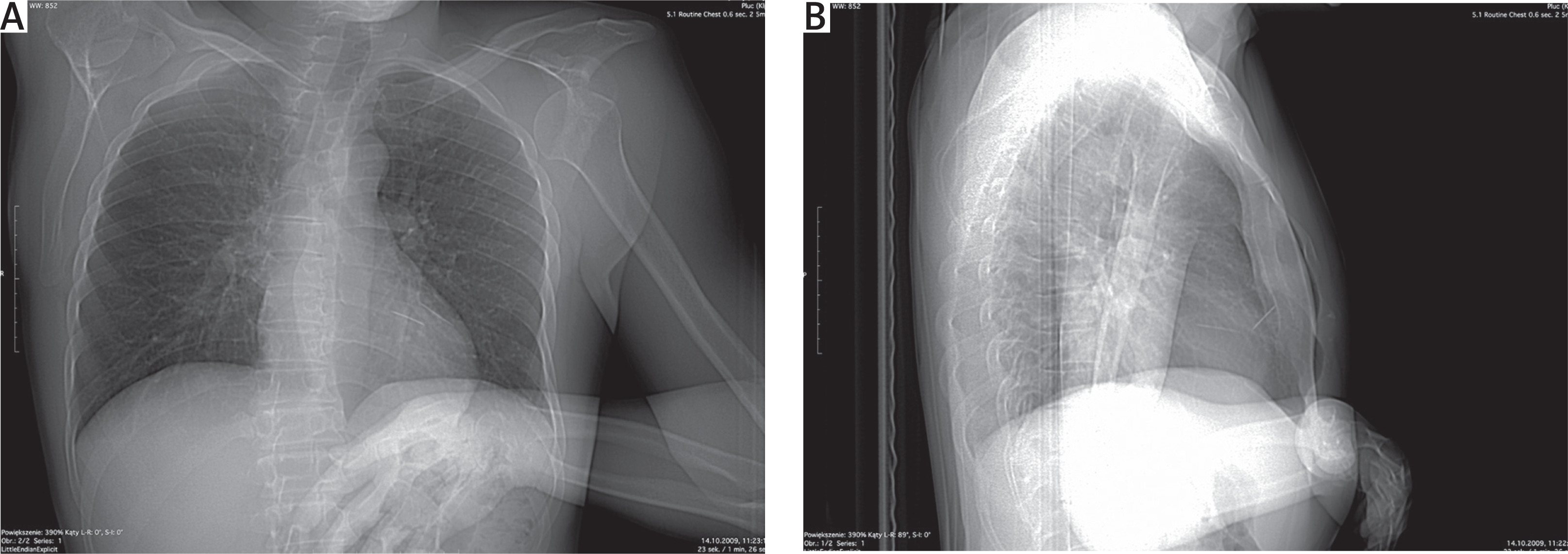
Figure 2
Chest radiographs obtained on 12.12.2024 in AP (A) and lateral (B) positions. Needle-like foreign bodies are visible in the same location as in 2009
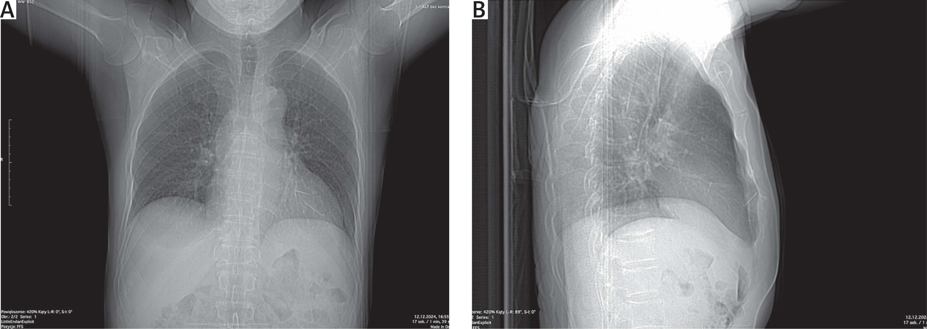
Figure 3
Computed tomography of the chest performed on 12.12.2024 showing a needle-like foreign body within the thoracic cavity
Penetrating chest trauma is rare; therefore, in suspected cases, thorough physical examination and imaging diagnostics are essential. The patient should be promptly monitored, and the physical examination should focus primarily on assessing cardiovascular and respiratory stability, wound depth, visibility of the foreign body, and the presence of Beck’s triad (jugular vein distension, muffled heart sounds, and hypotension). However, clinical symptoms may be nonspecific or absent, and the foreign body may not be visible on physical examination, highlighting the importance of imaging studies [1].
The gold standard for stable patients is computed tomography (CT), while transthoracic echocardiography (TTE) is preferred in hemodynamically unstable patients [2]. However, these modalities are not 100% reliable, as foreign bodies made of certain materials may not be visualized [2]. In our patient, advanced imaging was not feasible due to his refusal to consent, so only a chest X-ray was performed.
Self-inflicted chest injuries involving needle insertion are exceptionally rare, particularly in individuals without a documented history of psychiatric disorders. According to a literature review of a similar case, only 34 cases of self-injury involving needles have been reported since 1967 [3]. Additionally, Pfortmueller et al. [4] reported that the hospitalization of prisoners due to stabbing injuries is uncommon, especially in individuals over 60 years of age.
Considering these factors, as well as the patient’s repeated visits to medical facilities under similar circumstances and his consistent refusal of surgical treatment, this case raises the possibility that factors other than the treatment of the retained foreign bodies may have contributed to his healthcare-seeking behavior. Despite the presence of needle-like foreign bodies in the chest for nearly two decades, the patient did not exhibit any acute complications such as cardiac tamponade, sepsis, or hemodynamic instability. The prolonged clinical stability observed in this case, multiple hospitalizations, and refusal of treatment highlight the challenges associated with managing retained intracardiac foreign bodies in individuals who decline surgical intervention. In carefully selected asymptomatic patients without evidence of disease progression or acute complications, a conservative management strategy may represent a reasonable alternative when surgery is declined.