Introduction
Perimenopause is a phase of female reproductive aging that typically occurs in women between the ages 45–54 and is characterised by hormonal fluctuations that can last up to five years. This phase begins with changes in menstrual cycle length of more than seven days and progresses to a transition marked by amenorrhea lasting 60 days or more. It is associated with various vasomotor symptoms due to increased follicle-stimulating hormone (FSH) and decreased ovarian estradiol (E2) synthesis, leading to reduced quality of life. An estimated 80% of women experience symptoms such as hot flushes, nocturnal hyperhidrosis (night sweats) and sleep disturbances. These symptoms can also persist for some time after the last menstrual period [1–3].
A major consequence of oestrogen deficiency is the loss of intestinal barrier integrity, which leads to dysbiosis of the colonic microbiota [4, 5]. The bacteria within the microbiota involved in oestrogen metabolism are collectively referred to as the estrobolome. The composition of the estrobolome can be altered by factors such as diet, age, infectious and chronic degenerative diseases, and other environmental components [6, 7]. A critical molecule of the estrobolome is the enzyme β-glucuronidase, a glycosyl hydrolase that cleaves glycosidic residues and releases aglycones from glucuronides. This enzyme can catalyse both exogenous and endogenous substrates with glycosidic bonds. In the liver, oestrogens are glucuronidated and excreted via bile. Subsequently, the microbiota can reverse this conjugation through the action of β-glucuronidase, leading to enterohepatic recirculation of oestrogens. While about half of the excreted oestrogens are eliminated via bile, only 7% are found in the faeces, suggesting that the remainder is reabsorbed. This highlights the importance of β-glucuronidase in the hydrolysis of estrogenic compounds, facilitating their absorption [8–10].
Oestrogens are derived from C27 cholesterol and synthesised by cytochrome P450 aromatase and 17β-hydroxysteroid dehydrogenase (17β-HSD). Animal models have shown that supplementation with probiotics such as Lactobacillus acidophilus and Lactobacillus rhamnosus increases the expression of 17β-HSD and its isoforms, which in turn improves circulating oestrogen levels [11, 12]. In premenopausal women, E2 is the predominant oestrogen, whereas in postmenopausal women, estrone (E1) which is synthesised in peripheral tissues such as the skin and adipose tissue is the main oestrogen. Subsequently, oestrogens are metabolised in the liver, glucuronidated and excreted, serving as substrates for bacterial β-glucuronidase [12–15].
The β-glucuronidase from Lactobacillus rhamnosus shows reactivating activity with both endogenous and exogenous oestrogens, favouring E1-3-glucuronide over E2-17-glucuronide. Since E1 is the major oestrogen in menopausal women, the role of this bacterium in modulating their oestrogen levels is of great research interest [16].
The beneficial effects of probiotics in menopausal women include significant improvements in physical, psychosocial, vasomotor and sexual health [17]. Lim et al. [18] found that administration of 1 × 10⁸ colony- forming units (CFU) of Lactobacillus acidophilus for 12 weeks improved the quality of life of menopausal women by reducing associated symptoms without adverse effects.
Therefore, it is important to investigate the consumption of probiotics such as Lactobacillus acidophilus, Lactobacillus rhamnosus and Bifidobacterium longum for their effects on the homeostasis of the colonic microbiota, blood oestrogen concentration and β-glucuronidase levels, as well as their effects on the characteristic symptoms of this phase of female life [19, 20].
Material and methods
A supplement containing Lactobacillus acidophilus, Lactobacillus rhamnosus and Bifidobacterium longum was purchased from Suministros Herbolarios y Químicos, El Marqués, Querétaro, Mexico. Reagents for determining total cholesterol (TC) #4718917190, low-density lipoprotein cholesterol (LDL) #7005806190, high-density lipoprotein cholesterol (HDL) # 7528604190 and iron #5401658190 were purchased from Roche Diagnostics, Mannheim, Germany. ELISA kits for E2 #6001010, luteinizing hormone (LH) #6001002, FSH #6001003 and progesterone #6001009 were purchased from MexLab, Zapopan, Jalisco, Mexico. The QIAamp Fast DNA Stool Mini Kit #51604 was purchased from Qiagen, Valencia, CA, USA. The Master Mix SYBR™ Select #4472903 was purchased from Thermo Fisher Scientific, Waltham, MA, USA. β-D-glucuronide #P0501-25MG and phenolphthalein #105945-100G were purchased from Sigma- Aldrich, St. Louis, MO, USA. The estrone-3-glucuronide kit #501290 was purchased from Cayman Chemical, Ann Arbor, MI, USA.
Design study
This study was designed as a pilot, single-arm pre-post investigation, a choice primarily guided by ethical considerations. Consistent with single-arm trial principles, establishing a single experimental group ensures treatment equity, giving all participants equal opportunity to access a potentially beneficial intervention in this case, probiotic supplementation. Because the primary goal of this pilot phase was to evaluate underlying mechanistic effects rather than definitive clinical efficacy, a single-arm approach was considered a reasonable and ethically sound initial step [21]. A sample size of 20 participants was determined based on feasibility and strict recruitment constraints. Because perimenopause and vaginal/hormonal health are still often perceived as sensitive or taboo topics, enrolment faced cultural barriers and a lower-than-expected response rate during the recruitment period. Nevertheless, this sample size remains appropriate and sufficient for the exploratory aims of a pilot investigation. Nevertheless, the absence of a parallel placebo or control group remains an important limitation. Without a concurrent control arm, it is challenging to fully distinguish the observed hormonal and microbiological changes from the natural variability associated with perimenopause or from external confounding factors such as seasonal influences and spontaneous lifestyle modifications.
Ethical approval of the pilot study
Potential participants were invited through social media campaigns to take part in the study. After a detailed explanation of the objectives and methodology of the project, all participants provided written informed consent before the study began.
Participants in the study
From the pool of women invited to participate, 20 perimenopausal women aged 50–55 years were selected based on the following inclusion criteria: no antibiotic use within the past two months, not following a restrictive diet (e.g. ketogenic or vegetarian), not currently receiving hormone therapy, and being in apparent good health. Exclusion criteria included non-compliance with the supplementation protocol, illness or adverse reactions during the study period, or withdrawal from the study for personal reasons.
Intervention and supplementation
Volunteers received a probiotic supplement for 12 weeks. The supplement contained 109 CFU per day of Lactobacillus acidophilus, Lactobacillus rhamnosus and Bifidobacterium longum. All participants were explicitly instructed, and their adherence was monitored via verbal confirmation during follow-up visits, to maintain their usual diet and physical activity throughout the intervention period. Although no formal dietary records or food-frequency questionnaires were administered, this approach was intentionally selected based on our single-arm pre-post design. Implementing dietary modifications or active nutritional interventions would have introduced substantial confounding, masking the isolated mechanistic effects of the probiotic supplementation. Participants reported no adverse effects from the supplementation.
Symptom assessment
A survey was conducted at baseline and after 12 weeks of supplementation to assess the presence, absence and intensity of 11 common perimenopausal symptoms. The intensity of each symptom was quantified using the greene climacteric scale [22].
Anthropometric measurements
Body height was measured with an InBody BSM 170 stadiometer. Body weight, percent body fat, skeletal muscle mass (SMM), mineral content and phase angle were measured with an InBody 970 analyzer. To minimise inter-observer error, all measurements were performed by the same trained individual.
Biochemical analysis
At the beginning of the study and after 12 weeks of supplementation, venous blood samples were collected between 7:00–9:00 am after participants had fasted overnight for 8–12 hours. The blood samples were centrifuged at 4000 rpm for 10 minutes. Serum samples were then collected. The levels of TC, LDL, HDL and iron were analysed on the COBAS c111 (Roche Diagnostics, Mannheim, Germany) according to the manufacturer’s specifications in duplicate.
Hormonal analysis
Serum concentrations of E2, LH, FSH, and progesterone were determined in duplicate using commercially available ELISA kits according to the manufacturer’s instructions with ELISA techniques.
Analysis of specific gut bacteria and enzyme activity
Relative bacterial abundance (quantitative polymerase chain reaction – qPCR):
Fresh fecal samples were collected and stored at –70ºC until DNA extraction. DNA was extracted using the QIAamp Fast DNA Stool Mini Kit according to the manufacturer’s instructions.
For the identification of Lactobacillus and Bifidobacterium 16S rRNA genes, 5 ng of DNA was used, determined in triplicate from stool samples collected at baseline and post-intervention. Normalisation was performed using the total bacterial 16S rRNA gene. Quantitative PCR was performed using the following primers: Lactobacillus (5´-AGCAGTAGGGAATCTTCCA -3´) and (5´-CACCGCTACACATGGAG -3´), Bifidobacterium (5´- TCGCGTCYGGTGTGAAAG -3´) and (5´-CCACATCCAG CRTCCAC -3´),Total bacteria 16S rRNA gene (5´-CGGYCCAGACTCCTACGGG -3´) and (5´- TTACCGCGGCTGCTGGCAC -3´), using the QuantStudio 1 Real-Time PCR System (Thermo Fisher Scientific, Waltham, MA, USA). The Master Mix SYBR™ Select was used under the following thermal cycling conditions: 95°C for 2 min, followed by cycles of 95°C for 5 s, an annealing step (50°C, 55°C and 59.5°C respectively) for 20 s and 65°C for 10 s [23–25].
Assay for β-glucuronidase activity
This assay was also performed with stool samples collected at baseline and after the intervention. A 0.5 g aliquot of each sample was homogenised in phosphate-buffered saline, centrifuged at 2500 rpm and the resultingsupernatant was collected. The supernatant was thenincubated for 5 minutes with phenolphthalein β-D-lucuronide. Enzyme activity was quantified in triplicate using a standard curve prepared with phenolphthalein.
Estrone-3-glucuronide measurement
To quantify the substrate of β-glucuronidase, the concentration of E1-3-glucuronide in the sample super- natants above was measured in duplicate using an ELISA kit according to the manufacturer’s instructions.
Statistical analysis
Results are expressed as mean ± standard deviation. Data normality was formally assessed using the Shapiro-Wilk test; because the data followed a normal distribution, parametric tests were considered appropriate for subsequent analyses. Because this study was designed strictly as an exploratory pilot investigation, no a priori sample size calculation was performed, and the study was not initially powered to detect specific clinical effect sizes. However, to maintain statistical rigour and minimise the risk of Type I errors (false positives) arising from multiple simultaneous comparisons, a post hoc Bonferroni correction was applied. The statistical significance of differences between groups was therefore determined using a paired Student’s t-test. The specific significance threshold for this test was adjusted to p < 0.0028, ensuring that the observed biochemical and hormonal changes remain robust despite the exploratory nature of the study and the small cohort size. Additionally, changes in symptoms were evaluated with a χ2 test, and a Pearson correlation analysis was performed for the entire data matrix. All analyses were conducted using the GraphPad Prism 9 statistical software package, and unless otherwise specified by post hoc adjustments, statistical significance was established at p < 0.05.
Results
Anthropometric and biochemical parameters
The results of the body composition and biochemical analyses are presented in Table 1. No significant changes were observed in weight, SMM, percentage of fat mass, mineral content, or phase angle. The absence of changes in anthropometric parameters is consistent with previous findings, such as those by Hatakka et al. [26], who reported minimal weight changes after four weeks of supplementation with 2 × 10¹⁰ CFU of Lactobacillus rhamnosus and Propionibacterium freudenreichii. This suggests that the primary effect of the intervention was not on systemic energy homeostasis but on specific metabolic pathways.
Table 1
Body composition and biochemical analysis
Regarding blood biomarkers, no significant differences were found in iron or HDL cholesterol levels. However, a significant decrease in LDL cholesterol was observed (–21.12 ±24.97 mg/dl), representing a notable change in the participants’ lipid profile. This may be because certain Lactobacillus strains can deconjugate bile acids through their hydrolase activity, or because the production of short-chain fatty acids mediated by these bacteria can lower cholesterol levels by limiting its availability in the liver, which is. required for fatty acid and cholesterol synthesis [26, 27].
The significant decrease in LDL cholesterol is particularly important for the cardiovascular health of this population, given the commonly observed increase in this lipoprotein during the transition to menopause [28]. Some studies have reported the LDL-lowering effect of Bifidobacterium lactis, provided in enriched milk to obese patients at a dose of 80 ml (3.4 × 10⁸ CFU/ml) for 45 days [29]. Another example is from Rerksuppaphol et al. [27], who supplemented hypercholesterolemic patients three times daily for six weeks with Lactobacillus acidophilus (109 CFU) and Bifidobacterium bifidum (109 CFU). The difference between the control and experimental groups was –40.1 mg/dl for TC and –28.2 mg/dl for LDL. Other studies, in addition to supporting the anti- cholesterolemic effect, also suggest an impact on appetite reduction following supplementation with a synbiotic containing Bacillus subtilis [30]. Several mechanisms have been proposed; the main one involves the ability of some commensal bacteria to deconjugate bile acids through hydrolase activity. Deconjugated bile acids are poorly absorbed, causing them to interact with cholesterol, precipitate it, and facilitate its excretion. The incorporation of cholesterol into the cell membrane of Lactobacillus and Bifidobacterium and the conversion of cholesterol to coprostanol have also been studied as hypocholesterolemic mechanisms [31–33].
Hormone levels before and after supplementation
The changes in hormone levels are summarized in Table 2. A significant increase was observed in E2 levels, with a change of 6.86 ±5.20 pg/ml. In contrast, FSH and LH decreased (–8.91 ±4.48 mIU/Ml, –0.17 ±0.14 mIU/Ml, respectively). These values are statistically correlated (Pearson’s r = –0.5, p < 0.05) and may indicate a positive change associated with the hypothalamic-pituitary- ovarian axis mediated by the increase in E2 [34].
Table 2
Hormone determination before and after supplementation
In addition to a change associated with lipid metabolism, the intervention led to a significant hormonal shift, characterised by an increase in serum E2 and a corresponding decrease in FSH, with a negative correlation between the two. These outcomes are consistent with those of Zou et al. [35], who, in an analysis of National Health and Nutrition Examination Survey, data, found that probiotic consumption was associated with higher E2 levels in perimenopausal and menopausal women.
Similarly, an intervention study by Honda et al. [36] reported a rise in E1 and E2 levels in women who received a mixture of 84 bacterial strains (≥ 1 billion CFU/day) for 12 weeks, including Lactobacillus and Bifidobacterium longum. In that study, oestrogen levels were maintained in the probiotic group while they decreased in the placebo group, with significant differences in E1 and E2.
Specific gut bacteria Bifidobacterium and Lactobacillus
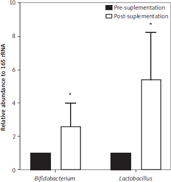
The changes in the abundance of specific bacteria were measured before and after the intervention, and the data were normalised to the 16S rRNA gene (Figure 1). A significant increase in the relative abundance of the main probiotic genera was observed. The relative abundance of Bifidobacterium increased by 2.59 ±1.4 fold and that of Lactobacillus by 5.43 ±2.8 fold. These results indicate a favourable modulation of specific gut bacteria after the intervention.
Figure 1
Relative abundance (fold changes) of Bifidobacterium and Lactobacillus
*Statistical significance was determined using a paired Student’s t-test (p < 0.05).
Data were normalised to total bacterial 16S rRNA gene. Results are presented as mean ± SD for 20 participants.
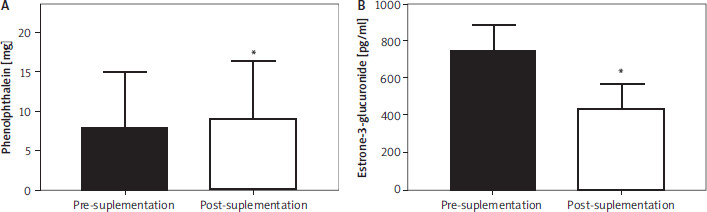
In the analysis of fecal samples, the activity of the enzyme β-glucuronidase and the concentration of its substrate E1 glucuronide were examined before and after the intervention (Figure 2). The results showed a significant increase in β-glucuronidase activity (1.21 ±1.15 mg phenolphthalein). At the same time, a decrease in E1 glucuronide concentration was observed (–260.8 ±139.8 pg/ml).
Figure 2
Comparison between groups. A) β-glucuronidase activity expressed in mg of phenolphthalein. B) Amount of estrone 3-glucuronide (pg/ml)
*Statistical significance was determined using a paired Student’s t-test (p < 0.05).
Data are presented as mean ± SD for 20 participants.
In this study, a negative correlation was found between conjugated E1 content and Bifidobacterium abundance, and a positive correlation between Bifidobacterium abundance and β-glucuronidase activity (Pearson’s r = –0.9 and 0.6, respectively; p < 0.05). Regarding the remarkably strong negative correlation (r = –0.9), it is worth noting that the low dispersion and tight alignment of these data points are intrinsic to the relative quantification scale of the normalised qPCR data, which compresses variance through the 2–∆∆Ct method relative to a baseline of 1. This highlights the role of this group of bacteria in hormone regulation in perimenopausal women.
Discussion
The proposed mechanism by which the micro- biota modulates oestrogen levels is through the activity of β-glucuronidase, which deconjugates estrogenic hormones that have been metabolised and excreted via bile, allowing for their reabsorption [37]. There are over 279 β-glucuronidase enzymes, divided into seven groups, and only some can reactivate oestrogens; their expression depends on the bacterial strain [38]. Some bacteria, such as Lactobacillus rhamnosus, a strain used as a probiotic in this study, exhibit oestrogen activating activity through β-glucuronidase. Another strain used, Lactobacillus acidophilus, has also been shown to possess β-glucuronidase activity, suggesting these strains could be modulating the observed E2 and E1 levels [39]. The activating capacity of the enzyme depends on its structure. Ervin et al. [40] analysed β-glucuronidase variants and found that enzymes from Loop 1, Mini-Loop 1, and flavin mononucleotide-binding classes can hydrolyse E1 and E2 conjugates. Loop 1 class β-glucuronidase enzymes exhibit high catalytic efficiency due to an “aromatic cage” in their active site that facilitates precise recognition and positioning of the oestrogen steroid core.
Beyond the enzyme’s structure, its activity level is crucial for activating oestrogens. Contradictory results on the modulation of β-glucuronidase are reported in the literature, which may be resolved by identifying the specific isoform being analysed [36]. Factors influencing the activity and expression of this enzyme include antibiotic use, diet, age, and sex. Some authors have reported a decrease in this enzyme’s activity with increasing age, with this decline being more notable in women [41]. However, other authors report that changes in this enzyme depend on the strain being analysed [42]. Older adult populations show higher β-glucuronidase activity from Lactobacillus but lower activity from Enterococcus. Furthermore, it has been observed that the activity from Lactobacillus increases with age, while that from Enterococcus decreases over the years [43].
This underscores the importance of studying whether the strains used as probiotics have oestrogen activating activity in menopausal women. When a probiotic is administered, it faces the challenge of passing through the gastrointestinal tract and encounters colonization resistance from both the host and commensal intestinal bacteria upon reaching the colon. Therefore, success depends on the probiotic strain’s ability to adhere to the intestinal mucosa [44].
In the present study, a notable increase in the relative abundance of Bifidobacterium and Lactobacillus was observed after supplementation. This was accompanied by hormonal modulation, providing a robust mechanistic link for these observations by identifying β-glucuronidase activity as the central driver connecting the microbial and hormonal shifts.
Symptoms
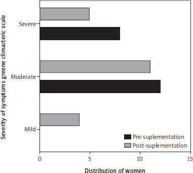
Of the 11 common symptoms measured by green’e climacteric scale, no significant changes were observed. However, the small number of participants limits statistical power (Figure 3).
Figure 3
Comparison of symptoms before and after supplementation according to the greene climacteric scale
Changes in symptoms are determined by a χ2 test (p = 0.07).
Symptoms commonly associated with menopause, such as skin dryness and mental fog, are closely linked to the drop in oestrogen, which also causes physiological changes leading to vaginal dysbiosis. The literature indicates that probiotics can help restore this balance and have positive effects on menopausal symptoms and anti-inflammatory effects [45, 46].
Notably, some studies, such as Lim et al. [18], have reported significant improvements in menopausal symptoms with probiotics, even without changes in hormone levels, suggesting that these benefits may occur through multiple pathways. While the current intervention did not improve reported symptoms, it is important to investigate this further due to the observed trend.
In this context, it is important to acknowledge the inherent limitations of this exploratory pilot study, particularly its small sample size and the methodological constraints on strict tracking of diet and physical activity, as these lifestyle factors are well known to influence gut microbiota composition. Moreover, this investigation focused exclusively on quantifying Lactobacillus and Bifidobacterium, which represent only a fraction of the vast and complex microbial ecosystem in the human colon. Despite these limitations, this work provides a valuable foundational framework and critical guidance for the design of future large-scale randomised controlled trials with larger cohorts of participants.
Conclusions
The preliminary results of this pilot study suggest a potential relationship between the balance of specific intestinal bacteria and hormone regulation during perimenopause. Supplementation with Lactobacillus acidophilus, Lactobacillus rhamnosus, and Bifidobacterium longum over a 12-week period was favourably associated with certain biochemical parameters and the overall well-being of the participants. These exploratory findings indicate that modulating specific gut microbiota through probiotic supplementation might influence oestrogen metabolism. This effect could be partly mediated by β-glucuronidase activity, an enzyme involved in oestrogen deconjugation by hydrolysing glucuronides, which theoretically allows excreted oestrogens to be reabsorbed through the enterohepatic circulation. While the observed shifts in β-glucuronidase activity and specific bacterial populations underscore the importance of the intestinal microenvironment during the perimenopausal transition, these data must be interpreted with caution due to the lack of a control group and the small cohort size. Ultimately, this pilot investigation provides a valuable foundational framework, demonstrating the potential relevance of probiotic interventions and offering essential methodological guidance for the design of future rigorous, large-scale randomised controlled trials.