Introduction
Intra-aortic balloon pump (IABP) therapy is a mechanical circulatory support method used to enhance coronary perfusion and reduce left ventricular afterload in patients with severe cardiac dysfunction. Introduced into clinical practice in the 1960s [1], IABP has been widely used in the management of cardiogenic shock, acute myocardial infarction, and perioperative cardiac surgery support. The device is typically implanted through the common femoral artery, though alternative access sites such as the subclavian and axillary arteries are increasingly used, especially in patients requiring prolonged support or those with peripheral artery disease [2–4]. IABP is particularly beneficial as hemodynamic support for patients with ischemic heart disease, severe heart failure, valvular dysfunction, and high-risk percutaneous coronary interventions (PCI), as well as a bridging therapy for patients awaiting orthotopic heart transplantation (OHT). It is also used in combination with other mechanical circulatory support devices, such as extracorporeal membrane oxygenation (ECMO), left ventricular assist devices (LVAD), or other ventricular pumps, to optimize cardiac function in critically ill patients [5–7]. The therapeutic effect of IABP is achieved through counterpulsation, where the balloon inflates during diastole, increasing coronary blood flow, and deflates just before systole, reducing afterload. This mechanism decreases myocardial oxygen demand and enhances cardiac output, which may lead to improvements in ejection fraction (EF) and overall left ventricular function.
Aim
In our study, we propose a novel approach to minimally invasive sheathless percutaneous implantation of IABP via the left subclavian artery under ultrasound guidance, as well as a non-surgical technique for balloon removal.
Material and methods
Between June 2024 and December 2024, we implanted IABP in 10 patients, 9 males and 1 female. All patients were in critical condition and undergoing evaluation for orthotopic heart transplantation due to end-stage heart failure. In addition to inflammatory or ischemic heart damage, they suffered from arrhythmias, valvular disease, diabetes, mild renal failure, and mild pulmonary hypertension. The overall condition of the patients was severely compromised, with a mean ejection fraction of 19.2% and a mean age of 58 years. Two patients had a history of cardiac arrest with successful resuscitation (Table I).
Table I
Baseline clinical characteristics of the study population (N = 10)
IABP implantation technique
During intraoperative ultrasound examination, the left subclavian artery was identified as distally as possible from the clavicle to avoid difficulties in case of complications and conversion to open surgery. Under ultrasound guidance, the puncture was performed using the Seldinger technique, and a 6F/11 cm introducer was placed. Control angiography was performed to identify the origin of the subclavian artery from the aorta, followed by the introduction of a pigtail catheter into the descending aorta. Another angiography was performed to visualize the celiac trunk and the superior mesenteric artery (Figure 1). A guidewire from the IABP set was advanced through the pigtail catheter. After removal of the 6F sheath, the balloon catheter was advanced over the guidewire and inserted without a sheath. In cases where advancement through the skin or arterial wall was difficult, a dilator from the 7.5F introducer set was used to facilitate insertion. Finally, the balloon markers were aligned according to the angiographic landmarks of the subclavian artery origin and visceral vessels. After activating the IABP and confirming its position, it was secured to the chest wall in a standard manner.
Figure 1
Percutaneous sheathless IABP implantation through the left subclavian artery (LSA). A – LSA puncture and sheath insertion; B – angiography of LSA orifice; C – IABP in working position; D – insertion site
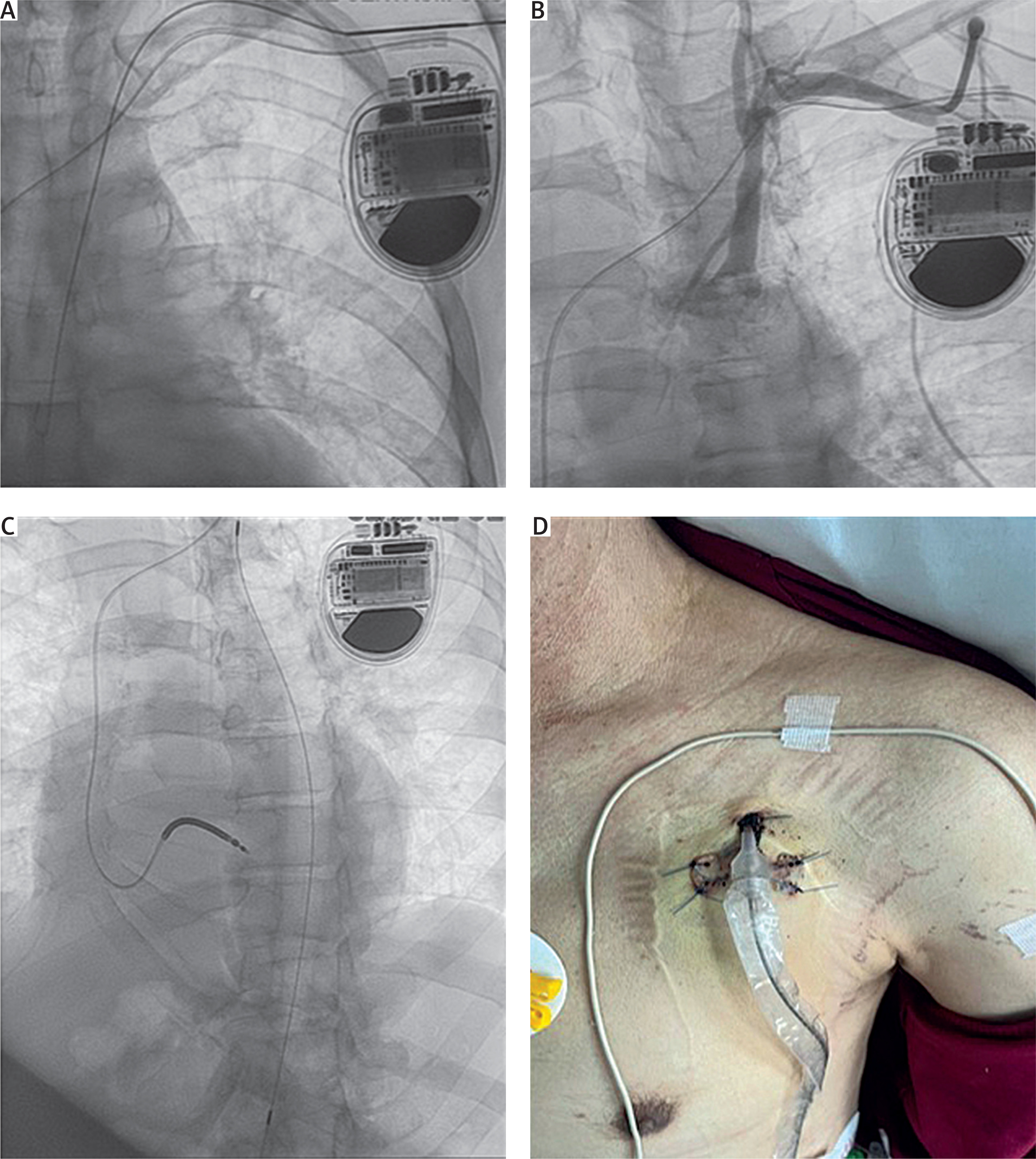
Sheathless implantation significantly reduces vascular trauma and allows for percutaneous closure of the vascular access site using the AngioSeal closure device instead of surgical closure. Maintaining the patency of the IABP working channel is crucial. It is achieved by flushing with saline to ensure that a guidewire can be introduced into the channel when removing the balloon.
Balloon removal technique
A 0.018” guidewire was introduced into the IABP working channel under fluoroscopic guidance, preferably using a stiff-type guidewire. The balloon was then removed, and a 6F, 11 cm introducer was advanced over the guidewire. Subsequently, the guidewire was exchanged for the AngioSeal 6F system wire, and standard vessel closure with the AngioSeal system was performed.
Results
Definitive therapy was achieved in 6 (60.0%) patients, including 5 who underwent OHT and 1 who received LVAD support. The remaining 4 patients died during the bridging period. Three patients died due to causes unrelated to IABP implantation, primarily due to progressive heart and multiorgan failure. In 1 patient with a fatal outcome, massive embolism of the visceral arteries occurred, along with signs of ischemic stroke, which may have been indirectly associated with IABP as a complication (Table II).
Table II
Characteristics, duration of IABP support, and clinical outcomes in 10 consecutive patients
The mean duration of IABP support in all patients was 21.6 days. In patients who underwent orthotopic heart transplantation or received a left ventricular assist device it was 29.5 days, with a median duration of 18.5 days (maximum 69 days). In contrast, among non-survivors the mean duration of IABP support was 9.75 days, and the median was 9.5 days (maximum 18 days).
No local complications such as bleeding or infection at the implantation site were observed, despite the prolonged duration of IABP support. However, in 2 patients with extended IABP use (ninth day of 69 and third of 21 days, respectively), mechanical complications occurred: 1 case involved balloon perforation, and the other involved perforation of the helium pressure line. In both patients, the IABP devices were percutaneously replaced without further complications.
Six patients who underwent orthotopic heart transplantation or received a left ventricular assist device had their intra-aortic balloon removed and the subclavian artery was closed using the Angio-Seal 6F vascular closure device This method facilitated effective closure without complications, even after prolonged IABP support, and has been effective in achieving rapid hemostasis following IABP removal.
Discussion
The concept of intra-aortic balloon pump implantation via percutaneous access was first introduced by Bregman et al., in 1979, who described the use of the femoral artery for percutaneous IABP insertion using the Seldinger technique. This approach marked a major advance, allowing rapid and less invasive deployment of the device without the need for surgical cutdown [2, 8].
The idea of using the subclavian artery for IABP implantation has been applied since 1989, with early work by McBride et al. exploring alternative arterial access routes [3]. In recent years, however, this technique has gained popularity, especially among patients requiring prolonged mechanical support [7, 9, 10]. Another alternative is IABP implantation through the axillary or brachial artery, which may offer improved patient mobility and reduced risk of infection compared to femoral access, which may have a beneficial effect on their general condition and prognosis [11, 12].
A growing number of reports indicate the effectiveness of multimodal prehabilitation including physical training, psychological support, and nutritional interventions [13–15]. This strategy not only improves post-transplantation outcomes but also does not increase treatment costs, supporting the idea of active preparation of the patient for OHT. In a study by Chen et al. [16] early rehabilitation of patients with femoral IABP used as a bridge to heart transplantation helped avoid the negative effects of prolonged immobilization. However, because of the limitations of femoral access, the authors used a special protocol with a tilt table to allow safe mobilization.
In 2022, Nishida et al. reported 10-year experience with intra-aortic balloon pump placement using a surgical access via a chimney graft implanted in the axillary artery. This approach allowed 90% of patients to remain mobile during support and led to successful achievement of the intended therapy (including heart transplantation) in nearly 87% of cases. The authors emphasized that axillary access enables effective circulatory support while maintaining patient mobility [17]. In our study, the access was completely percutaneous and minimally invasive, which simplified the procedure and may have reduced the risk of surgical complications. In most cases described in the literature, the subclavian approach requires surgical preparation, often involving the implantation of an artificial chimney graft. Such a procedure increases the risk of infection and may lead to subsequent difficulties in arterial reconstruction. The percutaneous puncture technique eliminates this problem, reducing vascular trauma as well as the risk of infectious and structural complications [18, 19].
Additionally, in patients with peripheral artery disease (PAD), including significant stenosis or occlusion of the iliac arteries, femoral access may be technically challenging or even impossible. According to data from the literature, PAD is a known risk factor for complications associated with IABP implantation. In such cases, IABP implantation via the subclavian artery provides a safer and more effective strategy [10, 11, 20, 21].
In patients eligible for orthotopic heart transplantation, both femoral accesses may be crucial for oxygenation pump, renal replacement therapy, or ECMO support. Keeping femoral access sites available ensures flexibility in managing advanced heart failure and enables rapid initiation of life-saving therapies, especially since these sites may already be occupied by ECMO cannulas or other implantable circulatory support devices. In the context of heart transplantation preparation, minimizing trauma to peripheral arteries is essential, and subclavian access may be the optimal solution for this patient group [22, 23].
The observed definitive therapy rate in our cohort (60%) is lower than reported in other studies of IABP as a bridge to transplantation, such as Estep et al. (84%), Nishida et al. (91.7%), and Barge-Caballero et al. (69%) [17, 19, 24]. This difference may be explained by the fact that our patients were in a more critical condition, with a markedly reduced mean ejection fraction (19.2%) and high Nt-proBNP levels and frequent comorbidities, including prior cardiac arrest. In contrast, other cohorts included more ambulatory patients, often supported via axillary access and capable of physical activity during bridging. Despite these limitations, our minimally invasive, fully percutaneous approach via the subclavian artery proved safe and effective even in this high-risk population.
Conclusions
The percutaneous, minimally invasive approach to IABP implantation via the subclavian artery appears to be an effective and safe method of mechanical circulatory support. One of its key advantages is the facilitation of early patient mobilization, which is particularly important in patients requiring prolonged support. Even in cases where IABP was used for more than 10 days, we did not observe any complications related to vascular access.
Both implantation and removal of the balloon resulted in less procedural trauma compared to open surgical access. This technique not only minimizes the risk of infection and arterial complications but also reduces the cumulative risk associated with invasive procedures during the bridging therapy in orthotopic heart transplantation.
Further studies on a larger cohort of patients are necessary to validate these findings and to establish the long-term benefits and safety of this technique in patients awaiting orthotopic heart transplantation or requiring prolonged mechanical support.