Introduction
Median sternotomy, introduced by Julian and colleagues in 1957, remains the gold standard surgical approach for coronary artery bypass grafting (CABG). It facilitates postoperative recovery and provides optimal visibility when appropriate sternal stability is achieved [1]. However, sternal complications, including dehiscence, malunion, and deep sternal wound infection (DSWI), occur in 0.5–8% of patients and lead to significant morbidity, prolonged hospital stays, and mortality rates approaching 10–47% [2, 3]. These complications constitute a significant healthcare burden, with estimated costs per episode exceeding $60,000 in developed countries [4].
Conventional sternal closure using a stainless-steel wire cerclage has demonstrated reliability over decades of clinical practice. However, biomechanical studies reveal substantial stress concentrations at the fiber-bone interfaces, particularly under physiological loading conditions such as coughing, which generate forces exceeding 200 Newtons [5]. These mechanical limitations become clinically relevant in high-risk populations characterized by deterioration in bone quality (osteoporosis), high body mass index (BMI ≥ 30 kg/m2), diabetes, chronic obstructive pulmonary disease (COPD), and advanced age, which increases the overall risk of dehiscence by 2.5- to 5-fold [6, 7]. Rigid plate fixation systems have emerged as biomechanically superior alternatives by distributing forces more evenly across the sternum and theoretically reducing micro-mobility, which impedes bone healing [7–9]. Similar to the evolution of cardiovascular implant technologies, such as drug-eluting stents designed to inhibit neointimal hyperplasia and multilayer flow modulators for aneurysm stabilization [10, 11], the improvement of sternal fixation methods reflects a broader trend toward precision-engineered, patient-specific devices in cardiovascular surgery. Finite element analysis (FEA) indicates that plate systems reduce maximum stress at fixation points by approximately 40% compared to wire cerclage under equivalent loading [12]. Clinical research has shown promising results, with decreased pain scores, improved lung function, and increased patient satisfaction with plate fixation in randomized controlled trials by Raman et al. [13] and Allen et al. [14]. However, these studies have predominantly examined high-risk cohorts, leaving uncertainty about optimal closure strategies for broader patient populations. Despite accumulating evidence, several critical questions remain unanswered: 1) Do plate systems provide meaningful clinical advantages in unselected CABG populations? 2) Which patient subgroups may derive greater benefit from plate fixation? 3) Do the additional operative time and cost associated with plate systems justify their routine application?
This study addresses these questions through a comparative analysis of consecutive CABG patients undergoing plate or wire sternal closure at a single tertiary referral center. We hypothesized that rigid plate fixation would demonstrate more advantageous outcomes in terms of sternal dehiscence, wound complications, and hospital resource utilization compared to traditional wire cerclage, and that these benefits would be most pronounced in high-risk patient subgroups. Secondary objectives include identifying predictive factors for successful closure and developing evidence-based recommendations for personalized closure strategy selection.
Material and methods
Study design and setting
This single-center retrospective cohort study was conducted at the Department of Cardiovascular Surgery, Faculty of Medicine, Hatay Mustafa Kemal University, a tertiary referral institution that performs approximately 300 cardiac surgical procedures annually. The study was conducted in accordance with the Strengthening the Reporting of Observational Studies (STROBE) guidelines [15].
Patient population and selection criteria
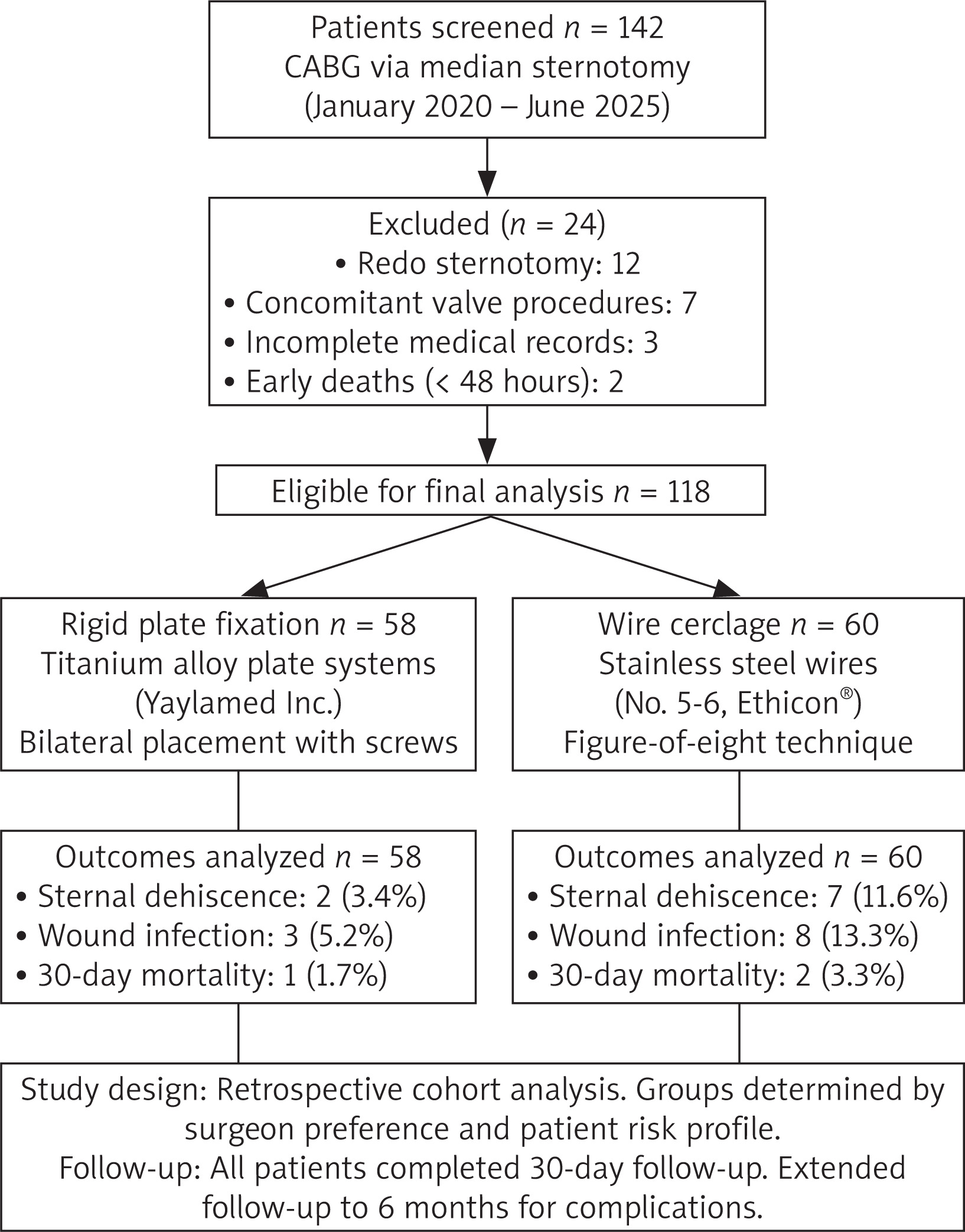
All consecutive adult patients (≥ 18 years of age) who underwent elective or emergency CABG via median sternotomy between January 2020 and June 2025 were screened for inclusion in the study. Inclusion criteria were: 1) isolated CABG or CABG with concurrent minor procedures; 2) primary sternotomy; 3) availability of complete perioperative and follow-up data. Exclusion criteria were: 1) repeat sternotomy; 2) emergency operations; 3) concurrent major cardiac procedures (valve replacement, aortic surgery); 4) preexisting sternal pathology (infection, tumor, radiation injury); 5) traumatic sternal injury; 6) incomplete medical records; 7) death within 48 hours of surgery (preventing outcome assessment). A final cohort of 118 patients was generated by excluding 24 of the 142 screened patients (12 repeat sternotomies, 7 concurrent valve procedures, 3 missing records, 2 early deaths). Patients were retrospectively stratified according to the sternal closure technique used: rigid plate fixation (Group 1, n = 58) or wire cerclage (Group 2, n = 60). The choice of closure method was based on surgeon preference and patient risk factor assessment, with the preference for plate fixation increasing in high-risk patients during the study period (Figure 1).
Figure 1
Flowchart of patient selection and group allocation. Flowchart showing screening, exclusion criteria, and final allocation of patients to rigid plate fixation (n = 58) and wire cerclage (n = 60) groups. Patients were excluded due to repeat sternotomy (n = 12), concurrent major cardiac procedures (n = 7), incomplete medical records (n = 3), or death within 48 hours of surgery (n = 2). A total of 118 patients were included in the final analysis
Surgical techniques and interventions
All procedures were performed by three experienced cardiovascular surgeons using standard techniques. Median sternotomy was performed with an oscillating saw, and the sternum was retracted using self-retaining retractors. CABG was performed using standard cardiopulmonary bypass (CPB) with moderate hypothermia (32–34°C) and intermittent antegrade blood cardioplegia. Depending on the target vessel anatomy and quality, both internal mammary artery and saphenous vein grafts were used as conduits.
For wire cerclage closure (Group 2), 6–8 stainless-steel wires (No. 5 or 6, Ethicon, Johnson & Johnson, USA) were used with alternating simple interrupted wires in a figure-of-eight pattern and penetrated the sternum in parasternal positions 1–2 cm from the midline. The wires were tightened sequentially from top to bottom using wire benders using standard torque.
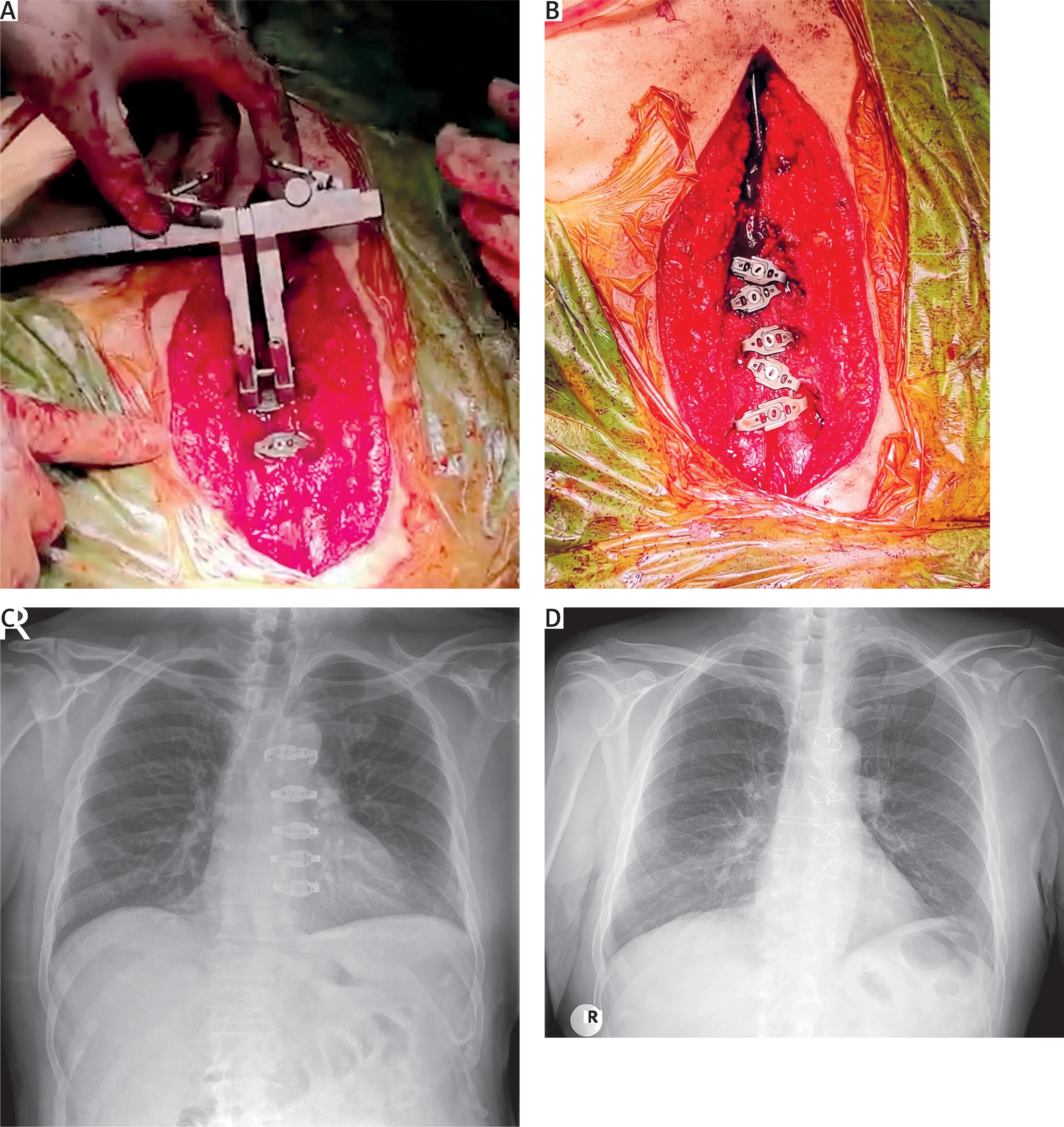
Titanium alloy plate systems (Peta Sternal Closure Plate, Invamed, Ankara, Turkey; distributed by Yaylamed A.Ş., Turkey) were used for rigid plate fixation in Group 1 patients. After sternal approximation with two temporary guidewires, intercostal spaces were measured using special calipers. Appropriately sized plates (usually with 4–6 holes) were selected and placed bilaterally along the sternal borders. Self-tapping titanium screws (3.5 mm in diameter, 10–14 mm in length) were inserted into the sternal cortex through predrilled holes in the plate, ensuring bi-cortical attachment where possible. Typically, 2–3 plates were used per sternal half and placed at the manubrial, mid-sternal, and lower sternal levels.
In both groups, meticulous hemostasis was achieved before closure. Two mediastinal drains (32 French) and one pericardial drain were routinely placed. The pre-sternal tissues were approximated with absorbable sutures (2-0 Vicryl), and skin closure was performed with subcuticular 3-0 or 4-0 monofilament absorbable sutures. The operative steps for both techniques are shown in Figure 2.
Figure 2
Sternal closure techniques: rigid plate fixation and wire cerclage. A – Intraoperative view after completion of coronary artery bypass grafting, with sternal width and intercostal distance measured using a caliper to guide selection of patient-specific plate size and position. B – Rigid plate fixation using bilateral titanium plates secured with self-tapping screws at the manubrial, mid-sternal, and lower sternal levels provides stable bicortical fixation. C – Early postoperative anteroposterior chest radiograph showing the final configuration of the rigid plate construct (or wire cerclage) in situ. D – Conventional stainless steel wire cerclage technique with six to eight parasternal wires placed in approximately figure-eight and simple interrupted configurations between the sternal halves
Perioperative management protocol
Standard perioperative protocols were implemented throughout the study. Antimicrobial prophylaxis consisted of 2 g of cefazolin administered intravenously 30 minutes before surgery, with additional doses every 4 hours during surgery and continued for 48 hours postoperatively. Glucose control was targeted to a blood glucose level of 140–180 mg/dl using continuous insulin infusion protocols. Weaning from mechanical ventilation began when hemodynamic stability and adequate gas exchange were achieved. Mediastinal drains were removed when blood flow decreased to less than 50 ml within 8 hours. Early mobilization protocols encouraged sitting on postoperative day 1 and walking on day 2.
Data collection and outcome measures
Comprehensive perioperative data were abstracted from electronic medical records and surgical databases. Baseline variables included age, sex, BMI, diabetes mellitus (fasting blood glucose ≥ 126 mg/dl or treatment), hypertension (blood pressure ≥ 140/90 mm Hg or treatment), chronic kidney disease (estimated glomerular filtration rate < 60 ml/min/1.73 m2), chronic obstructive pulmonary disease (forced expiratory volume in 1 s/forced vital capacity < 0.70), smoking history (current or within 1 year), and preoperative left ventricular ejection fraction.
Operative variables included the number of distal anastomoses, duration of CPB, aortic cross-clamp time, and increased operative time due to closure technique (measured from the beginning of sternal approximation to final skin closure).
Primary outcome: Sternal dehiscence was defined as palpable sternal instability (> 5 mm movement with manual pressure) on physical examination and confirmed by computed tomography (CT), with sternal separation ≥ 2 mm or radiographic evidence of hardware failure [1, 3].
Secondary outcomes: (1) DSWI, diagnosed as purulent drainage, positive culture, and infection involving deep tissues requiring surgical intervention according to CDC criteria [16]; (2) superficial wound infection, defined as cellulitis or drainage involving only the skin/subcutaneous tissues that responded to antibiotics; (3) total 72-hour postoperative mediastinal drainage volume; (4) length of intensive care unit (ICU) stay; (5) length of hospital stay; (6) reoperation requirement for any sternal complication; (7) 30-day all-cause mortality. All patients underwent routine follow-up at 2 weeks, 6 weeks, 3 months, and 6 months postoperatively, with additional follow-up visits performed as clinically indicated. Results were evaluated according to standardized definitions by independent raters not involved in surgical care.
Statistical analysis
Sample size calculations were performed retrospectively to assess study power. Based on literature reporting dehiscence rates of 2–4% with plates and 8–12% with wires, our sample of 118 patients provided 78% power to detect this difference with a two-sided α = 0.05.
Statistical analysis was performed using IBM SPSS Statistics version 26.0 (IBM Corp., Armonk, NY, USA). Continuous variables were assessed for normality using the Shapiro-Wilk test and visual inspection of quantile-quantile (Q-Q) plots. Normally distributed data were presented as means ± standard deviations and compared using independent samples t-tests. Non-normally distributed data were reported as medians [interquartile range (IQR)] and analyzed with the Mann-Whitney U test. Categorical variables were expressed as frequencies and percentages, compared using the χ2 test or Fisher’s exact test when expected cell frequencies were < 5.
Relative risks and 95% confidence intervals were calculated for the primary and key secondary outcomes. In subgroup analyses, treatment effects in high-risk populations (BMI ≥ 30 kg/m2, diabetes mellitus, age ≥ 70) were examined using interaction tests. Multivariate logistic regression analysis was planned to identify independent predictors of sternal dehiscence, but the limited number of events precluded reliable modeling. Statistical significance was defined as a two-sided p-value of < 0.05. Given the exploratory nature of the subgroup analyses, no adjustment was made for multiple comparisons.
Results
Baseline characteristics and operative variables
Between January 2020 and June 2025, 118 patients met the inclusion criteria and constituted the study cohort: 58 in the plate fixation group and 60 in the wire cerclage group. The mean age of the overall cohort was 61.2 ±8.9 years, with a male predominance (68%). Baseline demographic, clinical, and operative characteristics were well balanced between the groups (Table I), suggesting minimal selection bias. There were no significant differences in age (60.8 ±9.2 vs. 61.6 ±8.7 years; p = 0.68), gender distribution (67.2% vs. 68.3% male; p = 0.91), or BMI (29.4 ±3.8 vs. 29.9 ±4.1 kg/m2; p = 0.54). The prevalence of comorbidities was similar between the groups. Diabetes mellitus affected 36.2% of the plate group and 38.3% of the wire group (p = 0.81). Hypertension was present in 51.7% and 55.0% of the wire group (p = 0.70). Obesity (BMI ≥ 30 kg/m2) affected 28.9% and 31.6% of the patients (p = 0.72). Chronic kidney disease, COPD, and smoking history were similarly distributed. Surgical characteristics were equivalent between the groups. The mean number of distal anastomoses was 3.4 ±0.9 compared to 3.2 ±0.8 (p = 0.24). Cardiopulmonary bypass time (108 ±32 minutes vs. 112 ±28 minutes; p = 0.51) and aortic cross-clamp time (76 ±24 minutes vs. 79 ±22 minutes; p = 0.48) were not significantly different. Mean closure time was 12 minutes longer in the plate group (38 ±8 minutes vs. 26 ±6 minutes; p = 0.01), representing an approximately 46% increase in closure time.
Table I
Baseline demographic and clinical characteristics. Data are presented as mean ± standard deviation or n (%)
Primary and secondary clinical outcomes
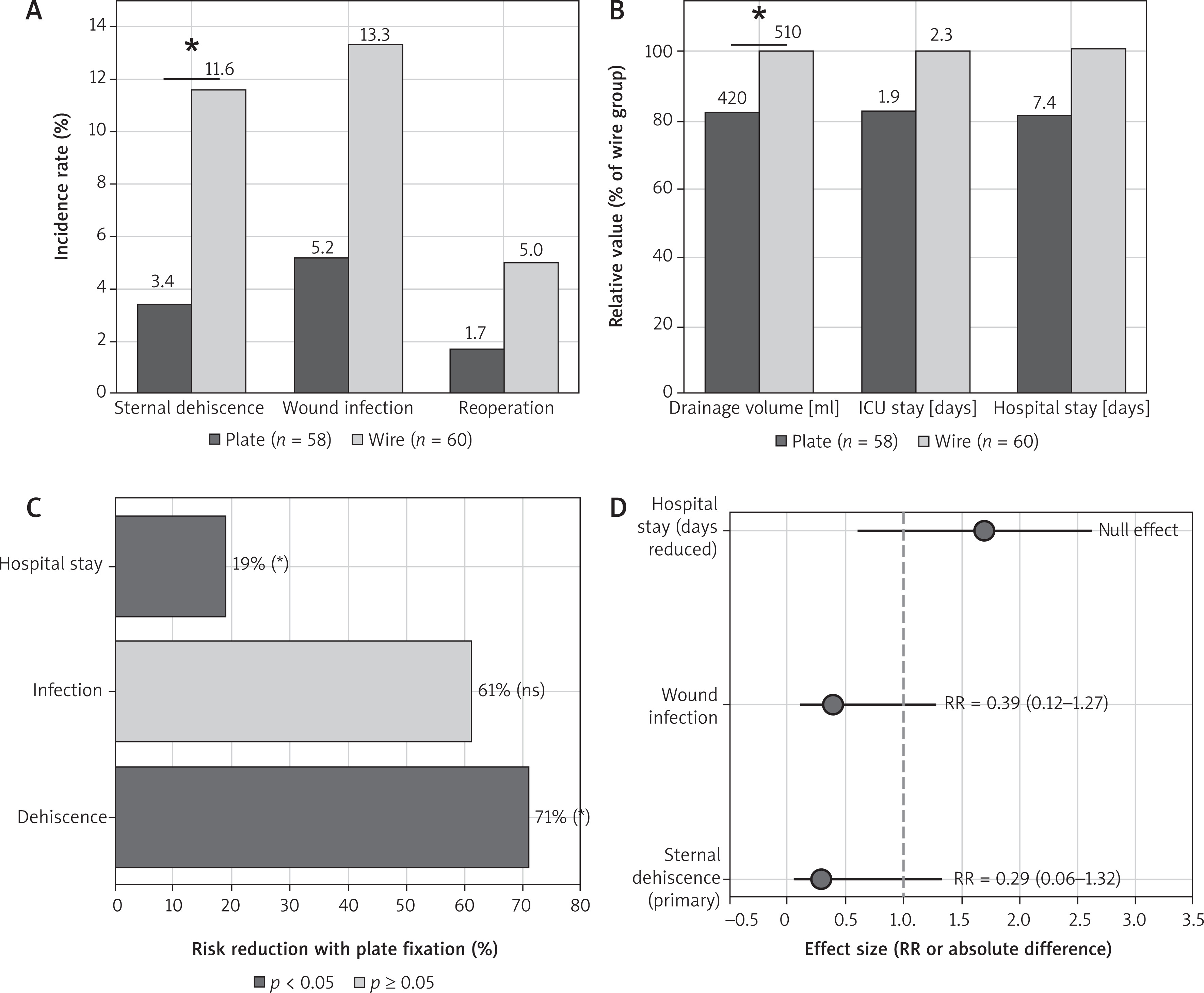
The primary outcome of sternal dehiscence occurred significantly less frequently in the plate fixation group compared to wire cerclage: 2 (3.4%) patients and 7 (11.6%) patients, respectively (p = 0.04; relative risk 0.29, 95% CI: 0.06–1.32). This represents a 71% relative risk reduction and an 8.2% absolute risk reduction with plate fixation. The number of patients needed to treat to prevent one episode of dehiscence was 12.
Secondary outcomes demonstrated consistent advantages with plate fixation (Table II). Deep sternal wound infection occurred in 1 (1.7%) patient in the plate group compared to 3 (5.0%) patients in the wire group (p = 0.32). When superficial and deep infections were combined, total wound infection rates were 5.2% (3/58) in the plate group and 13.3% (8/60) in the wire group (p = 0.08). Reoperation for sternal complications was required in 1.7% of patients in the plate group and 5.0% of patients in the wire group (p = 0.29).
Table II
Primary and secondary clinical outcomes
Postoperative drainage volumes were significantly reduced with plate fixation. The mean total 72-hour mediastinal drainage in the plate group was 420 ±160 ml compared to 510 ±180 ml in the wire group (mean difference –90 ml, 95% CI: –150 to –30 ml; p = 0.03), representing an 18% reduction.
The use of hospital resources favored plate fixation. While ICU stay was non-significantly shorter (1.9 ±0.7 vs. 2.3 ±0.8 days; p = 0.07), total hospital stay was significantly shorter in the plate group (7.4 ±2.1 vs. 9.1 ±2.6 days; p = 0.02), representing a 19% reduction.
Thirty-day mortality did not differ significantly between groups: 1 death (1.7%) in the plate group and 2 deaths (3.3%) in the wire group (p = 0.56). All deaths occurred in patients with multiple comorbidities and were attributed to cardiac causes unrelated to sternal complications.
These findings are summarized in Table II and Figure 3.
Figure 3
Comparison of clinical outcomes between plate and wire fixation groups. A – Complication rates showing a significantly lower incidence of sternal dehiscence and non-significantly less wound infection with plate fixation. B – Resource utilization parameters (postoperative drainage, intensive care unit stay, hospital stay) were normalized for the wire cerclage group and showed a reduction in drainage and shorter hospital stay with plate fixation. C – Relative risk reduction for primary outcomes: 71% reduction in dehiscence and 19% reduction in hospital stay (p < 0.05). D – Effect sizes and 95% confidence intervals (RR = relative risk) for primary and secondary endpoints
Subgroup analysis in high-risk populations
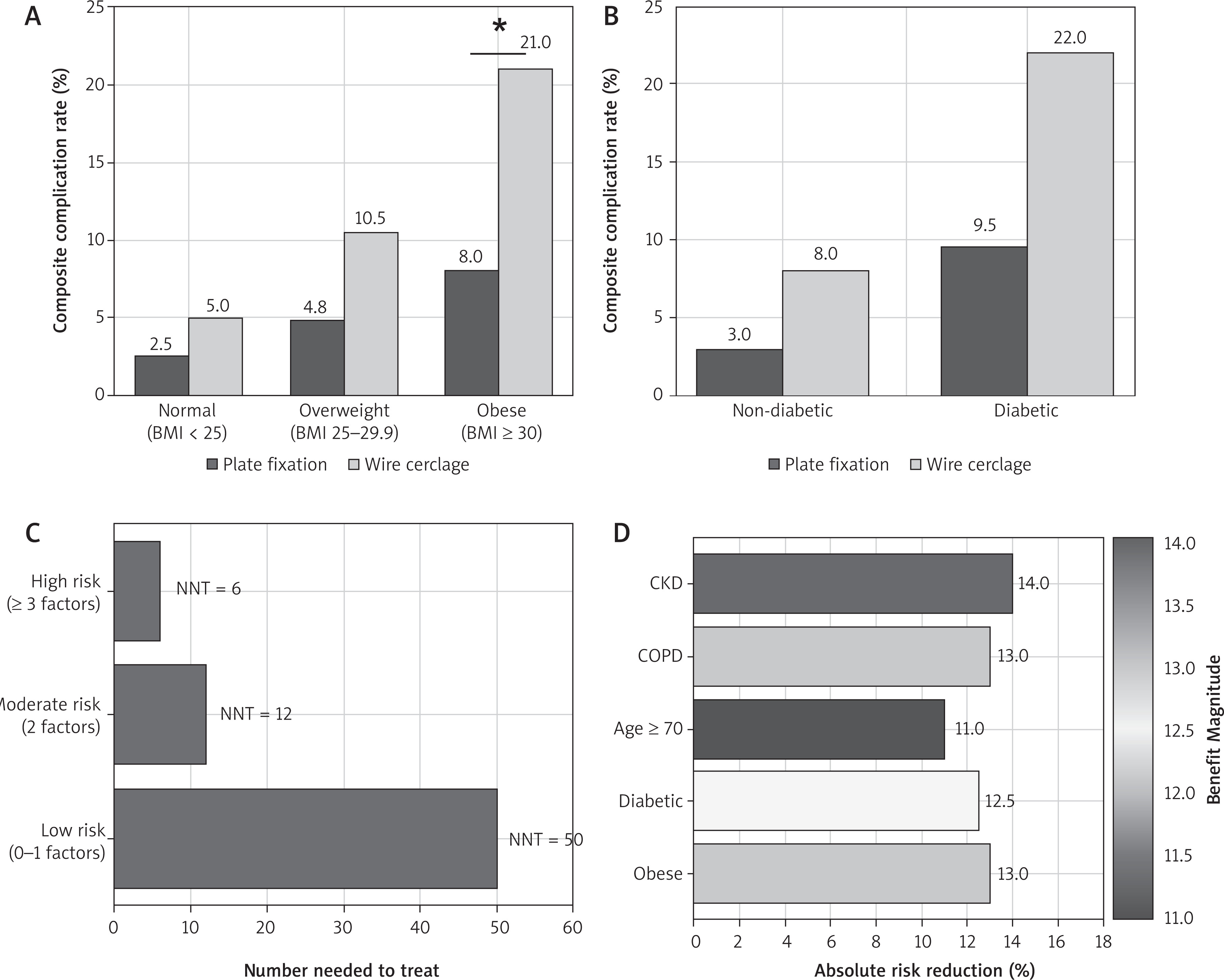
In obese patients (BMI ≥ 30 kg/m2), plate fixation was associated with lower complication rates. The composite complication rate (dehiscence or infection) was 8% (2/17) in the plate group compared to 21% (4/19) in the wire group (p = 0.04), representing a 62% relative risk reduction. In diabetic patients, complication rates were 9.5% (2/21) compared to 22% (5/23) (p = 0.08). In elderly patients (≥ 70 years, n = 28), complication rates were 7% in the plate group and 18% in the wire group (p = 0.06). In patients with multiple risk factors (obesity, diabetes, age ≥ 70), complications occurred in 12% (2/16) in the plate group and 31% (5/16) in the wire group (p = 0.02). Due to small subgroup sizes, formal interaction testing was not performed, and these findings should be interpreted cautiously.
Subgroup-specific results showing the effects of obesity, diabetes, and cumulative risk factors are shown in Figure 4.
Figure 4
Subgroup analysis showing the clinical benefits of plate fixation in high-risk populations. A – The effect of obesity on the composite complication rate, showing lower complication rates among obese patients treated with plate fixation (p < 0.05). B – The effect of diabetes mellitus, with consistently lower complication rates in the plate fixation group. C – Treatment efficacy according to the overall risk profile expressed as the number needed to treat (NNT); lower values indicate greater benefit. D – Magnitude of the absolute risk reduction by individual risk factors (obesity, diabetes, older age, COPD, CKD)
Discussion
This retrospective cohort study of 118 consecutive CABG patients suggests that rigid plate fixation may provide clinically significant advantages over traditional wire cerclage for sternal closure. Plate fixation was associated with a lower incidence of sternal dehiscence, reduced postoperative drainage, and a shorter hospital stay. Favorable trends were also observed in several high-risk subgroups, including obese patients. Overall, these findings support consideration of an individualized, risk-stratified approach to selecting a sternal closure technique.
The potential clinical advantages of plate fixation observed in our study are consistent with biomechanical principles established by finite element analysis and cadaveric studies. Wire cerclage creates point loads at the bone-wire interfaces, creating stress concentrations that can exceed the yield strength of bone, particularly during forceful coughing (peak forces of 200–300 N) [12, 17]. In contrast, plate systems distribute forces over a larger surface area through multiple fixation points, reducing peak stress by approximately 40% [12]. This mechanical advantage allows for reduced micro-mobility at the osteotomy site and earlier bone union.
Cadaver studies indicate that plate constructs provide 2–3 times greater stiffness and fracture load compared to wire cerclage [18]. Our clinical results confirm these laboratory findings, with dehiscence rates (3.4% vs. 11.6%) being quite similar to those reported in biomechanical estimates. The consistency between laboratory and clinical data strengthens confidence in the mechanical basis of plate superiority.
Our findings are broadly consistent with previous randomized controlled trials examining plate fixation. Raman et al. reported a 73% reduction in sternal complications with plates compared to wires in a multicenter randomized controlled trial (RCT) of 236 high-risk patients (1.3% vs. 4.6%; p = 0.07). Allen et al. demonstrated similar benefits with improvements in pain scores and lung function in the plate group in their study of 236 patients [14]. The present study extends these observations to an unselected CABG population and suggests that plate fixation may be associated with lower rates of sternal complications, reduced postoperative drainage, and shorter hospital stay.
Recent meta-analyses support the superiority of plates. A 2019 systematic review by Cataneo et al., analyzing 12 studies with 1,618 patients, found that rigid fixation reduced sternal complications (OR = 0.42, 95% CI: 0.26–0.68) [6]. More recently, a comprehensive systematic review and meta-analysis by Shrestha et al. including 2,452 patients across 14 comparative studies showed that rigid fixation was associated with significantly better sternal healing at 3 and 6 months, approximately 1 day shorter hospital stay, and less early postoperative pain compared to wire closure [19]. Our 3.4% dehiscence rate with plates falls within the 1–5% range reported in high-quality studies, while the 11.6% rate in our wire group reflects the high risk inherent in real-world practice, including in intermediate-risk patients.
Our exploratory subgroup analyses suggested that the potential benefits of plate fixation may be greater in selected high-risk populations. Recent observational data from Japan also support the use of rigid plate fixation in high-risk cohorts; Nakamura et al. reported a 2.1% rate of deep sternal wound infection and progressive computed tomography-documented sternal fusion in 48 high-risk patients treated with rigid plate fixation [20]. Obese patients (BMI ≥ 30 kg/m2) showed a 62% reduction in complications with plates, a finding that has important clinical implications given that the prevalence of obesity in contemporary cardiac surgery populations is approaching 40% [21]. Obesity impairs wound healing through several mechanisms: increased mechanical stress on the sternum from adipose tissue weight, decreased tissue oxygenation, and proinflammatory adipokine secretion. Plate fixation partially mitigates these factors through improved mechanical stability.
The overall wound infection rate observed in the wire cerclage group (13.3%) may appear higher than that reported in some recent series; however, this finding is consistent with real-world cohorts enriched with high-risk patients. Large observational studies and registry-based analyses have reported total sternal wound complication rates ranging from 8% to 20% in populations with high prevalence of obesity, diabetes, chronic obstructive pulmonary disease, and advanced age. In particular, Silverborn et al. [22] and Kubota et al. [23] have demonstrated that non-infectious sternal dehiscence and wound complications are common in routine clinical practice despite standardized preventive protocols. The relatively high infection rate in the wire cerclage group in our study may reflect the cumulative burden of patient-related risk factors rather than technical failure, reinforcing the need for improved mechanical stabilization strategies in vulnerable populations.
Similarly, diabetic patients showed significant benefits (9.5% vs. 22% complications). Diabetes impairs bone healing through advanced glycation end-product accumulation, decreased osteoblast function, and microvascular disruption [24]. The improved mechanical stability derived from plates may compensate for these biological shortcomings. Older patients (≥ 70 years) and those with multiple risk factors also experienced significant benefits, but sample sizes limited statistical power in these analyses. In addition to traditional plate systems, hybrid closure concepts have also been investigated. Miazza et al. presented combined band and plate fixation as a specific option for patients at high risk for sternal complications, reporting low morbidity and excellent early stability in a single-center experience [25].
Cost-effectiveness remains an important consideration in choosing a closure technique. Plate systems typically cost $800–$1,500 more than wire fixation and add 10–15 minutes of operative time. However, sternal complications impose significant economic burdens. Allen et al. reported that, despite higher upfront costs, plate fixation reduced total episode costs per patient by $3,352 due to complication prevention and shorter hospital stays [26]. Besides reducing sternal complications and length of stay, rigid plate fixation has also been integrated into enhanced recovery pathways. In a large single-center cohort of 608 patients, Gerdisch et al. reported that rigid plate fixation combined with an enhanced recovery protocol reduced opioid use in the hospital and at discharge to nearly zero without worsening pain scores or increasing readmissions [27]. At a typical cost of $2,000–$3,000 per day, the reduction in hospital stay we observed by 1.7 days suggests a potential savings of $3,400–$5,100 per patient, significantly exceeding the costs of the plate system.
Cost-effectiveness likely varies by risk profile. Routine plate use may not be cost-neutral in low-risk patients with dehiscence rates below 2%. Conversely, in high-risk patients with complication rates approaching 20–30%, plates offer compelling value. Risk-stratified approaches that reserve plates for high-risk patients optimize both clinical and economic outcomes.
Based on our findings and the existing literature, we recommend a risk-stratified approach to sternal closure: (1) High-risk patients (≥ 2 risk factors: obesity (BMI ≥ 30), diabetes, age ≥ 70, COPD, chronic kidney disease, osteoporosis, long-term mechanical ventilation): Rigid plate fixation is recommended as a first-line approach. (2) Moderate-risk patients (1 risk factor): Consider patient preferences, institutional expertise, and cost constraints. Plate fixation is reasonable but not mandatory. (3) Low-risk patients (no risk factors): Wire cerclage remains the acceptable standard approach, with plate retention based on surgeon preference or patient request.
Several limitations warrant consideration. First, the retrospective observational design introduces potential selection bias and confounding factors. While baseline characteristics were well balanced, unmeasured factors may influence the results. The choice of closure technique was not randomized but was based on surgeon preference and perceived risk, increasing the likelihood that plates would be preferred in high-risk patients. However, the direction and magnitude of any resulting selection bias cannot be determined with certainty and should be considered when interpreting the findings.
Second, the single-center design may limit generalizability. Institutional protocols, surgical expertise, and patient populations vary across centers. However, the characteristics of our center (tertiary referral, moderate case volume, standard protocols) are representative of many cardiac surgery programs and may support the generalizability of the findings.
Third, sample size limitations, particularly in subgroup analyses, reduced statistical power to detect smaller effect sizes. The study achieved 78% power for the primary outcome but was insufficient for rare events such as death. Larger studies are needed to confirm the subgroup findings and identify additional effect modifiers.
Fourth, follow-up was limited to 30 days for mortality assessment and 6 months for other outcomes. Late sternal complications occurring beyond this timeframe were not recorded. However, most dehiscence events occur within 2–4 weeks after surgery, suggesting that the follow-up period was adequate to capture the outcomes of interest. Fifth, we did not collect patient-reported outcomes such as pain scores, quality of life, or satisfaction, which are important patient-centered endpoints increasingly valued in surgical research. Future studies should include these measures.
A significant limitation of this study is the lack of a formal cost or cost-effectiveness analysis based on our institutional cohort. While economic impacts were discussed using data from the literature, we did not directly record device costs, operating room expenses, or post-discharge healthcare utilization. Consequently, the conclusions regarding the financial impact of rigid plate fixation should be interpreted cautiously. Future prospective studies including detailed cost accounting and life-year adjusted analyses are needed to definitively determine the economic value of rigid sternal fixation strategies in different healthcare systems. However, the observed reductions in dehiscence and length of hospital stay observed in our cohort suggest that rigid plate fixation may have the potential to offset its higher initial costs in selected high-risk patients.
Clinical perspective
What is known: Sternal dehiscence after median sternotomy leads to significant morbidity and healthcare costs. Rigid plate fixation offers biomechanical advantages over wire cerclage, but its use is limited.
What the study adds: This study found that rigid plate fixation was associated with a lower incidence of sternal dehiscence, shorter hospital stay, and reduced postoperative drainage compared with wire cerclage. Exploratory subgroup analyses suggested that these associations may be more pronounced in certain high-risk patient groups.
Clinical implications: These findings support consideration of a risk-stratified approach to sternal closure selection. However, larger prospective studies are required before specific recommendations regarding patient selection or routine use of rigid plate fixation can be made.
Conclusions
Several research priorities emerge from this study. First, prospective randomized trials with adequate power for subgroup analyses will definitively determine the most appropriate closure strategies for specific patient populations. Second, comparative effectiveness studies should examine newer fixation technologies (bioabsorbable plates, hybrid wire-plate constructs, cable systems) to determine whether they offer additional advantages. Third, the development and validation of clinical prediction models that incorporate multiple risk factors will enable precisely individualized risk stratification and closure technique selection. Fourth, cost-effectiveness analyses from various healthcare systems will inform resource allocation decisions and reimbursement policies. As evidence supporting rigid fixation continues to accumulate, its role in selected patient populations may warrant further consideration in future guideline development.
In conclusion, rigid plate fixation was associated with a lower incidence of sternal dehiscence, reduced postoperative drainage, and a shorter hospital stay compared to traditional wire cerclage for sternal closure after CABG. Exploratory subgroup analyses suggested that these associations may be more pronounced in certain high-risk patient groups; however, these findings should be interpreted cautiously because of the limited subgroup sample sizes. Although the economic implications of rigid plate fixation were not formally evaluated, the observed reductions in postoperative complications and hospital stay warrant further investigation. Taken together, these findings support consideration of a risk-stratified approach to sternal closure selection.
Larger prospective randomized trials are needed to confirm these observations, clarify the role of rigid plate fixation in specific patient populations, and inform future clinical practice.