Prosthetic arteriovenous grafts for hemodialysis are associated with a higher risk of infection if placed in the lower limbs. This is usually performed when upper-arm access options have been exhausted. When graft infection is complicated by graft thrombosis, this constitutes a clinical scenario that needs a special treatment strategy.
A 62-year-old female patient on dialysis presented with an infected left saphenofemoral arteriovenous graft, placed 8 years before her current admission. Her chronic medication regimen was unremarkable for the management of vascular access, except for antiplatelet therapy with clopidogrel 75 mg once daily. She had a history of gradual loss of kidney function and subsequent end-stage kidney disease due to complications after surgical treatment for morbid obesity that was performed 17 years ago. She had failed kidney transplantation on the right side, 4 years ago, which was attributed to poor implant quality and hypotension. Vascular access history included a left brachiocephalic fistula, an early failed right brachial-axillary graft, and a left brachial-axillary graft which remained functional for 9 years before the construction of the saphenofemoral graft. Four sessions of percutaneous graft thrombectomy were performed during this interval. The saphenofemoral graft underwent eight sessions (one session per year) of percutaneous thrombectomy with stent implantation in the venous anastomosis in previous years. Our patient presented to the emergency department due to a high fever (39.1°C) after the midweek dialysis session.
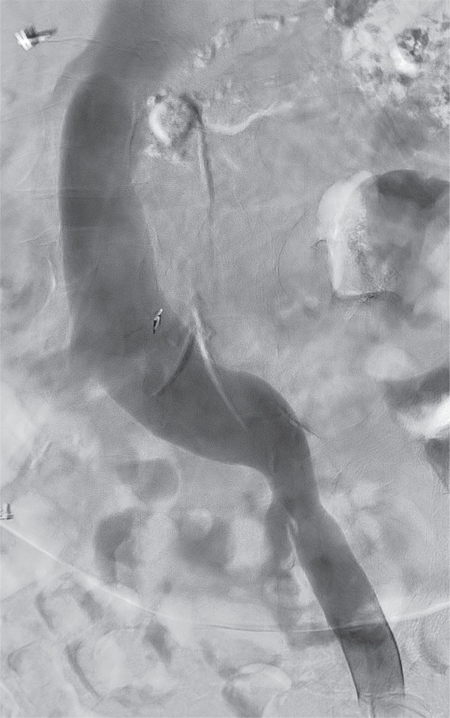
Computed tomography angiography at presentation revealed normal graft patency (Figure 1) with pus accumulation at the lateral aspect of the graft (Figure 2). No distant infectious loci were detected. A left femoral temporary hemodialysis catheter (THC) was inserted as no other options for cannulation were available due to venous obstruction of the right femoral and subclavian veins, as multiple catheters had been placed in the past (Figure 3). The left iliac axis and the inferior vena cava were patent (Figure 4).
Figure 1
Computed tomography angiography (CTA) revealed normal graft patency (red arrow: arterial anastomosis, blue arrow: venous anastomosis, green arrow: pus accumulation, green arrows: venous stent)
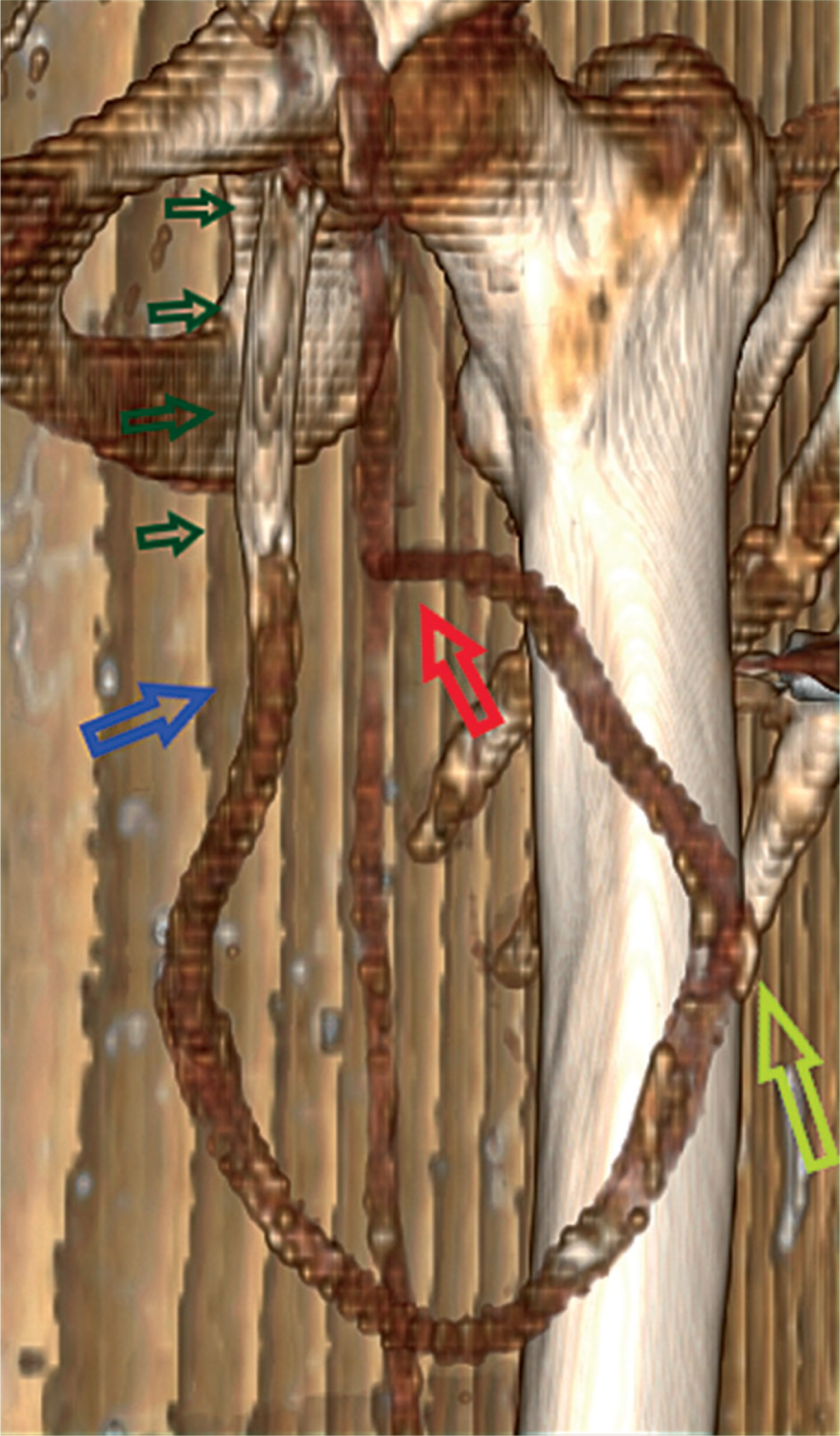
Figure 2
A – Pus accumulation was detected in the CTA around the locally destroyed graft from multiple cancellations. B – The locally infected graft
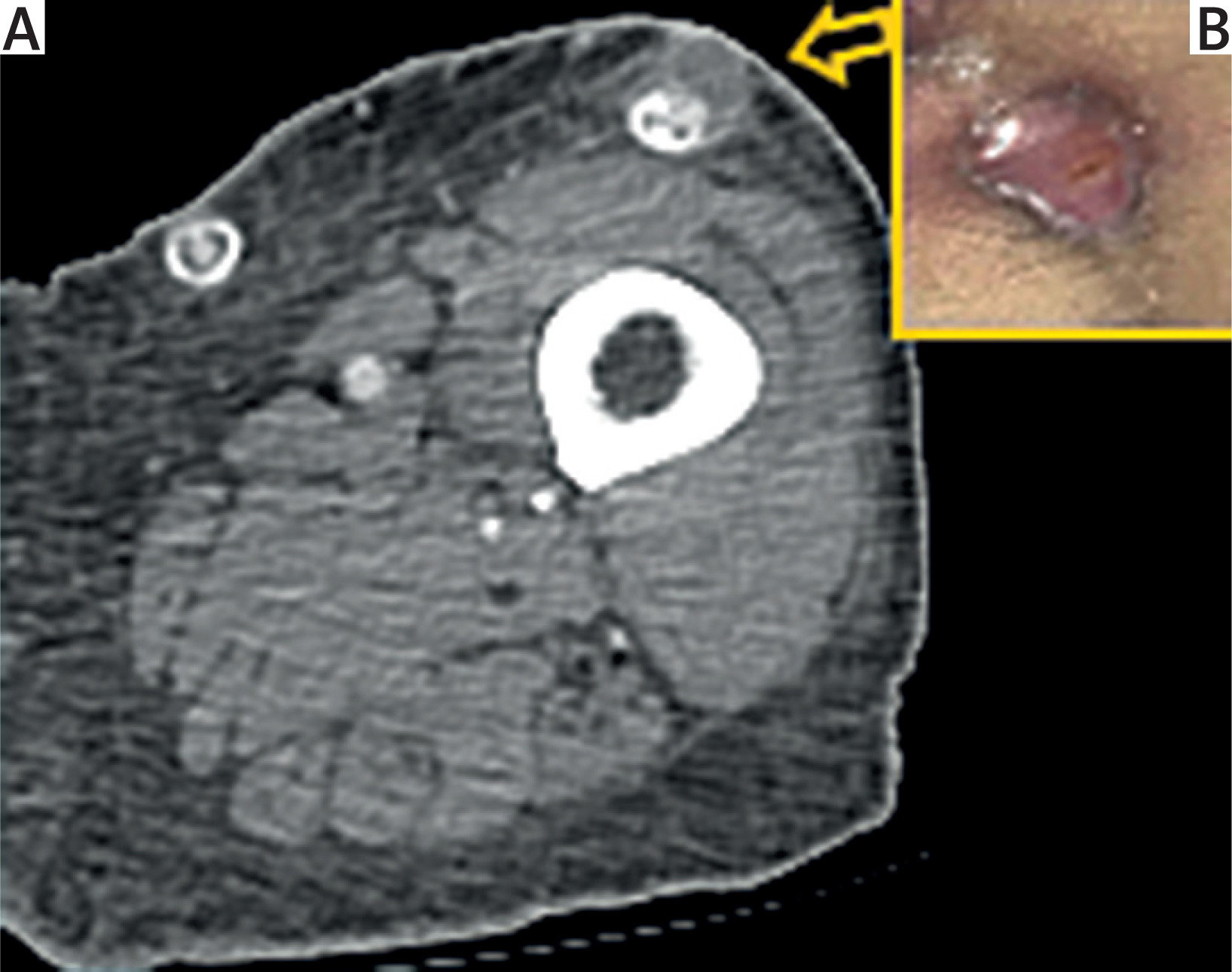
Figure 3
A left femoral temporary hemodialysis catheter (THC) was inserted (purple arrow) just proximal to a pre-existing venous stent (green arrows)
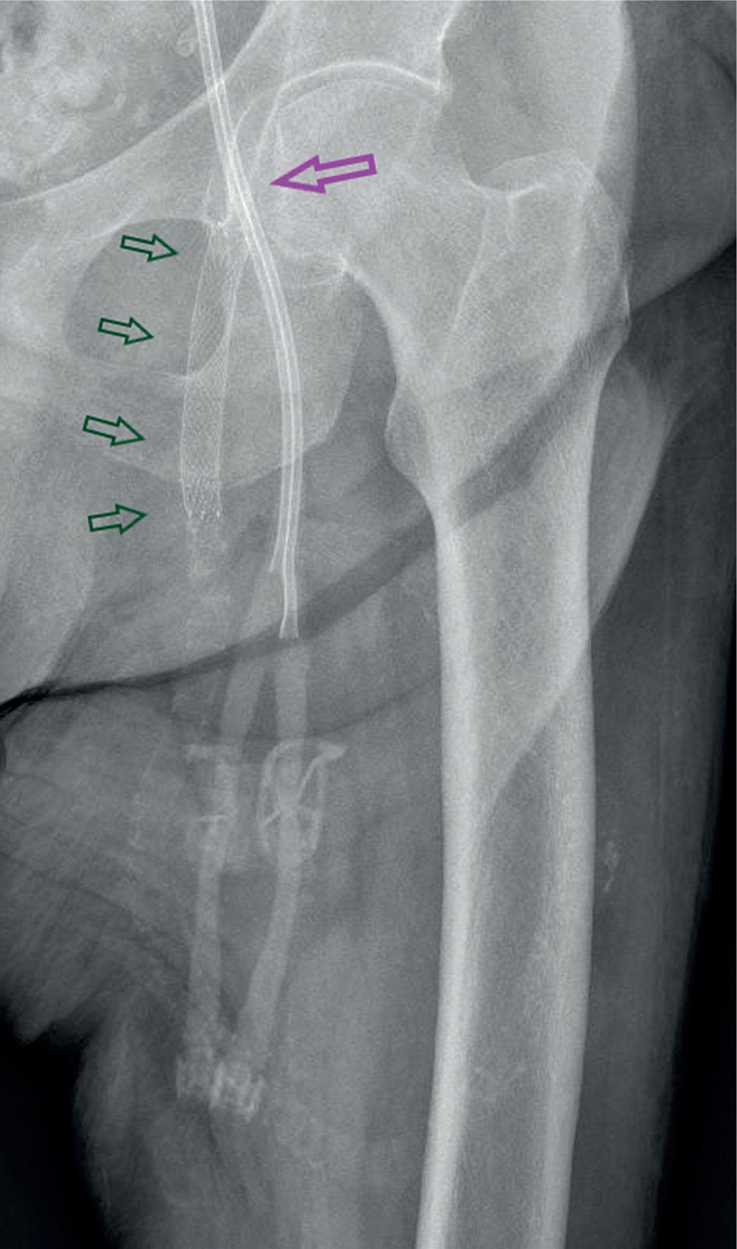
Figure 4
Digital subtraction phlebography revealed normal patency of the left iliac axis where the THC was inserted
Under general anesthesia, she underwent a bypass procedure with a 7 mm graft around the infected area in uninfected tissue planes, using a sterile drape to cover the skin, and afterward, the infected part of the graft was removed. Although the culture of the pus and the excised graft were negative, Staphylococcus aureus was detected in blood cultures. She received appropriate antibiotic treatment with daptomycin 750 mg q.o.d, rifampin 600 mg q.o.d., and amikacin sulfate 250 mg q.o.d. for 4 weeks. The wound was left open, and daily wound care was performed with dressings changes (Figure 5).
Figure 5
A – After removal of the infected segment the wound was left open (green arrow). The ipsilateral THC was seen medially (yellow arrow). B – The wound edges were approached with silk sutures 2 weeks later. After partial graft excision (gray arrows), the new graft (light blue arrows) was anastomosed (red arrows) to the old graft (light green arrows)
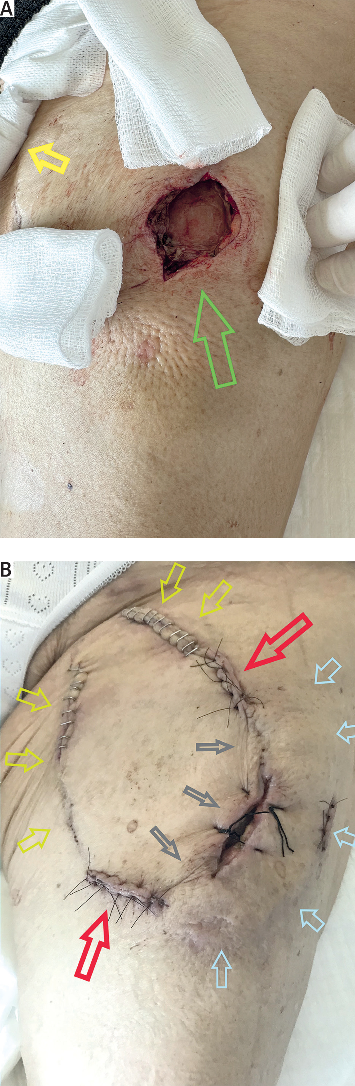
On the sixth postoperative day, the graft was thrombosed. The presence of an ipsilateral catheter may have contributed to graft thrombosis. Subsequently, the femoral THC was removed, and a new one was placed in the left subclavian vein after percutaneous thrombectomy of an existing venous stent. Consequently, an urgent Fogarty thrombectomy was performed in the saphenofemoral graft. Normal pulsation was achieved, but unfortunately, the graft was thrombosed again after 1 hour. We decided not to re-intervene due to the presence of infection and concerns about the presence of stenosis at the anastomosis sites or even more proximally, which may have been responsible for the re-thrombosis and thus would have made Fogarty thrombectomy impossible. We planned to recanalize the graft in the Interventional Radiology Suite (IRS) after regression of the infection and wound healing. Two weeks later, the wound edges came closer with the aid of two silk sutures (Figure 5).
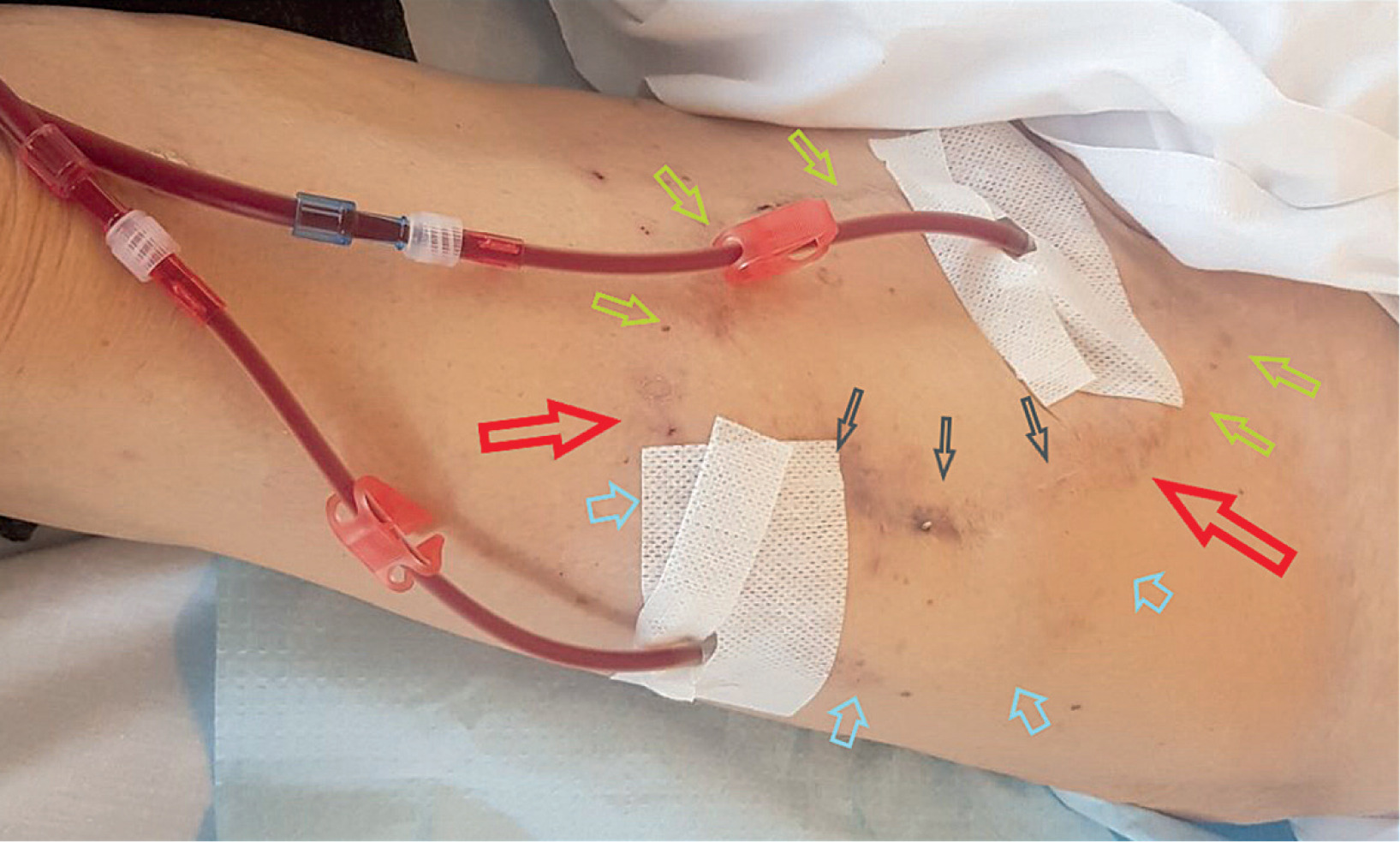
Sixty-eight days after the re-thrombosis, complete recanalization was achieved percutaneously in the IRS (Figure 6). She continued her antiplatelet treatment with clopidogrel 75 mg q.d. The patient receives satisfactory hemodialysis through the graft at 3 months follow-up (Figure 7).
Figure 6
Completion digital subtraction angiography after the percutaneous thrombectomy (orange arrow: proximal graftgraft anastomosis, red arrow: previous arterial anastomosis, blue arrow: previous venous anastomosis with the great saphenous vein, green arrows: venous stent)
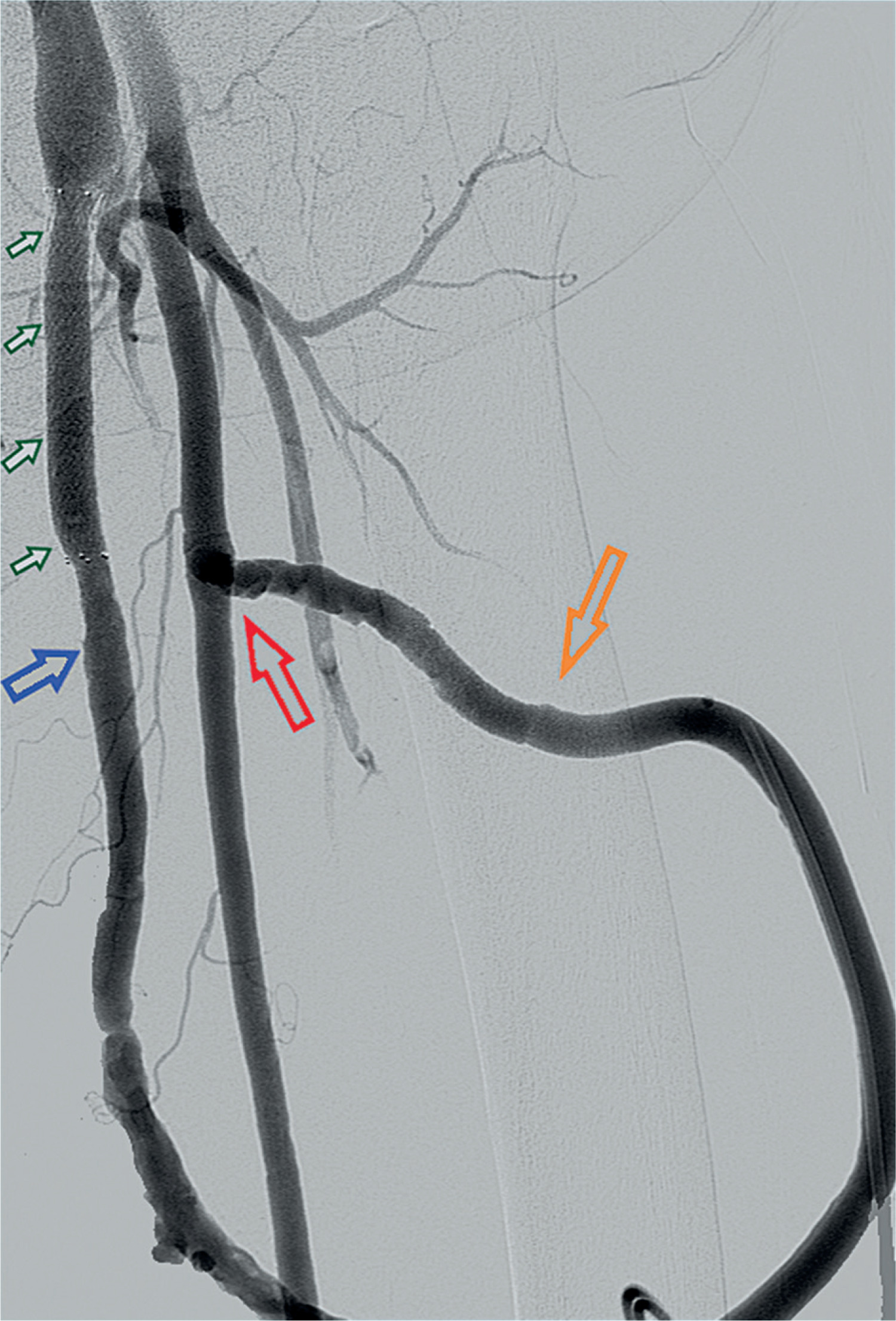
Figure 7
Successful graft cannulation. The new graft (light blue arrows) is connected (red arrows) with the old graft (light green arrows). The route of the excised part is shown (gray arrows)
The chronological sequence of infection, surgical management, postoperative thrombosis, and delayed endovascular recanalization is summarized in Table I. Prosthetic arteriovenous grafts carry a risk of about 10% for infection in contrast to 5% of native arteriovenous fistulas [1, 2]. Diagnosis is often established based on clinical signs such as erythema, skin break, purulence, and exposed graft fabric. Lower extremity location is a risk factor for graft infection among others including poor personal hygiene, diabetes mellitus, increased age, graft duration, repeated cannulation, buttonhole technique, and number of hospitalizations [3].
Table I
Timeline of clinical events and interventions
Most infections are local and caused by Gram-positive bacteria, mostly by Staphylococcus aureus, followed by Gram-negative organisms, and to a lesser extent by polymicrobial species [4]. Lower extremity graft infections are usually due to Gram-negative bacteria and may more often spread at other sites generating a remote infection [1, 5]. Thus, whole-body imaging is usually performed with computed tomography angiography, positron emission tomography, or nuclear scintigraphy [6, 7]. Ultrasound may detect local infection and complications such as fluid collections or pseudoaneurysm formation [8].
Bacteremia may be apparent in more than 50% of infected grafts, as in our patient, and local bleeding is present in more than 30% of cases [9]. Broad-spectrum antibiotics are regularly used followed by targeted antibiotic treatment after culture results [1].
Graft salvage is normally feasible when the infected part is remote to anastomoses and the treatment strategy involves a stepwise approach like the one we described concerning our patient [10]. If it involves the anastomoses, total graft excision is required with venous patch angioplasty of the femoral artery or extra-anatomic bypass after arterial ligation. In cases with mild infections, solely medical management could be sufficient [11, 12]. A lateral approach has been described to avoid groin infection with promising results [13]. Negative pressure wound therapy may be used to improve healing after debridement [14]. On the other hand, dysfunctional fistulas and grafts, as well as graft thrombosis, are nowadays treated by endovascular means [15, 16]. Thrombectomy devices may be used, or alternative techniques such as lysis-assisted balloon (LAB) thrombectomy, as we used in our patient [16]. This procedure includes initial lysis with 5 mg of recombinant tissue plasminogen activator (rt-PA) followed by thrombectomy with a high-pressure balloon to macerate the thrombus. The “facing sheaths” technique is employed [16].
Although pharmacological thrombolysis is generally considered ineffective in chronic thrombosis due to thrombus organization and fibrosis, the use of rt-PA in this case was not intended to achieve standalone chemical thrombolysis. Instead, rt-PA was administered as part of a LAB thrombectomy strategy, aiming to soften and partially fragment the organized thrombus and to facilitate subsequent mechanical disruption by high-pressure balloon angioplasty. In addition, local thrombolytic administration may reduce residual thrombotic burden and decrease the risk of early re-thrombosis following endovascular recanalization. Therefore, rt-PA was used as an adjunct to mechanical intervention rather than as a primary thrombolytic therapy, which is consistent with contemporary endovascular practice in chronically thrombosed arteriovenous grafts. A stent graft is electively used if stenosis persists [17, 18].
When graft infection is complicated with postoperative thrombosis a special problem arises, and a particular strategy is required. Open and percutaneous thrombectomy carries the risk of spreading the infection in the sterile graft segments, the bloodstream, and remote organs. A stepwise approach which includes treatment of the infection first and treating thrombosis at a later stage is feasible. Although early graft decluttering is preferable, the latter procedures may also be successful based on the current equipment and expertise. In our patient, we tried initially an open thrombectomy to avoid later endovascular techniques, but we finally chose to delay treatment until complete wound healing was achieved. Moreover, we realized that Fogarty thrombectomy would be insufficient as inaccessible proximal lesions would be apparent, amenable only to endovascular techniques. Another option would be to act earlier, but after the establishment of sterile blood and wound cultures and open wound, granulation was ongoing. This case highlights the process to salvage an infected and failed arteriovenous access when the patient does not have other access options. A staged approach and hybrid treatment with open surgery and percutaneous techniques may prove valuable.