The distribution of human cases of alveolar echinococcosis (AE) represents a significant public health concern in several regions, including central and eastern Europe, the Near East, Russia, China, and northern Japan [1]. AE is a parasitic disease that primarily affects the liver, with a predilection for the right lobe. The larva of the Echinococcus multilocularis parasite infiltrates the liver, and over time, it spreads to other organs through infiltration or metastasis formation [2].
AE is characterized by a prolonged asymptomatic incubation period, which typically lasts between 5 and 15 years. Following the latent phase the disease progresses chronically [1]. The clinical presentation of AE varies, with symptoms manifesting as cholestatic jaundice in approximately one-third of cases, and abdominal pain in another one-third of cases. In some patients, the diagnosis of AE is incidental, discovered during the investigation of various non-specific symptoms such as fatigue, weight loss, hepatomegaly, or abnormalities detected through ultrasound or routine laboratory tests [3]. The multifaceted clinical features and the insidious nature of AE present significant challenges for early detection and appropriate management.
This case report describes an unusual presentation of advanced AE with an asymptomatic course and underscores the significance of considering this parasitic infection as a potential differential diagnosis in regions where it is endemic. We present a case of Echinococcus multilocularis infection in a 39-year-old female patient. She was initially admitted to the Pulmonary and Tuberculosis Department following the incidental detection of a lung mass in the right hemithorax during a work-related X-ray. The mass measured 96 mm in diameter and was localized in the lower regions of the right lung. Notably, the patient remained asymptomatic throughout the disease.
Subsequent computed tomography (CT) imaging revealed a multi-chambered, thick-walled fluid area with calcifications originating from IV and VIII of the liver, exerting pressure on the diaphragmatic dome. Consequently, the patient was referred to the Department of Parasitology and Tropical Medicine for further evaluation. Magnetic resonance imaging (MRI) confirmed the existence of a polycyclic structure measuring 150 × 112 × 126 mm in the right lobe of the liver, significantly impacting the diaphragmatic dome and protruding into the thoracic cavity. The lesion demonstrated the involvement of the inferior vena cava, accompanied by an adjacent fragment of collapsed lung parenchyma. Additionally, irregular cystic foci, possibly representing progenitor cysts, were identified within the lung parenchyma.
A CT scan of the head was performed to exclude cerebral invasion and yielded negative results. Serological tests confirmed the diagnosis of Echinococcus multilocularis infection, consistent with the imaging findings. Albendazole therapy was initiated, and the patient was transferred to the Department of General and Transplant Surgery for a scheduled surgical intervention involving two body cavities. A right posterolateral thoracotomy incision was performed, followed by resection of segments of the middle and lower right lung lobes. Intraoperatively, pericardial infiltration was observed. The lesion was excised, and a pericardial patch was sutured in place. Subsequently, a portion of the infiltrated diaphragm was resected to facilitate access to the liver lesion. The liver lesion was excised, and suturing of the diaphragm was performed, followed by the insertion of a chest tube into the right pleural cavity.
The surgical procedure was complicated by bleeding from a chest vessel, necessitating the intraoperative transfusion of packed red blood cells. Following the procedure, the patient was transferred to the intensive care unit (ICU). A follow-up CT scan performed 4 days later revealed right-sided edema measuring 75 × 13 mm, for which the chest tube was retained to drain air and residual fluid. No further complications were observed. The subsequent pathological examination, consistent with the clinical presentation, confirmed AE involving the liver, lung, and pericardium. After a recovery period, the patient was discharged home in good condition (Figure 1).
Figure 1
A – Echinococcal cyst originating from the right lobe of the liver (segments VIII and IV) and invading the mediastinum. B – Cystic lesions with calcifications measuring approximately 125 × 89 mm originating from the right hepatic lobe and extending through the esophageal hiatus into the mediastinum and pericardium; an adjacent lesion of similar appearance in the right pleural cavity measuring 92 × 60 mm. Atelectasis due to compression in the surrounding lung parenchyma. C – Follow-up MRI. A residual fluid space measuring 39 × 11 mm in segment IVa of the liver; no features of a parasitic cyst were found. D – Follow-up CT scan 1 year after surgery. Scarring in segment VIII of the liver
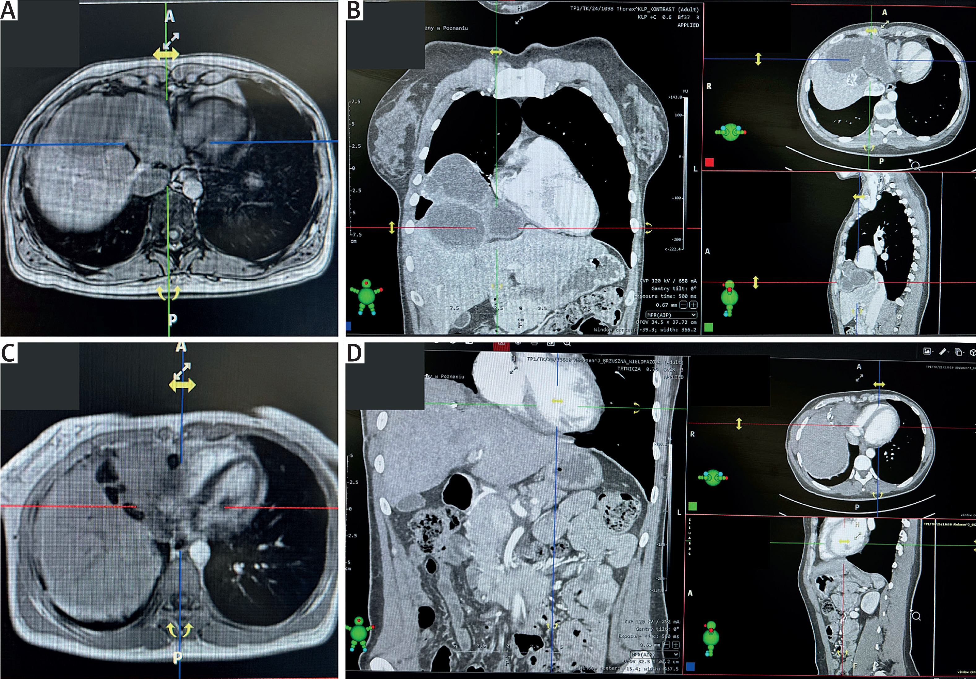
A 2-month follow-up CT scan demonstrated satisfactory healing, absence of edema, and no free fluid in the abdomen or thorax. However, the scan revealed the presence of a fluid-filled space in segment VIII of the liver, raising suspicion of residual echinococcal disease. The administration of albendazole was continued, and the patient remained under regular monitoring and care at the Parasitology and Tropical Medicine outpatient clinic.
This particular case presents a unique diagnostic pathway for AE. Unlike the typical diagnostic pathway, the diagnosis in this case was incidental, stemming from a work-related X-ray examination of the lungs. In the typical course of a disease, ultrasonography (US) examination plays a pivotal role in the diagnosis of AE. The clinical criteria for diagnosing AE emphasize the importance of conducting an initial radiological examination to rule out pulmonary and cerebral involvement [1]. This step is crucial in the diagnostic process to ensure a comprehensive evaluation of the disease and guide appropriate treatment strategies. While AE predominantly affects the liver and has a propensity for metastasis to other organs, such as the lungs [3], the involvement of the pericardium is relatively rare [4].
This emphasizes the diverse nature of AE and the potential for unexpected disease dissemination, necessitating a comprehensive evaluation and tailored treatment approach. Treatment planning in AE necessitates a multidisciplinary discussion, incorporating all available pre-treatment imaging findings. Radical surgery and chemotherapy remain the preferred treatment approach for AE cases that are amenable to complete lesion resection [1]. The primary therapeutic objective is complete (R0) resection, in accordance with oncological surgical principles. In untreated or inadequately treated cases of AE, the prognosis is exceptionally grave. Studies have shown that the mortality rate in such cases exceeds 90% within 10 to 15 years of diagnosis [5]. Following the initiation of any form of treatment for AE, it is essential to establish a comprehensive long-term follow-up plan. This includes regular monitoring using US at shorter intervals and CT and/or MRI at intervals of 2 to 3 years [1].