Coronary artery disease (CAD) is a major cause of morbidity and mortality among patients with end-stage renal disease (ESRD), accounting for approximately 39–45% of all deaths [1]. Evidence suggests that coronary artery bypass grafting (CABG) offers superior outcomes in this population compared to medical therapy or percutaneous coronary intervention (PCI) [2]. However, the selection of conduits for CABG in dialysis-dependent patients is limited. The radial artery is typically unsuitable once an arteriovenous fistula (AVF) has been created for hemodialysis access. In addition, long-standing renal dysfunction often leads to significant atherosclerosis of the ascending aorta, making proximal aortic anastomosis technically challenging and increasing the risk of cerebral embolization.
Bilateral internal mammary artery (BIMA) grafting presents an appealing option as it avoids manipulation of the aorta. However, concerns persist about the risk of coronary steal when using an in-situ internal mammary artery (IMA) graft on the same side as the AVF to revascularize the left anterior descending (LAD) artery [2].
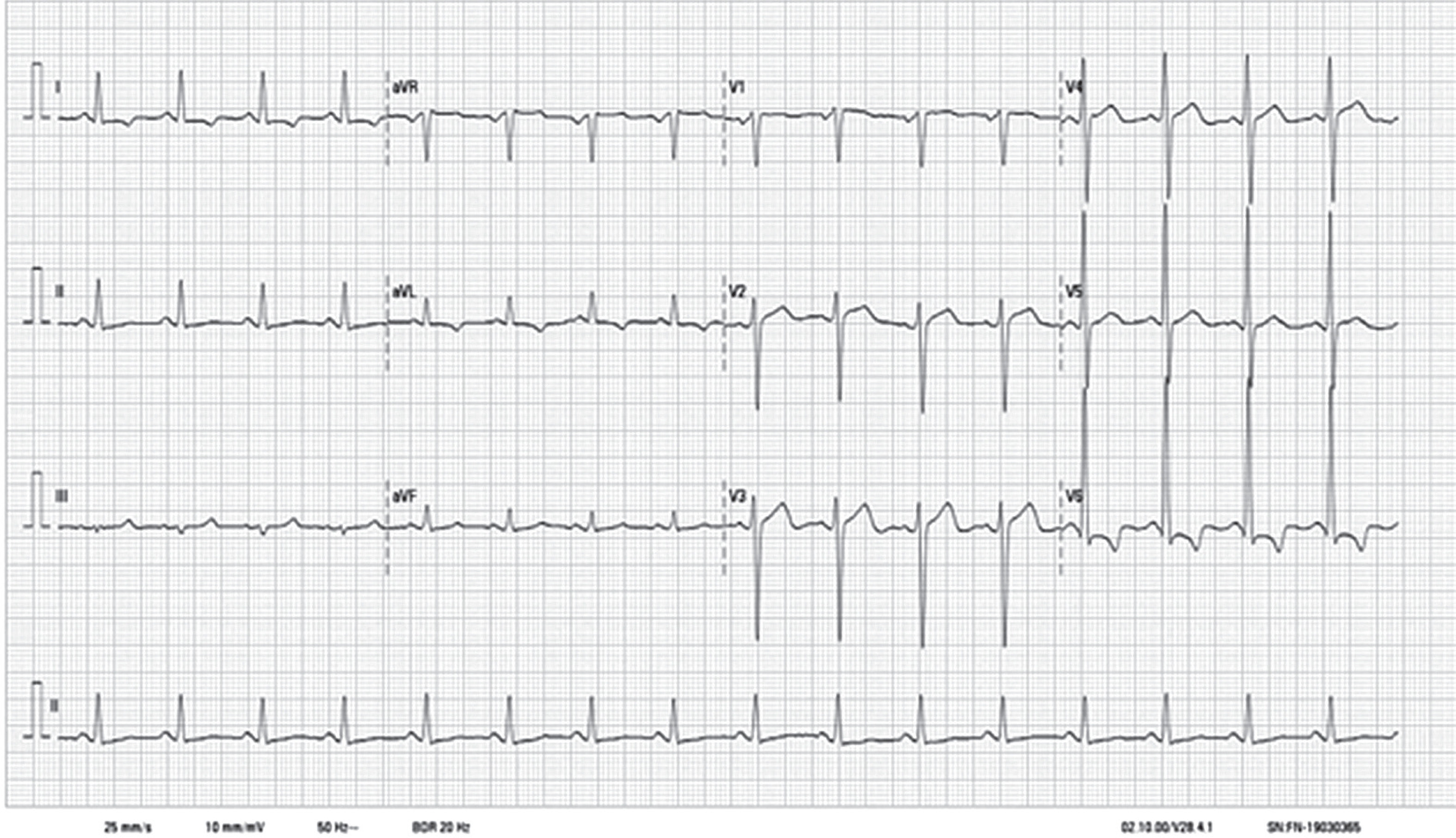
We present 3 cases of patients with chronic kidney disease on maintenance hemodialysis, all diagnosed with triple-vessel coronary artery disease, who underwent off-pump, total arterial CABG. In each case, an in-situ IMA graft ipsilateral to the AVF was used as the sole inflow conduit. All patients had uneventful postoperative recoveries, and their clinical characteristics, operative details, and outcomes are summarized in Table I (Figures 1 and 2).
Table I
Summary of case characteristics, operative details, and clinical outcomes
| Case | Age/sex | Comorbidities | Dialysis access | LV function (EF) | Angiography findings | Surgical strategy (OPCAB) | Post-operative course | Follow-up outcome |
|---|---|---|---|---|---|---|---|---|
| 1 | 50/M | Diabetes, HTN, CKD | Left forearm Radiocephalic AVF (immature); Permcath initially | Severe dysfunction (25%) | Triple-vessel CAD; no SAS (Figure 1) | LIMA–LAD; LIMA–RIMA–Y to Ramus and OM (PDA not grafted) | Uneventful; permcath dialysis until AVF maturation; discharged on POD 12. Received thrice weekly dialysis through AVF during postoperative period | DSE at 3 months: EF 42%, improved LV function: Improved ventricular contraction on incremental-dose dobutamine infusion; stable dialysis via AVF |
| 2 | 59/M | Diabetes, HTN, CKD V | Left forearm radiocephalic AVF | Preserved (65%) | Triple-vessel CAD; no SAS | LIMA–LAD; LIMA–RIMA–Y sequential to OM and PDA | Extubated on day of surgery; discharged on POD 4; 2 cycles of dialysis via AVF during post-operative period | 6 weeks: Stable, asymptomatic; listed for renal transplant |
| 3 | 59/M | HTN, CKD (2 years on MHD) | Left upper arm brachiocephalic AVF | Moderate dysfunction (45%) | Triple-vessel CAD; R ICA stenosis; no SAS | LIMA–LAD; LIMA–RIMA–Y to OM (PDA not grafted | Extubated on POD 1, discharged on POD 5. underwent 2 dialysis session through AVF, with no ischemic changes observed in ECG during dialysis (Figure 2) | At 6 weeks, stable, asymptomatic, uneventful dialysis through AVF |
[i] HTN – hypertension, CKD – chronic kidney disease, CKD V – Stage V chronic kidney disease, MHD – maintenance hemodialysis, AVF – arteriovenous fistula, Permcath – permanent catheter, LV – left ventricle, EF – ejection fraction, CAD – coronary artery disease, SAS – subclavian artery stenosis, CABG – coronary artery bypass grafting, LIMA – left internal mammary artery, RIMA – right internal mammary artery, LAD – left anterior descending artery, OM – obtuse marginal artery, OPCAB – off-pump coronary artery bypass, PDA – posterior descending artery, Ramus – ramus intermedius artery, POD – postoperative day, R ICA – right internal carotid artery, DSE – dobutamine stress echocardiography.
Figure 1
A, B – preoperative imaging of CT angiography neck and brain vessels showing normal neck and cerebral vessels
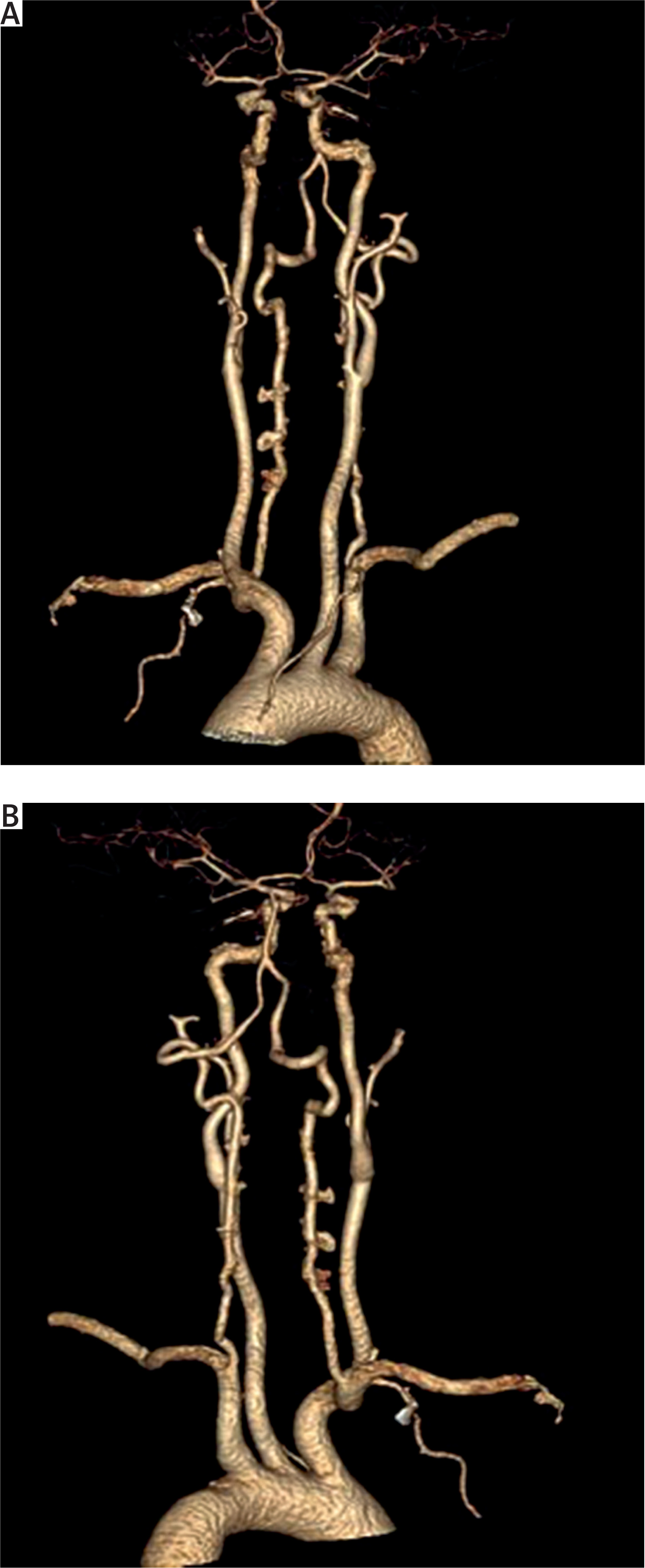
Figure 2
Electrocardiogram obtained during dialysis in one of the study patients, showing no ischemic changes
Cardiac complications are the leading cause of death in patients on maintenance hemodialysis, with about 15% due to myocardial infarction and most others from left ventricular dysfunction [2]. Screening for CAD is crucial before renal transplantation, as untreated CAD increases the risk of graft failure. Revascularization is recommended for significant CAD, but the ideal approach remains debated. While PCI is more commonly performed, studies show that CABG offers better long-term outcomes in ESRD patients [1, 2]. However, prognosis remains poor due to widespread atherosclerosis, comorbidities, and often reduced cardiac function [2].
Selecting the appropriate graft conduit for surgical revascularization, particularly for the LAD artery, is a matter of ongoing debate. Radial arteries are often unavailable due to prior use for dialysis access, and saphenous vein harvesting may be limited in patients with peripheral vascular disease. Moreover, atherosclerosis in the ascending aorta complicates proximal anastomosis and raises the risk of cerebral embolism [2]. Consequently, the IMA is often the preferred conduit.
However, when an AVF is present on the left side (radiocephalic or brachiocephalic), the use of an in-situ left IMA as the sole inflow source raises concerns about the possibility of coronary steal. The concept of “coronary steal” remains controversial. Some studies have reported that using an in-situ IMA ipsilateral to the AVF may divert blood away from the coronary circulation [3], while others have not found any association [1, 4]. Hachiro et al. [1] demonstrated that using an ipsilateral in-situ IMA in dialysis-dependent patients was not associated with adverse outcomes.
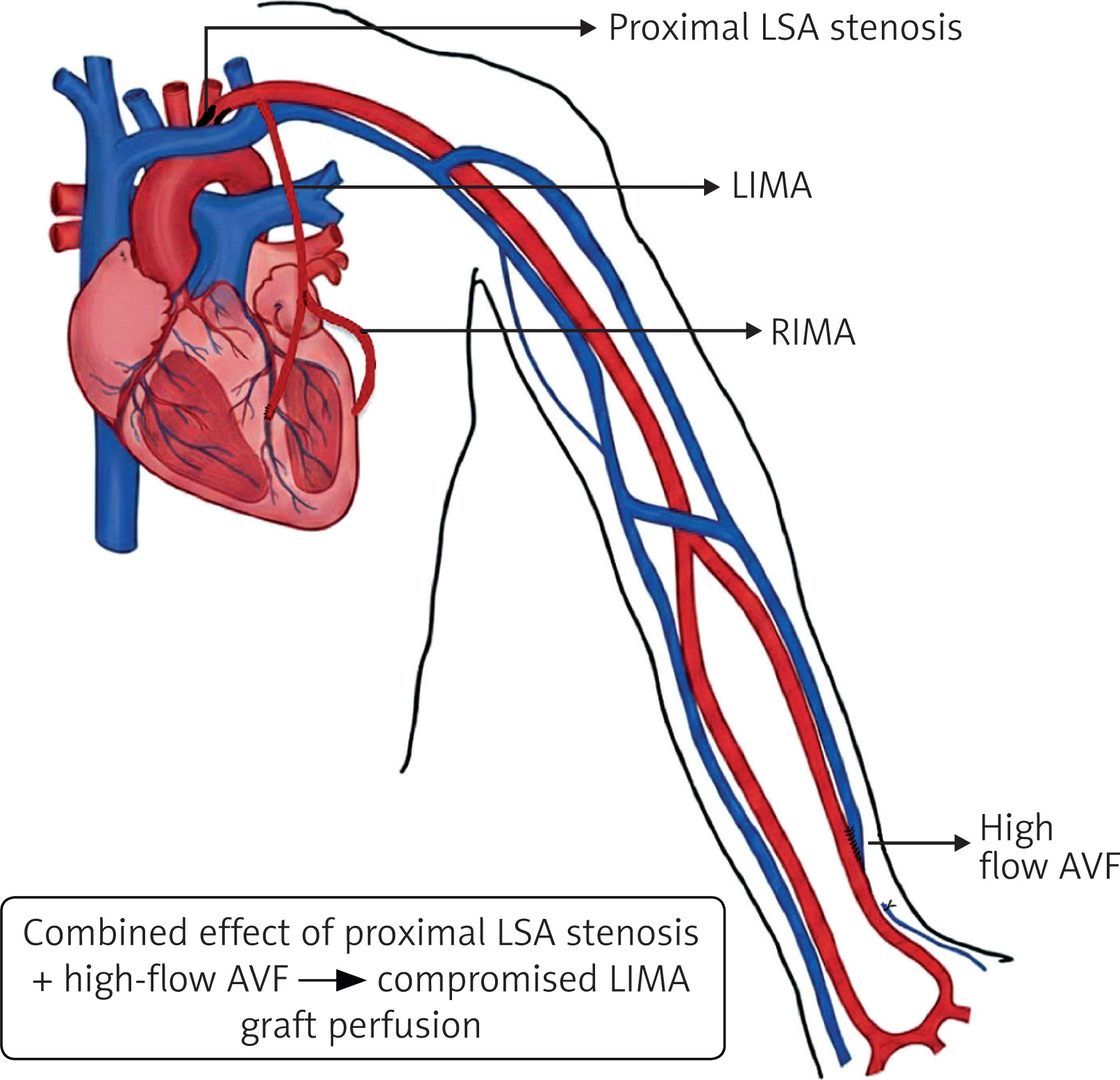
Importantly, coronary steal is more likely when there is significant stenosis of the ipsilateral subclavian artery [3]. In such cases, blood flow may preferentially divert into the AVF rather than the coronary circulation (Figure 3). Therefore, it is recommended to measure blood pressure in both upper limbs and avoid using the IMA on the side with a pressure difference greater than 15 mm Hg, which may indicate significant subclavian artery stenosis [3]. Other risk factors for coronary steal include abnormal IMA anatomy, a proximally located AVF, and progressive increases in AVF flow over time [1, 5].
Figure 3
Schematic illustration of the hypothesized LIMA graft hypoperfusion in the setting of proximal left subclavian artery stenosis and ipsilateral arteriovenous fistula, demonstrating competitive flow steal affecting a LIMA–RIMA composite Y graft
LIMA – left internal mammary artery, RIMA – right internal mammary artery, LSA – left subclavian artery, AVF – arteriovenous fistula.
Despite these concerns, evidence suggests that using an in-situ IMA ipsilateral to the AVF is safe in appropriately selected patients. BIMA grafting, although associated with improved long-term outcomes, carries a higher risk of sternal wound complications [2], particularly in diabetic or chronic renal failure patients. To mitigate this, skeletonized harvesting of the IMA is recommended to preserve sternal blood supply [2].
The location of the AVF also influences the likelihood of coronary steal. Proximally located, high-flow AVFs are more likely to result in steal phenomena than distal ones, especially in the presence of subclavian artery stenosis [6–8]. Agarwal et al. [8] reported a case of symptomatic subclavian steal syndrome due to a high-output AVF and subclavian stenosis, where AVF revision was required. In our case series, all our patients had an AV shunt in the left forearm and proximal arm, with no subclavian artery stenosis.
Narita et al. [9] reported a case of severe cardiogenic shock following CABG in a dialysis-dependent patient, where an in-situ IMA graft was used on the same side as an AVF as the sole source of coronary inflow. The patient required postoperative support with extracorporeal membrane oxygenation (ECMO) and an intra-aortic balloon pump (IABP). Hemodynamic status improved following ligation of the AVF. However, the condition of the subclavian artery – an important independent risk factor for coronary steal – was not assessed or reported in the case.
We found that total arterial revascularization using composite BIMA grafts, with the in-situ IMA on the side of the AVF as the sole inflow source, was both feasible and safe. This approach avoids aortic manipulation, thereby reducing stroke risk, improves graft patency, eliminates leg wound complications, and supports total arterial revascularization. Nonetheless, careful preoperative evaluation, including assessment of subclavian artery patency and AVF flow characteristics, is essential for optimal outcomes.
The study is restricted by its very small sample size, short follow-up, and absence of objective graft patency or AVF flow measurements. The findings are therefore preliminary and should be considered as hypothesis-generating.
In conclusion, conduit selection for CABG in dialysis-dependent patients with an ipsilateral AVF remains a clinical challenge due to concerns about coronary steal. In this small case series, total arterial revascularization using an in-situ IMA ipsilateral to the AVF as the sole inflow source was technically feasible and associated with uneventful recovery, preservation of dialysis access, and no ischemic complications during hemodialysis. These findings suggest that, with careful preoperative assessment of subclavian patency and AVF flow, ipsilateral IMA use may represent a safe and effective revascularization strategy in selected patients. Larger studies with longer-term follow-up are required to confirm its safety and durability.