eISSN: 1644-4124
ISSN: 1426-3912
Central European Journal of Immunology
Current issue Archive Manuscripts accepted About the journal Special Issues Editorial board Abstracting and indexing Subscription Contact Instructions for authors
Editorial System
Submit your Manuscript
SCImago Journal & Country Rank
3/2023
vol. 48
 
Share:
Share:
Experimental immunology

Novel inflammatory markers in patients with severe COVID-19 and a pulmonary thrombotic event

Jarosław Bakiera
1
,
Karolina Strzelec-Pawełczak
2
,
Katarzyna Czarnek
3
,
Ida Osuchowska-Grochowska
2
,
Jacek Bogucki
4
,
Agnieszka Markiewicz-Gospodarek
2
,
Aleksandra Górska
2
,
Zuzanna Chilimoniuk
2
,
Sebastian Radej
2, 3
,
Mateusz Szymański
2
,
Piero Portincasa
5
,
Cezary Grochowski
2, 3

1.
Department of Laboratory Diagnostics, Coagulation and Microbiology, Stefan Wyszyński Regional Specialist Hospital, Lublin, Poland
2.
Department of Human Anatomy, Medical University of Lublin, Lublin
3.
Institute of Health Sciences, The John Paul II Catholic University of Lublin, Lublin, Poland
4.
Department of Organic Chemistry, Medical University of Lublin, Lublin, Poland
5.
Clinica Medica “A. Murri”, Department of Biomedical Sciences & Human Oncology, University of Bari “Aldo Moro” Medical School, Bari, Italy
Cent Eur J Immunol 2023; 48 (3): 167-173
Online publish date: 2023/09/21
Article file
- Novel inflammatory.pdf  [0.18 MB]
Get citation
 
PlumX metrics:
 

Introduction

Venous thromboembolism (VTE), clinically manifested as deep vein thrombosis (DVT) or acute pulmonary embolism (PE), is the third most common acute cardiovascular syndrome following myocardial infarction and stroke [1]. The annual incidence of PE is between 39 and 115 per 100,000 inhabitants. The incidence of VTE is almost eight times higher in people aged 80 and older than in the fifth decade of life [2]. Therefore, with the incre- asing age of the population, long-term studies show an increasing tendency in the annual incidence rates of PE over time [3-6]. A recent analysis from the World Health Organization (WHO) mortality database (2000-2015) found an average of 38,929 PE-related deaths each year in 41 countries of the WHO European Region (including Central Asia) representing a population of approximately 651 million [7]. Between 2000 and 2015, the annual age-standardized death rates associated with PE decreased by almost 50% (from 12.7 to 6.5 deaths per 100,000 inhabitants) with no significant gender differences. Despite this overall positive trend, the study also found that PE-related mortality continues to increase exponentially with age, reaching or even exceeding 80 deaths per 100,000 elderly population, and that PE also remains a relatively important cause of (in comparison with other causes) deaths among younger women, in whom it accounted for up to 13 cases per 1000 deaths [7, 8].

COVID-19 first occurred in Wuhan (Hubei province) and the cause of the infection was soon identified as a coronavirus [1]. However, a great number of cases occurred without any in the Huanan animal market, which suggested human-to-human transmission. Since the outbreak of coronavirus disease 2019 (COVID-19), clinicians have struggled with the attempt to diagnose and manage the severe and fatal complications of COVID-19 appropriately. Several reports have described significant procoagulatory events, including life-threatening pulmonary embolism in these patients [9-58]. SARS-CoV-2 invades the host cell by using the superficial S protein complex (spike protein complex). The complex consists of two subunits: S1 and S2. The S1 domain binds the appropriate receptor on the host cell surface, and, at the same time, the S2 domain is responsible for membrane fusion [10]. Several studies have pointed to the ACE2 host cell receptor as a crucial molecule in SARS-CoV-2 invasion.

While ACE2 mRNA is found in all organs, ACE2 protein expression is detected in the heart, kidney, testis, lung type I and type II alveolar epithelial cells, nasal, and oral mucosa and nasopharynx (basal layer of the non- keratinizing squamous epithelium). Additional tissues include smooth muscle cells and endothelium of vessels from the stomach, small intestine and colon, in smooth muscle cells of the muscularis mucosae and the muscularis propria, in enterocytes of all parts of the small intestine including the duodenum, jejunum, and ileum (but not the colon), skin (basal cell layer of the epidermis extending to the basal cell layer of hair follicles, smooth muscle cells surrounding the sebaceous glands, cells of the eccrine glands), endothelial, and smooth muscle cells of the brain.

Affinity to the infected tissue shown by coronaviruses (CoVs) is dependent on surface cell ACE-2 expression. Recent publications suggest stronger affinity to ACE-2 presented by SARS-CoV-2 than to other known CoVs [11-13, 47-49]. The activity of S1 and S2 domains is controlled with the assistance of many enzymes such as cathepsin L, furin and transmembrane protease serine 2 (TMPRSS2). All of these enzymes are present in various tissues of the intestine, liver and lungs. It proves that SARS-CoV-2 could invade different systems and organs. A two-step model of cleavage was proposed: first separating S1 and S2, second activating the S2 site. S proteins could be cleaved by one of the specific proteases mentioned above. Co-expression of TMPRSS2 and ACE2 was found in pneumocytes, indicating the importance of TMPRSS2 in SARS-CoV-2 invasion. TMPRSS2 as a transmembrane enzyme cleaves the S1/S2 complex and activates the S2 domain. TMPRSS2 acts in the lumen of the viral endocytic vesicle. Interestingly, TMPRSS2 cleaves S proteins in many sites, creating molecules of 85, 110, 150, 45 and 55 kDa. Cleavage controlled by TMPRSS2 corresponded to SARS-CoV-2 infectivity [21, 45-48].

Material and methods

We performed a retrospective study on a group of 226 COVID-19 patients and selected group of patients who experienced an episode of pulmonary embolism. The analyzed group consisted of 136 men and 90 women with a mean age of 70 years (26-83). The two groups of patients were divided based on the percentage of the involved pulmonary tissue. Analyzed patients were hospitalized in Provincial Specialist Hospital Cardinal Stefan Wyszyński in Lublin.

All components were classified in accordance with the applicable regulations on sensitive data. Several selected inflammatory markers such as C-reactive protein, D-dimer, lymphocyte count, platelet count, as well as novel inflammatory biomarkers such as neutrophil to lymphocyte ratio and platelet to lymphocyte ratio, were analyzed. The neutrophil to lymphocyte ratio is calculated by dividing the absolute count for neutrophils by the absolute count for lymphocytes, while the platelet to lymphocyte ratio is calculated by platelet count divided by absolute lymphocyte count.

Each patient underwent high resolution computed tomography. The images were assessed by an experienced radiologist.

The study achieved full approval of the Medical University of Lublin Ethics Committee. Every single stage of the performed study was carried out in compliance with the Helsinki Declaration and national legislation.

Statistics

The data collected in this way were compiled and analyzed using Statistica TIBCO® Data Science software version 13.3 (http://statistica.io.) and an Excel spreadsheet.

Results

Two groups were distinguished among analyzed patients. The first group consisted of patients with < 50% of lung capacity changes seen in high resolution tomography scan, which covered 167 patients. The second group, with long capacity changes over > 50%, consisted of 79 patients. Among those there were 136 men (60%) and 90 women (39%) with the mean age of 70 years.

Previously mentioned inflammatory parameters were measured. A detailed descriptive analysis is presented in Tables 1-3.

Table 1

Combined presentation of gathered data. N is the number of performed analyses of selected parameters. Some of the parameters were measured more than once

VariableCombined data
nMeanMedianMinimumMaximumSDStandard error
D-dimer (µg/l)6323922.3401181.0000.07000128000.011239.11447.068
CRP (mg/l)81382.23942.1000.050002285.0165.325.798
Neutrophils (103/µl)7697.0346.1500.0000024.64.350.157
Lymphocytes (103/µl)7691.5000.9800.03000107.55.970.215
Platelet count (103/µl)769224.210212.0009.00000931.0107.073.861
Interleukin 6682756.74976.6002.0300054253.011169.051354.446
Neu/Lym ratio7699.6266.0000.0000068.310.030.362
PLT/Lym ratio769272.109212.0691.235681773.1219.467.914
Table 2

Presentation of gathered data in patients with < 50% of lung capacity changes. N is the number of performed analyses of selected parameters. Some of the parameters were measured more than once

Variable  < 50% of lung capacity changes
nMeanMedianMinimumMaximumSDStandard error
D-dimer (µg/l)4523312.9611156.0000.07000122705.010060.32473.197
CRP (mg/l)58384.12938.1000.050002285.0189.497.848
Neutrophils (103/µl)5506.4785.5250.0000024.54.280.182
Lymphocytes (103/µl)5501.1881.0500.030007.00.790.034
Platelet count (103/µl)550219.336205.0009.00000931.0114.594.886
Interleukin 6414369.145158.0008.6000054253.014207.222218.794
Neu/Lym ratio5508.1755.0240.0000054.78.650.369
PLT/Lym ratio550241.751186.68026.114651773.1192.798.221
Table 3

Presentation of gathered data in patients with < 50% of lung capacity changes. N is the number of performed analyses of selected parameters. Some of the parameters were measured more than once

Variable  > 50% of lung capacity changes
nMeanMedianMinimumMaximumSDStandard error
D-dimer (µg/l)1805452.5611368.000225.0000128000.013674.461019.234
CRP (mg/l)23077.44755.1500.4100324.074.974.944
Neutrophils (103/µl)2198.4317.9501.530024.64.220.285
Lymphocytes (103/µl)2192.2850.7700.2100107.511.100.750
Platelet count (103/µl)219236.452230.00065.0000533.084.285.695
Interleukin 627308.29532.5002.03003849.0780.49150.206
Neu/Lym ratio21913.2709.5260.077168.312.140.820
PLT/Lym ratio219348.351297.6001.23571695.2260.6017.609

Presented data were analyzed using the Spearman correlation coefficient, which revealed a statistically significant result regarding analyzed parameters, as presented in Table 4 (p < 0.05). A positive correlation was observed for C-reactive protein, neutrophil levels as well as neutrophil to lymphocyte ratio and platelet to lymphocyte ratio. A negative correlation was observed for lymphocyte count. Interestingly, there was no correlation for D-dimer, platelet count or interleukin 6 (IL-6) level.

Table 4

Spearman correlation coefficient analysis results

Variable% lung capacity changes
D-dimer (µg/l)0.077
CRP (mg/l)0.230*
Neutrophils (103/µl)0.338*
Lymphocytes (103/µl)–0.216*
Platelet count (103/µl)0.034
Interleukin 6–0.171
Neu/Lym ratio0.344*
PLT/Lym ratio0.288*

In this study the analysis of both groups was performed, using the Mann-Whitney U-test, which showed statistically significant results between groups. The group that consisted of patients with < 50% of lung capacity changes had higher parameter values for each analyzed parameter. Especially, the statistical significance of novel inflammatory markers was much higher than standard inflammatory markers such as C-reactive protein and IL-6 (Table 5).

Table 5

Mann-Whitney U-test statistical analysis

Parameter< 50% of lung capacity changes  > 50% of lung capacity changesp< 50% of lung capacity changes (n)> 50% of lung capacity changes (n)
D-dimer (µg/l)138725.061303.00.036452180
CRP (mg/l)229436.5101454.50.0092583230
Neutrophils (103/µl)192877.5103187.50.000000550219
Lymphocytes (103/µl)222506.573558.50.0001550219
Platelet count (103/µl)203332.592732.50.0024550219
Interleukin 61578.0768.00.044127
Neu/Lym ratio192616.0103449.00.000000550219
PLT/Lym ratio193853.0102212.00.000000550219

Discussion

The incidence of PE in hospitalized COVID-19 patients is approximately 1.9-8.9% [36, 40, 50, 51]. The retrospective nature of the analyzed cohorts and relatively short observation periods could have underestimated the actual incidence of PE. Critical COVID-19 patients requiring admission to the ICU seem to be at higher risk of thromboembolic complications, especially PE, which may occur in up to 26.6% of these patients [43]. In a prospective observational study involving 150 patients admitted to four ICU wards in two French hospitals, despite antithrombotic prophylaxis, the occurrence of PE was observed in 16.7% of treated patients [38]. The authors also reported that thromboembolic events occurred more frequently in patients with acute respiratory distress syndrome (ARDS) in COVID-19 patients compared to the historic ARDS cohort of a different etiology, underlining the unique procoagulatory effect of COVID-19 compared to other etiologies of ARDS. In a retrospective cohort of 184 COVID-19 patients admitted to the ICU in three hospitals in the Netherlands, it was found that 13.6% of patients developed PE despite anticoagulation [39]. Interestingly, the incidence of PE increased to 33.3% when the follow-up period was extended from 1 to 2 weeks [46], at a time when increased awareness of the frequent occurrence of PE could lead to a higher rate of suspicion and extended diagnosis to detect this complication. Similarly, Poissy et al. reported that 20.6% of patients admitted to a French ICU had pulmonary embolism on average 6 days after admission to the ICU despite the use of anticoagulants [41]. These authors also found that the incidence of PE in COVID-19 patients was twice as high as in patients admitted to the ICU as a control group and in 40 patients admitted to the ICU due to severe influenza. Abnormalities in various coagulation parameters have been frequently reported [49, 50] and were associated with poor prognosis [51].

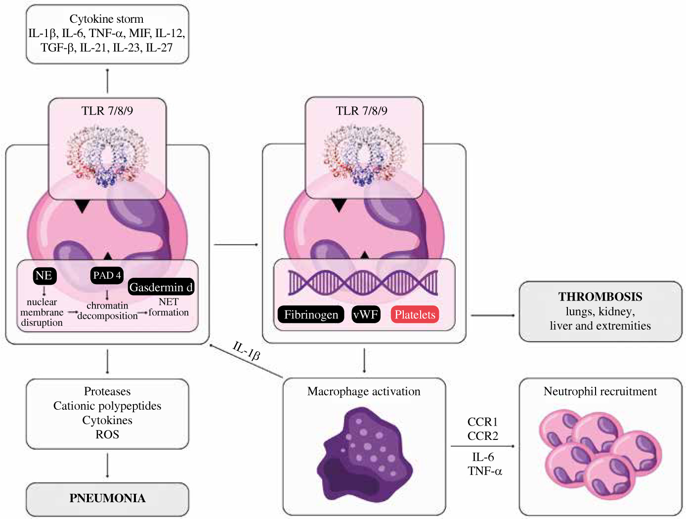
Platelet to lymphocyte ratio (PLR), a novel marker of inflammation, was identified in several studies as an increased inflammatory response marker, suggesting worse prognosis and higher intensity of cytokine storm [59] (Fig. 1).

Fig. 1

The role of neutrophils and platelets in the cytokine storm during COVID-19 infection. The activation of neutrophils induces selected cytokine production, which contributes to the cytokine storm and thus results in ARDS. Interaction of SARS-CoV-2 with TLR 7/8/9 results in the formation of neutrophil extracellular traps (NET), which are web-like chromatin structures produced by neutrophils. The interaction of fibrin and platelets with NET can cause thrombosis in several organs, especially the lungs

/f/fulltexts/CEJOI/51445/CEJI-48-51445-g001_min.jpg

A meta-analysis performed by Simadibrata et al. showed that a high PLR value was associated with severe COVID-19 [60]. Chung et al. claim that increased platelet activation occurs in acute PE, a higher RV/LV ratio reflecting a larger PE burden that may correspond to increased platelet consumption and therefore a smaller PLR [61]. This statement was confirmed by Phan et al., who found that PLR was significantly lower in patients with massive PE compared to patients with low-risk PE [62]. In this study platelet-to-lymphocyte ratio (PLR) was much higher in the group with < 50% of lung capacity changes in HR CT, which would be consistent with the hypothesis of Phan et al. Moreover, they also analyzed neutrophil-to-lymphocyte ratio and found out that a decrease in lymphocyte count coupled with minimal change in neutrophil and platelet counts influenced the increase in neutrophil-to-lymphocyte ratio (NLR) and PLR in nonsurvivors and the increased NLR and PLR were correlated with proinflammatory state. This study shows a similar positive correlation of NLR and negative correlation of lymphocyte count compared to the level of lung tissue involvement in HR CT.

Interleukin 6 is a soluble pro-inflammatory and immunoregulatory cytokine that contributes to host defense by stimulating acute phase responses, hematopoiesis and immune responses. Previous studies have shown significantly increased expression of IL-6 in both DVT patients and animal models. Moreover, blockade of IL-6 in a mouse study with the use of a specific antibody inhibited the formation and development of thrombosis [54-58]. In addition, the diagnosis of pulmonary embolism uses the level of D-dimers. In a study by Kara et al., compared to patients who did not have pulmonary embolism, patients with pulmonary embolism had a significantly higher mean level of D-dimers (pulmonary embolism, 6 ±7 µg/ml; no pulmonary embolism, 1 ±1 µg/ml, p = 0.001) [63]. Although statistically significant, this study shows that common inflammatory and thrombotic markers such as D-dimer level and IL-6 level have rather weak significance compared to novel inflammatory biomarkers when analyzing pulmonary embolism in COVID-19.

Conclusions

This study underlines the role of novel inflammatory biomarkers such as neutrophil to lymphocyte ratio and platelet to lymphocyte ratio in patients with pulmonary embolism in COVID-19. We suggest that these biomarkers may have higher assessment value compared to routinely used biomarkers.

Limitations

There are some limitations of this study that have to be acknowledged. Large population studies should be carried in order to assess the importance and clinical usefulness of selected biomarkers. In this study a clinical control group would enhance the usefulness of selected biomarkers in COVID-19. In addition, for all the patients recruited, the registration of chronic conditions was not possible, due to the heterogeneity of data collections in the different wards.

Notes

[1] Conflicts of interest The authors declare no conflict of interest.

References

1 

Raskob GE, Angchaisuksiri P, Blanco AN, et al. (2014): Thrombosis: a major contributor to global disease burden. Arterioscler Thromb Vasc Biol 2014; 34: 2363-2371.

2 

Wendelboe AM, Raskob GE (2016): Global burden of thrombosis: epidemiologic aspects. Circ Res 2016; 118: 1340-1347.

3 

de Miguel-Diez J, Jimenez-Garcia R, Jimenez D, et al. (2014): Trends in hospital admissions for pulmonary embolism in Spain from 2002 to 2011. Eur Respir J 44: 942-950.

4 

Dentali F, Ageno W, Pomero F, et al. (2016): Time trends and case fatality rate of in-hospital treated pulmonary embolism during 11 years of observation in Northwestern Italy. Thromb Haemost 2016; 115: 399-405.

5 

Lehnert P, Lange T, Moller CH, et al. (2018): Acute pulmonary embolism in a national Danish cohort: increasing incidence and decreasing mortality. Thromb Haemost 118: 539-546.

6 

Keller K, Hobohm L, Ebner M, et al. (2019): Trends in thrombolytic treatment and outcomes of acute pulmonary embolism in Germany. Eur Heart J 41: 522-529.

7 

Barco S, Mahmoudpour SH, Valerio L, et al. (2020): Trends in mortality related to pulmonary embolism in the European Region, 2000–15: analysis of vital registration data from the WHO Mortality Database. Lancet Respir Med 8: 227-287.

8 

Danzi GB, Loffi M, Galeazzi G, Gherbesi E (2020): Acute pulmonary embolism and COVID-19 pneumonia: a random association? Eur Heart J 41: 1858.

9 

Cellina M, Oliva G (2020): Acute pulmonary embolism in a patient with COVID-19 pneumonia. Diagn Interv Imaging 101: 325-326.

10 

Ullah W, Saeed R, Sarwar U, et al. (2020): COVID-19 complicated by acute pulmonary embolism and right-sided heart failure. JACC Case Rep 2: 1379-1382.

11 

Casey K, Iteen A, Nicolini R, Auten J (2020): COVID-19 pneumonia with hemoptysis: acute segmental pulmonary emboli associated with novel coronavirus infection. Am J Emerg Med 38: 1544.e1-3.

12 

Foch E, Allou N, Vitry T, et al. (2020): Pulmonary embolism in returning traveler with COVID-19 pneumonia. J Travel Med 27: taaa63.

13 

Rotzinger DC, Beigelman-Aubry C, von Garnier C, Qanadli SD (2020): Pulmonary embolism in patients with COVID-19: time to change the paradigm of computed tomography. Thromb Res 190: 58-59.

14 

Fabre O, Rebet O, Carjaliu I, et al. (2020): Severe acute proximal pulmonary embolism and COVID-19: a word of caution. Ann Thorac Surg 110: e409-e411.

15 

Sulemane S, Baltabaeva A, Barron AJ, et al. (2020): Acute pulmonary embolism in conjunction with intramural right ventricular thrombus in a SARS-CoV-2-positive patient. Eur Heart J Cardiovasc Imaging 21: 1054.

16 

Audo A, Bonato V, Cavozza C, et al. (2020): Acute pulmonary embolism in SARS-CoV-2 infection treated with surgical embolectomy. Ann Thorac Surg 110: e403-e404.

17 

Le Berre A, Marteau V, Emmerich J, Zins M (2020): Concomitant acute aortic thrombosis and pulmonary embolism complicating COVID-19 pneumonia. Diagn Interv Imaging 101: 321-322.

18 

Jafari R, Cegolon L, Jafari A, et al. (2020): Large saddle pulmonary embolism in a woman infected by COVID-19 pneumonia. Eur Heart J 41: 2133.

19 

Griffin DO, Jensen A, Khan M, et al. (2020): Pulmonary embolism and increased levels of D-dimer in patients with coronavirus disease. Emerg Infect Dis 26: 1941-1943.

20 

Martinelli I, Ferrazzi E, Ciavarella A, et al. (2020): Pulmonary embolism in a young pregnant woman with COVID-19. Thromb Res 191: 36-37.

21 

Lushina N, Kuo JS, Shaikh HA (2020): Pulmonary, cerebral, and renal thromboembolic disease associated with COVID-19 infection. Radiology 289: E181-183.

22 

Harsch IA, Skiba M, Konturek PC (2020): SARS-CoV-2 pneumonia and pulmonary embolism in a 66-year-old female. Pol Arch Intern Med 130: 438-439.

23 

Ueki Y, Otsuka T, Windecker S, Raber L (2020): ST-elevation myocardial infarction and pulmonary embolism in a patient with COVID-19 acute respiratory distress syndrome. Eur Heart J 41: 2134.

24 

Ioan AM, Durante-Lopez A, Martinez-Milla J, et al. (2020): Pulmonary embolism in COVID-19. When nothing is what it seems. Rev Esp Cardiol 73: 665-667.

25 

Bruggemann R, Gietema H, Jallah B, et al. (2020): Arterial and venous thromboembolic disease in a patient with COVID-19: a case report. Thromb Res 191: 153-155.

26 

Perez-Girbes A (2020): Acute pulmonary embolism and Covid-19: a common association in seriously ill patients? Arch Bronconeumol 56: 34.

27 

Khodamoradi Z, Boogar SS, Shirazi FKH, Kouhi P (2020): COVID-19 and acute pulmonary embolism in postpartum patient. Emerg Infect Dis 26: 1937-1939.

28 

Poggiali E, Bastoni D, Ioannilli E, et al. (2020): Deep vein thrombosis and pulmonary embolism: two complications of COVID-19 pneumonia? Eur J Case Rep Intern Med 7: 001646.

29 

Marsico S, Espallargas Gimenez I, Carbullanca Toledo SJ, et al. (2020): Pulmonary infarction secondary to pulmonary thromboembolism in COVID-19 diagnosed with dual-energy CT pulmonary angiography. Rev Esp Cardiol 73: 672-674.

30 

Schmiady MO, Sromicki J, Kucher N, Ouda A (2020): Successful percutaneous thrombectomy in a patient with COVID-19 pneumonia and acute pulmonary embolism supported by extracorporeal membrane oxygenation. Eur Heart J 41: 3107.

31 

Polat V, Bostanci GI (2020): Sudden death due to acute pulmonary embolism in a young woman with COVID-19. J Thromb Thrombolysis 50: 239-241.

32 

Ahmed I, Azhar A, Eltaweel N, Tan BK (2020): First Covid-19 maternal mortality in the UK associated with thrombotic complications. Br J Haematol 190: e37-38.

33 

Molina MF, Al Saud AA, Al Mulhim AA, et al. (2020): Nitrous oxide inhalant abuse and massive pulmonary embolism in COVID-19. Am J Emerg Med 38: 1549.e1-2.

34 

Vitali C, Minniti A, Caporali R, Del Papa N (2020): Occurrence of pulmonary embolism in a patient with mild clinical expression of COVID-19. Thromb Res 192: 21-22.

35 

Grillet F, Behr J, Calame P, et al. (2020): Acute pulmonary embolism associated with COVID-19 pneumonia detected by pulmonary CT angiography. Radiology 296: E186-188.

36 

Leonard-Lorant I, Delabranche X, Severac F, et al. (2020): Acute pulmonary embolism in COVID-19 patients on CT angiography and relationship to D-dimer levels. Radiology 296: E189-191.

37 

Helms J, Tacquard C, Severac F, et al. (2020): High risk of thrombosis in patients with severe SARS-CoV-2 infection: a multicenter prospective cohort study. Intensive Care Med 46: 1089-1098.

38 

Klok FA, Kruip M, van der Meer NJM, et al. (2020): Incidence of thrombotic complications in critically ill ICU patients with COVID-19. Thromb Res 191: 145-147.

39 

Lodigiani C, Iapichino G, Carenzo L, et al. (2020): Venous and arterial thromboembolic complications in COVID-19 patients admitted to an academic hospital in Milan, Italy. Thromb Res 191: 9-14.

40 

Llitjos JF, Leclerc M, Chochois C, et al. (2020): High incidence of venous thromboembolic events in anticoagulated severe COVID-19 patients. J Thromb Haemost 18: 1743-1746.

41 

Poissy J, Goutay J, Caplan M, et al. (2020): Pulmonary embolism in COVID-19 patients: awareness of an increased prevalence. Circulation 14: 184-186.

42 

Beun R, Kusadasi N, Sikma M, et al. (2020): Thromboembolic events and apparent heparin resistance in patients infected with SARS-CoV-2. Int J Lab Hematol 42: 19-20.

43 

Middeldorp S, Coppens M, van Haaps TF, et al. (2020): Incidence of venous thromboembolism in hospitalized patients with COVID-19. J Thromb Haemost 18: 1995-2002.

44 

Wichmann D, Sperhake JP, Lutgehetmann M, et al. (2020): Autopsy findings and venous thromboembolism in patients with COVID-19: a prospective cohort study. Ann Intern Med 173: 268-277.

45 

Klok FA, Kruip M, van der Meer NJM, et al. (2020): Confirmation of the high cumulative incidence of thrombotic complications in critically ill ICU patients with COVID-19: an updated analysis. Thromb Res 191: 148-150.

46 

Bompard F, Monnier H, Saab I, et al. (2020): Pulmonary embolism in patients with Covid-19 pneumonia. Eur Respir J 56: 2001365.

47 

Thomas W, Varley J, Johnston A, et al. (2020): Thrombotic complications of patients admitted to intensive care with COVID-19 at a teaching hospital in the United Kingdom. Thromb Res 191: 76-77.

48 

Poyiadi N, Cormier P, Patel PY, et al. (2020): Acute pulmonary embolism and COVID-19. Radiology 297: E335-E338.

49 

Galeano-Valle F, Oblitas CM, Ferreiro-Mazon MM, et al. (2020): Antiphospholipid antibodies are not elevated in patients with severe COVID-19 pneumonia and venous thromboembolism. Thromb Res 192: 113-115.

50 

Stoneham SM, Milne KM, Nuttal E, et al. (2020): Thrombotic risk in COVID-19: a case series and case-control study. Clin Med 20: e76-81.

51 

Lax SF, Skok K, Zechner P, et al. (2020): Pulmonary arterial thrombosis in COVID-19 with fatal outcome: results from a prospective, single-center, clinicopathologic case series. Ann Intern Med 173: 350-361.

52 

Roumen-Klappe EM, den Heijer M, van Uum SH, et al. (2002): Inflammatory response in the acute phase of deep vein thrombosis. J Vasc Surg 35: 701-706.

53 

Tatò F (2014): Deep vein thrombosis–advances in diagnosis and treatment. MMW Fortschr Med 156 Spec no 2: 59-63, quiz 64.

54 

Matos MF, Lourenço DM, Orikaza CM, et al. (2011): The role of IL-6, IL-8 and MCP-1 and their promoter polymorphisms IL-6-174GC, IL-8-251AT and MCP-1-2518AG in the risk of venous thromboembolism: a case-control study. Thromb Res 128: 216-220.

55 

Vormittag R, Hsieh K, Kaider A, et al. (2006): Interleukin-6 and interleukin-6 promoter polymorphism (-174) G > C in patients with spontaneous venous thromboembolism. Thromb Haemost 95: 802-806.

56 

Mahemuti A, Abudureheman K, Aihemaiti X, et al. (2012): Association of interleukin-6 and C-reactive protein genetic polymorphisms levels with venous thromboembolism. Chin Med J (Engl) 125: 3997-4002.

57 

Wu J, Zhu H, Yang G, et al. (2017): IQCA-TAVV: To explore the effect of P-selectin, GPIIb/IIIa, IL-2, IL-6 and IL-8 on deep venous thrombosis. Oncotarget 8: 91391-91401.

58 

Nosaka M, Ishida Y, Kimura A, et al. (2015): Immunohistochemical detection of intrathrombotic IL-6 and its application to thrombus age estimation. Int J Legal Med 129: 1021-1025.

59 

Yang AP, Liu JP, Tao WQ, et al. (2020): The diagnostic and predictive role of NLR, d-NLR and PLR in COVID-19 patients. Int Immunopharmacol 84: 106504.

60 

Simadibrata DM, Pandhita BAW, Ananta ME, Tango T (2022): Platelet-to-lymphocyte ratio, a novel biomarker to predict the severity of COVID-19 patients: A systematic review and meta-analysis. J Intensive Care Soc 23: 20-26.

61 

Chung T, Connor D, Joseph J, et al. (2007): Platelet activation in acute pulmonary embolism. J Thromb Haemost 5: 918-924.

62 

Phan T, Brailovsky Y, Fareed J, et al. (2020): Neutrophil-to-lymphocyte and platelet-to-lymphocyte ratios predict all-cause mortality in acute pulmonary embolism. Clin Appl Thromb Hemost 26: 107602961990054.

63 

Kara H, Bayir A, Degirmenci S, et al. (2014): D-dimer and D-dimer/fibrinogen ratio in predicting pulmonary embolism in patients evaluated in a hospital emergency department. Acta Clin Belg 69: 240-245.

Copyright: © 2023 Polish Society of Experimental and Clinical Immunology This is an Open Access article distributed under the terms of the Creative Commons Attribution-NonCommercial-ShareAlike 4.0 International (CC BY-NC-SA 4.0) License (http://creativecommons.org/licenses/by-nc-sa/4.0/), allowing third parties to copy and redistribute the material in any medium or format and to remix, transform, and build upon the material, provided the original work is properly cited and states its license.
 
Quick links
© 2024 Termedia Sp. z o.o.
Developed by Bentus.