


Introduction
The most common chest wall deformities are pectus excavatum and carinatum. Surgical repair for these deformities is based on placing a metallic pectus bar via minimally invasive technique [1, 2]. The technique is well defined by our center’s experience and existing medical literature [3, 4]. Beside the successfully performed intervention, placing the bar, time for bar stay, bar removal process, possible complications in the period and ways to prevent them have been debated over the years and still it is not concluded [5, 6]. Along with this growth in bar removing surgery numbers, there is increasing concern over complications. The increased number of interventions and recent developments for pectus deformities repair underlined the need for definition and marking more clear medical borders of the bar removal timing and process itself.
Aim
In order to shine a spotlight on this subject we looked back at our patients and clinical experience and aimed to determine the outcomes and negative factors in the bar removal process. This would help the literature to gain further understanding of pectus bar removal surgery timing and pitfalls of the surgical procedure.
Material and methods
Patients
We have reviewed our prospectively recorded clinical database from 2006 to 2021 and analyzed it retrospectively. There were 1032 patients with a bar removal procedure. Patients’ demographics, medical and family history, Haller index before placing bar, placed bar number, body mass index, use of stabilizer and wire, length of hospital stay, time until bar removal, incision side and complications overall were analyzed.
After placing the bar with our classical procedure for surgery the patients were discharged and had single outpatient clinic control. If they did not have any complaints they were asked for a reminder of their situation after 3 years of bar placement. To ensure this period an additional reminder is placed in the patient’s file for bar removal timing and closely followed by a database analyst.
Surgical procedure for bar removal
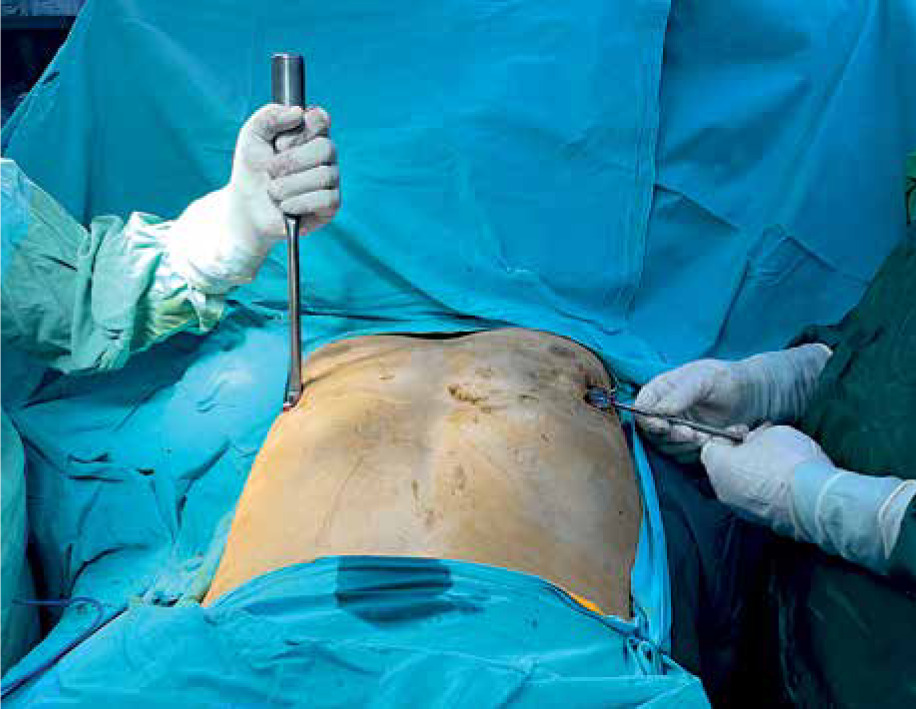
As the time for removal has been decided by either mechanism mentioned above, the patient has a standard evaluation for general anesthesia. Routine blood test, chest X-ray, ECG (electrocardiogram) and additional evaluation regarding the patient’s medical background are ordered. The bar (Zimmer Biomet Inc., Warsaw, Indiana, USA) removal procedure is performed under general anesthesia using a single-lumen intubation with a standard supine position with both arms opened wide. The patient was placed close to the left side of the surgical table (Photo 1). The surgical incisions used for the previous surgery were located and the left side (or if necessary both sides) was opened (Photo 2). After dissecting subcutaneous tissue and fibrotic soft tissue the pectus bar(s) (Zimmer Biomet Inc., Warsaw, Indiana, USA) and – if used – fixation tools like a stabilizer and steel wire were identified. After cutting them and removal of the wire(s) with all fragments and the stabilizer(s), the pectus bar was then mobilized bilaterally (Photo 3). Some cases, especially those with prolonged bar stay, require additional intervention such as using an osteotomy instrument or rongeur to set the bar free from bone and hard fibrotic and calcific healing tissue.


Photo 3
After cutting of the wire(s) with all fragments and removing the stabilizer(s), the pectus bar was then mobilized bilaterally
The bar bender (Zimmer Biomet Inc., Warsaw, Indiana, USA) was used to straighten both ends to allow a safe and smooth removal through the tunnel. Then the bar was carefully moved either direction gently without pulling to be sure there were no remaining strong adhesions or structures blocking the bar from an easy path.
The bar was then pulled out of the tunnel with care to avoid getting stuck or causing injury along the way. After the removal the residue tunnel was inspected to verify that there was no active bleeding which may indicate any mild or massive damage to the intrathoracic structures. After checking that there is no contact with the pleural space, if there is any doubt, a temporary air evacuation system or a chest tube is placed to remove the free air. After the irrigation with povidone iodine, the incisions were closed. It is important to suture subcutaneous planes carefully to avoid any remaining large space (which may cause seroma in early postoperative days) or peri-incisional skin contractions.
After the bar removal procedure as the patient was transferred to the ward a chest X-ray was taken within a hour to see any free air, or fluid within the pleural cavity and any other potential clinical situation. High flow nasal oxygen inhalation in case of limited and small amount of pneumothorax or chest tube insertion for larger ones was performed if necessary. If patients were stable they were discharged on the same day and followed up after 2 weeks as an outpatient clinic protocol.
Results
The patients were between 4 and 47 years old (mean age: 20 years). Predominantly 920 of the patients were male (90.8%), 112 of the patients were female (9.2%), male to female ratio was 7.35. 784 patients had pectus excavatum (76.1%), 248 patients had pectus carinatum (23.9%).
The mean time for pectus bar maintenance was 34.2 months (1–55). Grouping according to age is stated in Table I.
Table I
Distribution of mean bar removal time by age range
| Group | Mean bar remain time |
|---|---|
| 1 (4–12 years) | 36.7 months |
| 2 (13–19 years) | 32.3 months |
| 3 (20 and older) | 36.1 months |
For 594 (57.5%) patients the bar was removed earlier than planned (mean: 28.1 months, 0–35). The main reason for early bar removal was bar replacement, wound infection and patients’ wishes. In 69 (6.68%) patients the bar was removed less 2 years after surgery, in 111 (10.7%) patients the bar was removed between 24–36 month after surgery, in 387 (37.5%) patients the bar was removed between 36–48 months after surgery. A total of 57 (5.52%) patients underwent bar removal more than 4 years because of having concern that the repair could be compromised after bar removal. Recurrence after early bar removal occurred in 12 (1.16%) patients because of intolerable pain and bar replacement. The reasons for early bar removal are shown in Table II.
Table II
Indication of early pectus bar removal
| Indication | Case number, total = 1032 (57.5%) |
|---|---|
| Patient’s wish | 561 (54.3%) |
| Bar displacement | 18 (1.74%) |
| Wound chronic seroma/infection/dehiscence | 9 (0.87%) |
| Persistent pain | 6 (0.58%) |
| Total | 594 (57.5%) |
Mean operation time was 41 min (15–100 min), 609 (59%) patients had unilateral incision, 423 (41%) patients had bilateral incision for bar removal. Main length of hospital stay was 0.46 days (0–3). Some patients stayed more than a day for wound infection and hematoma control. Also some patients needed observation for 1 day because of intrathoracic bar removal. 528 (51%) patients had single bars, 504 (49%) patients had double bars. Mean BMI was 19.7 (10.38–32,23). The demographic and perioperative clinical features of groups 1, 2 and 3 are shown in Table III. Regarding significant differences, bar staying time was statistically significantly shorter in Group 2 (p = 0.006). Height was statistically significantly shorter in Group 1 (p < 0.001). Body mass index was statistically significantly higher in Group 1 (p = 0.003). The duration of surgery was statistically significantly higher in Group 3 (p = 0.027) (Table III).
Table III
Parametric data for groups 1-2-3 (mean values given) (ANOVA test was used)
The complication rate was 2.61%, The most common postoperative complication after bar removal was wound infection and/or seroma for 15 (1.45%) patients and the second most common complication after bar removal was pneumothorax in 10 (0.96%) patients. Bleeding after removing occurred in 6 (0.0058%) patients (5 minor which was stopped by compressing, 1 bleeding required major thoracotomy). This single patient is recorded as a major morbidity. The bleeding was from the internal mammary artery and the patient was managed by anterior thoracotomy, discharged 3 days later, and in the follow-up there was no additional problem. There has been no mortality or fatal complication related to bar removal.
The number of bars, steel wires, the presence of stabilizers, and bilateral incisions were statistically significantly lower in the first group (p = 0.028, p = 0.002, p < 0.001, p = 0.046, respectively) (Table IV). The formation of callus tissue was statistically significantly higher in Group 3 (p = 0.034) (Table V).
Table IV
Comparison of non-parametric data for groups 1-2-3 (percent values given) (Kruskal-Wallis test was used)
Discussion
Several reports have shown that pectus bar removal is a final stage of repair of pectus deformity, defined as an easy and practical surgical intervention. Our routine approach is to keep the bar in position for 2.5 years at least. Prior studies have noted that the importance of the Nuss repair has gained wide acceptance because it does not require tissue removal and osteotomies, it is quick, and it requires fewer scars than the conventional open approach.
In the early times of our experience with this procedure all patients were routinely planned to have their bars removed after 3 years, but some patients wanted early removal because of a prickling sensation and some patients wanted more time because of having concern that the repair could be compromised after bar removal and some patients have concerns because of the COVID-19 pandemic. The appropriate time was determined by meeting with the patient and their relatives. Our data show that a large proportion of subjects did not experience any significant discomfort and no destructive effect was associated with remaining pectus bars regardless of bar stay time. As a different point of view, we have seen that the bar removal process, which was postponed due to the COVID pandemic, can be safely performed even though it has extended the stay of the bar.
Another limitation lies in the possibility of timing problems: a subset comparison between patients who had their implants still in place after a standard or extended bar stay time was carried out.
The bar bender was used to straighten both ends to allow a safe and smooth removal through the tunnel. Then the bar was carefully rotated without pulling to be sure there were no remaining strong adhesions or structures blocking the bar from an easy path. This technique allows hassle-free and simple removal, as confirmed by others [7].
Both lateral incisions are opened to remove fixation sutures or wires. The substernal bar is simply grabbed with a bone hook or Kocher forceps and can be removed with even a towel clip and pulled through with the natural curvature of the bar without any bending. In general, reopening both lateral incisions is used for bar placement, as described by several authors. Furthermore, unbending and straightening of the pectus bar before starting its mobilization is recommended [8]. In our series, we also released the ends of the bar using a bilateral incision with a similar method, and then we removed it by bending it.
One interesting finding is callus formation around the bar, especially at the ends of the bar [9, 10]. It has been observed that submuscularly placed bars have a greater propensity for callus formation than subcutaneous bars [11]. As a result dissecting the bar out of the callus can be routine in some patients. In most patients who had a callus we had to use a Luer bone rongeur to free the bar from bony tissue. Park et al. described a technique for dissection of the bar from bony tissue [12]. In our series 69 (6.68%) patients had callus tissue and we had to use a Luer bone rongeur for callus release, similarly.
The duration of surgery was statistically significantly higher in Group 3 (p = 0.027) (Table III). Also callus tissue was statistically significantly higher in Group 3 (p = 0.034). When we examined the surgery time and callus tissue, the surgery time was found to be significantly longer in those with callus tissue (p = 0.002) (Table V). These relationships may partly be explained by difficulty of releasing the bar from callus tissue and dissection of bar tips from bony tissue. These results agree with the findings of other authors [10–12]. Proper bar placement is important during pectus repair to prevent severe bone formation.
Patients with a higher BMI (> 22 kg/m2) were more likely to have a longer operative time [13]. Our data showed no significant relation between BMI and surgery time, p = 0.748.
The other factors showed no significant difference including the age groups and the number of pectus bars removed.
It may be argued that retaining pectus bars longer than necessary could lead to an increase in bone overgrowth around bar tips, longer operating time for bar removal and an increased risk of complications. In reviewing the literature, no data were found on the association between callus tissue and bar remain time [6, 14]. In our data there was no association between callus tissue and bar remain time, similarly.
Cho et al. [15] observed a recurrence in 9.1% of 99 children with a mean age of 9 ±5, after a mean follow-up of 16 ±4 months. Correlatively, Kang et al. [16] reported on a pectus recurrence rate of 7.9% in 63 children with a mean age of 7 ±3 (range: 3–13). Kuyama et al. [17] observed a recurrence in 21 out of 42 pediatric patients (50%) aged 3–9 in whom the bars had been removed by the age of 12 and who had been followed for 5 years with serial assessments of the radiographic Haller index. Five of them (12%) were considered for – or received – redo surgery. In our series recurrence after early bar removal occurred in 12 (1.16%) patients because of intolerable pain and bar replacement. When people are evaluated according to the age of bar removal, some recurrences may have been underestimated.
In pectus bar, allergy development, local reaction, infection, replacement, etc., carry all the risks as with all metal implants. It can cause erosion of the heart or sternum and damage to the internal mammary artery [18]. In our series there were no lesions because of erosion of the sternum or other tissues.
The complication rate was 2.61%; the most common complication after bar removal was pneumothorax (10 patients, 0.96%). This was similar to other studies with a large number of patients [5, 13, 19]. Pneumothorax mainly occurred due to air entering the pleural space during dissection for bar removal. It can be resolved without drainage if there are no clinical symptoms. If the pneumothorax is due to clinical problems, pleural drainage should be done. Bleeding from the intercostal artery injury and from the internal mammary artery injury was reported as the most common cause of acute intraoperative hemorrhage during pectus bar removal [8]. Packing the bar tract and applying local pressure might be successful to control such bleeding. However, depending on the source and volume of hemorrhage, emergency sternotomy and/or thoracotomy might be necessary [8]. In our series bleeding after removal occurred in 6 patients (5 minor which stopped by compressing, 1 thoracotomy due to major bleeding). There has been no mortality or fatal complication related to bar removal. In order to manage the mentioned complications, it is recommended that the procedure be performed in a well-equipped center with cardiac surgery. The patient who did not develop any complications in the follow-ups can be discharged on the same day.
Reviewing the current literature, there are no evidenced-based guidelines supporting or disputing the common practice of elective implant removal after fracture healing. In the adult population, the recommendation related to hardware removal might be age dependent [8].
Bar removal is normally a quick and safe procedure [8, 19] and those bars would be an obstacle with cardiopulmonary resuscitation maneuvers [8] or with emergency sternotomy if needed. Another source of uncertainty is the unclear long-term effects of trace metals released into the bloodstream [20]. Therefore bar removal is recommended.
While there is no evidence to date that extending the bar remain time will improve long term results of the Nuss repair in every case, older pectus patients who are anxious about long term results may benefit from this approach psychologically, while patients operated on early in their lifetime may be safely left to grow out of adolescence with their bars in place in hope of averting any risk of recurrence.
These findings suggest that pectus bars can be left in place for a shorter time than the standard 3-year interval without any additional recurrence risk and without compromising quality of life. Our routine plan for bar removal was to wait 3 years but there were cases of earlier removal as a result of patients’ requests and clinical follow-ups. Therefore, removing the bar before 3 years may be preferred in clinical practice.
Apart from this opinion, as we mentioned before, it has been seen that there is no problem other than callus tissue in patients who have postponed due to the COVID-19 pandemic and whose bar has been stopped for a long time.
Conclusions
Patients with persistent pain after pectus repair should be well evaluated for the possibility of life-threatening complications during bar removal. There are still many unanswered questions about less bar remain time and the effect of long bar remain time on metal release in blood. Research questions that could be asked include bar remain time and benefit with respect to recurrence. These may require more detailed followed cohorts with a randomized decision of time for bar removal.