eISSN: 1644-4124
ISSN: 1426-3912
Central European Journal of Immunology
Current issue Archive Manuscripts accepted About the journal Special Issues Editorial board Abstracting and indexing Subscription Contact Instructions for authors
Editorial System
Submit your Manuscript
SCImago Journal & Country Rank
4/2022
vol. 47
 
Share:
Share:
Clinical immunology

The analysis of Fe-dependent serum enzymes in severe COVID-19 with a pulmonary thrombotic event

Jarosław Bakiera
1
,
Cezary Grochowski
2
,
Karolina Strzelec-Pawełczak
3
,
Ida Osuchowska-Grochowska
1
,
Mateusz Szymański
4
,
Aleksander Michalski
5
,
Kinga Kulczycka
6
,
Katarzyna Czarnek
6
,
Jacek Bogucki
7
,
Eliza Blicharska
8

1.
Department of Laboratory Diagnostics, Coagulation and Microbiology, Stefan Wyszyński Regional Specialist Hospital, Lublin, Poland
2.
Department of Human Anatomy, Medical University of Lublin, Lublin, Poland
3.
Neonatology Department, Independent Public Clinical Hospital No. 1 in Lublin, Poland
4.
Department of Anesthesiology, Stefan Wyszyński Regional Specialist Hospital, Lublin, Poland
5.
1st Military Hospital, Lublin, Poland
6.
Institute of Health Sciences, The John Paul II Catholic University of Lublin, Lublin, Poland
7.
Department of Organic Chemistry, Medical University of Lublin, Lublin, Poland
8.
Department of Analytical Chemistry, Medical University of Lublin, Lublin, Poland
Cent Eur J Immunol 2022; 47 (4): 293-298
Online publish date: 2023/01/13
Article file
- The analysis.pdf  [0.15 MB]
Get citation
 
PlumX metrics:
 

Introduction

Venous thromboembolism (VTE), clinically manifested as deep vein thrombosis (DVT) or acute pulmonary embolism (PE), is the third most common acute cardiovascular syndrome following myocardial infarction and stroke [1]. The annual incidence of PE is between 39 and 115 per 100,000 inhabitants. The incidence of VTE is almost eight times higher in people aged 80 and older than in the fifth decade of life [2]. As a consequence, with the age of the population, long-term studies show an increasing tendency in the annual incidence rates of PE [3-6] over time. A recent analysis of relevant registration data from the World Health Organization (WHO) mortality database (2000-2015) found an average of 38,929 PE-related deaths each year in 41 countries of the WHO European Region (including Central Asia) representing a population of approximately 651 million [7]. Between 2000 and 2015, the annual age-standardized death rates associated with PE decreased by almost 50% (from 12.7 to 6.5 deaths per 100,000 inhabitants) with no significant gender differences. Despite this overall positive trend, the study also found that PE-related mortality continues to increase exponentially with age, reaching or even exceeding 80 deaths per 100,000 elderly population, and that PE also remains a relatively important cause of (in comparison with other causes) deaths among younger women, in which it accounted for up to 13 cases per 1000 deaths [8]. Since the outbreak of coronavirus disease 2019 (COVID-19), clinicians have struggled with the attempt to diagnose and manage the severe and fatal complications of COVID-19 appropriately. Several reports have described significant procoagulatory events, including life-threatening pulmonary embolism in these patients [9-51].

The incidence of PE in hospitalized COVID-19 patients is approximately 1.9-8.9% [36, 40, 50, 51]. The retrospective nature of the analyzed cohorts and relatively short observation periods could have underestimated the actual incidence of PE. Critically, COVID-19 patients requiring admission to the ICU seem to be at higher risk of thromboembolic complications, especially PE, which may occur in up to 26.6% of these patients [43]. In a prospective observational study involving 150 patients admitted to four ICU wards in two French hospitals, despite antithrombotic prophylaxis, the occurrence of PE was observed in 16.7% of treated patients [38]. The authors also reported that thromboembolic events occurred more frequently in patients with acute respiratory distress syndrome (ARDS) in COVID-19 patients compared to the historic ARDS cohort of a different etiology, underlining the unique procoagulatory effect of COVID-19 compared to other etiologies ARDS. In a retrospective cohort of 184 COVID-19 patients admitted to the ICU in three hospitals in the Netherlands, it was found that 13.6% of patients developed PE despite anticoagulation [39]. Interestingly, the incidence of PE increased to 33.3% when the follow-up period was extended from 1 to 2 weeks [46], at a time when increased awareness of the frequent occurrence of PE could lead to a higher rate of suspicion and extended diagnosis to detect this complication. Similarly, Poissy et al. found that 20.6% of patients admitted to the French ICU had pulmonary embolism on average 6 days after admission to the ICU despite the use of anticoagulants [41]. These authors also found that the incidence of PE in COVID-19 patients was twice as high as in patients admitted to the ICU as a control group and in 40 patients admitted to the ICU due to severe influenza.

Abnormalities in various coagulation parameters were frequently reported [49, 50] and were associated with poor prognosis [51]. It is common knowledge that all the time in cells there occur oxidation and reduction reactions which are connected with conversion of oxidation states of metals. Iron and nickel are able to generate free radicals through Fenton or Haber-Weiss reactions. These reactive oxygen species (ROS) are a potential danger to biological macromolecules and when defense mechanisms are insufficient, cytotoxic and genotoxic effects in cells can be observed. The study confirmed that iron can donate or accept an electron from neighboring molecules. This situation leads to damage of cellular components or to generation of reactive oxygen species. The Fenton and Haber-Weiss reactions, Fe2+ + H2O2 Fe3+ + HO. + OH- and Fe3+ + H2O2 Fe3 + HOO. + H+, describe the reactions of free iron with free radicals within a cell in order to generate more toxic radicals. These radicals can react with biomolecules such as DNA and cause them damage. Iron is involved in a number important metabolic processes including mitochondria transpiration, xenobiotic biotransformation, metabolism of lipids and proteins and DNA synthesis [52, 53]. Nickel can induce production of reactive oxygen species (ROS) and interact with nucleic acids, causing DNA damage. Unfortunately, little is known about the epidemiology and pathophysiological mechanisms underlying COVID-19-related PE due to the lack of extensive prospective studies in this context. Understanding these aspects is critical to the early diagnosis and appropriate management of this potentially fatal complication.

Material and methods

The retrospective study covered a group of 226 COVID-19 patients with division of the group of patients into groups with > 50% and < 50% lung tissue involvement, including patients with a radiologically confirmed pulmonary thrombotic event. The analyzed group consisted of 136 men and 90 women with mean age of 70 years. The groups were divided based on the percentage of the involved pulmonary tissue. Analyzed patients were hospitalized in the Provincial Specialist Hospital Cardinal Stefan Wyszyński in Lublin, 1st Military Hospital in Lublin, Agricultural Medicine Institute in Lublin.

All components were cataloged in accordance with the applicable regulations on sensitive data. Fe-dependent serum enzymes such as hemoglobin, ferritin, as well as zinc-related enzymes (C-reactive protein – CRP, D-Dimer) were analyzed. Each patient underwent high resolution computed tomography. The images were assessed by an experienced radiologist. The data collected in this way were compiled and analyzed using statistical tools. All statistical calculations were performed using the TIBCO Software Inc. statistical package. (2017). Statistica (data analysis software system), version 13. http://statistica.io. and an Excel spreadsheet. The study received full approval of the Medical University of Lublin Ethics Committee. Every single stage of the performed study was carried out in compliance with the Helsinki Declaration and national legislation.

Results

Two groups were distinguished among analyzed patients. The first group consisted of patients with < 50% of lung volume changes seen in the high resolution tomography scan, which covered 167 patients. The second group, with long volume changes over > 50, consisted of 79 patients.

Several different trace element dependent parameters were measured, a detailed descriptive analysis of which is presented in Tables 1 and 2.

Table 1

Presentation of gathered data in patients with < 50% of lung volume changes. N is the number of performed analyses of selected parameters. Some of the parameters were measured more than once

Variable< 50% of lung volume changes
NMeanMedianMinimumMaximumSDSE
D-dimer4523312.9611156.0000.07000122705.010060.32473.197
CRP58384.12938.1000.050002285.0189.497.848
RBC5503.9984.0302.3100013.10.780.033
Hemoglobin (g/dl)55012.13412.2005.7000038.72.320.099
Hematocrit (%)55035.52435.90018.3000090.06.140.262
Ferritin1571061.506608.00015.7000021407.01891.77150.980
Table 2

Presentation of gathered data in patients with < 50% of lung volume changes. N is the number of performed analyses of selected parameters. Some of the parameters were measured more than once

Variable> 50% of lung volume changes
NMeanMedianMinimumMaximumSDSE
D-dimer1805452.5611368.000225.0000128000.013674.461019.234
CRP23077.44755.1500.4100324.074.974.944
RBC2194.1014.1602.01005.80.720.048
Hemoglobin (g/dl)21912.51112.6006.700017.12.040.138
Hematocrit (%)21936.55037.10019.200048.35.870.396
Ferritin661300.2951206.0001.45003820.0775.7895.492

Presented data were analyzed using the Spearman correlation coefficient, which revealed statistically significant results regarding analyzed parameters, which are presented in Table 3 (p < 0.05). In all of the presented parameters, a positive correlation was observed, except in the level of D-dimers. On the other hand, ferritin showed the strongest correlation among the presented parameters (Fig. 1).
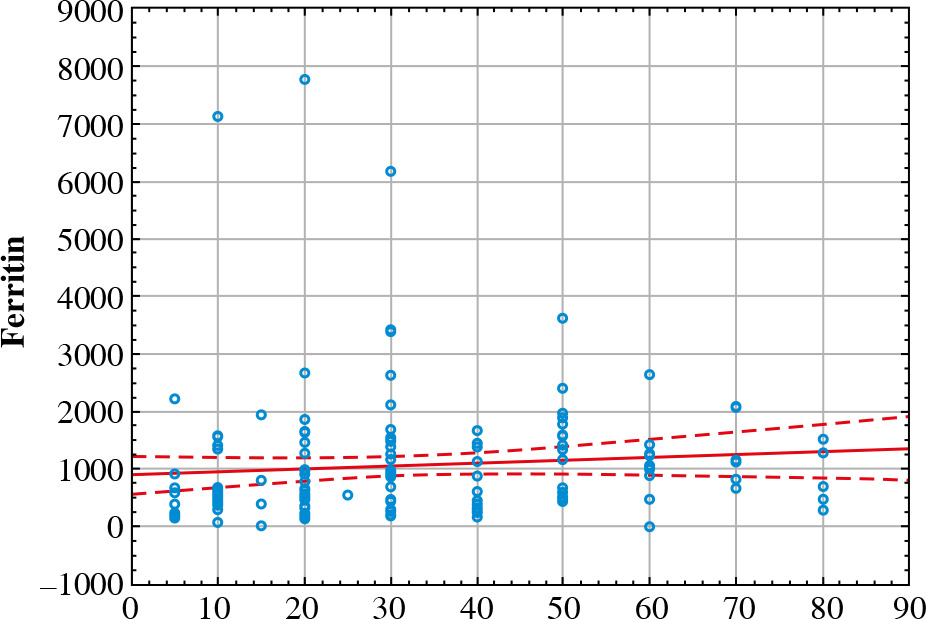
Fig. 1

Spearman correlation coefficient analysis of ferritin presented as a chart

/f/fulltexts/CEJOI/49887/CEJI-47-49887-g001_min.jpg
Table 3

Spearman correlation coefficient analysis results

Variable% lung volume changes
D-dimers0.077
CRP0.230
RBC0.138
Hemoglobin (g/dl)0.166
Hematocrit (%)0.169
Ferritin0.271

In the next step the analysis of both groups was performed, using the Mann-Whitney U-test, which showed statistically significant results between groups. The group consisting of patients with < 50% of lung volume changes had higher parameter values in each analyzed parameter, except RBC (p < 0.05). Especially, the level of ferritin was much higher in the first group (p = 0.000008). Elevated ferritin levels were observed in all patients with COVID-19 involvement of lung tissue.

Discussion

Iron plays a very important role due to its biological activity by regulating enzymatic activity and oxidation and reduction reactions [52]. Iron is involved in a number important metabolic processes including mitochondria transpiration, xenobiotic biotransformation, metabolism of lipids and proteins and DNA synthesis [53]. Iron crosses membranes as the ferrous form (Fe2+) and in this process a few enzymes and a divalent metal transporter are involved. This metal is carried by transferrin in ferric form but in the cell it is stored within ferritin. The ferric ions need reduction to the ferrous form through the Fenton reaction and then they can be released in cells. This process is connected with generation of ROS which cause oxidative stress in cells such as damaged cellular membranes and nuclei. Free forms of iron are toxic and detrimental for cells but binding by molecules is involved in that [54]. This metal is linked in plasma by transferrin, which has two iron-binding sites. In addition, iron in the cytosol is stored inside the ferritin which, when necessary, can release metal ions.

Ferritin was previously described as an acute phase factor as well as an indicator of dysregulation of the immune system in COVID-19 patients. Moreover, its role was underlined in the cytokine storm process. Ferritin was found to moderate the expression not only of the pro-inflammatory but also the anti-inflammatory cytokines. Moreover, Marcinkiewicz et al. suggested that interleukin (IL)-6, along with other cytokines, drives an acute phase response that elevates serum ferritin [55]. In the study performed by Carubbi et al. higher ferritin levels as well as D-dimer levels were linked to severity of pulmonary involvement but not associated with the outcome [56]. They speculated on the role of ferritin in pathogenesis of lung damage but underlined the need for further research supplemented with radiological analysis. Moreover, different studies reported higher ferritin levels in patients with pulmonary embolism caused by COVID-19 compared to non-pulmonary embolism patients [57, 58]. In this study patients with severe COVID-19 complicated with pulmonary embolism were analyzed. Based on the lung volume involved, assessed in the HR CT scan, two groups were distinguished. Interestingly, contrary to the previously mentioned study, the first group with lung volume involvement < 50% turned out to have higher ferritin (p = 0.000008) and D-dimer (p = 0.036) levels compared to the second group (Table 4).

Table 4

Comparison of results in both analyzed groups

< 50% of lung volume changes> 50% of lung volume changesUZpN < 50% of lung volume changesN > 50% of lung volume changes
D-dimers138725.061303.036347.00–2.091570.036452180
CRP229436.5101454.559200.50–2.601030.0092583230
RBC206449.089616.054924.00–1.906780.056550550219
Hemoglobin (g/dl)205077.090988.053552.00–2.400290.016550219
Hematocrit (%)205086.590978.553561.50–2.396870.016550219
Ferritin15617.59358.53214.50–4.471350.00000815766

In the study performed by Mulder et al. no associations were observed for higher D-dimer, higher CRP or higher ferritin concentration and clinical pulmonary thromboembolism. However, they observed a lower ferritin concentration, in patients with, compared to those without, clinical pulmonary thromboembolism [59]. Contrary to those results, in this study elevated levels of each parameter were observed; however, those parameter were higher in the second group.

Conclusions

The study demonstrated that elevated levels of several inflammatory and thrombotic parameters such as ferritin, D-dimer, CRP as well as hemoglobin do not correlate with the degree of lung tissue involvement in the computed tomography image. The study compared patients with a known thrombotic pulmonary event. The study did not compare the ferritin levels in patients with pulmonary tissue involvement in the group with and without known pulmonary embolism. This area of research should be deepened in view of the role of ferritin as a predictor of pulmonary embolism.

Notes

[1] Conflicts of interest The authors declare no conflict of interest.

References

1 

Raskob GE, Angchaisuksiri P, Blanco AN, et al. (2014): Thrombosis: a major contributor to global disease burden. Arterioscler Thromb Vasc Biol 34: 2363-2371.

2 

Wendelboe AM, Raskob GE (2016): Global burden of thrombosis: epidemiologic aspects. Circ Res 118: 1340-1347.

3 

de Miguel-Diez J, Jimenez-Garcia R, Jimenez D, et al. (2014): Trends in hospital admissions for pulmonary embolism in Spain from 2002 to 2011. Eur Respir J 44: 942-950.

4 

Dentali F, Ageno W, Pomero F, et al. (2016): Time trends and case fatality rate of in-hospital treated pulmonary embolism during 11 years of observation in Northwestern Italy. Thromb Haemost 115: 399-405.

5 

Lehnert P, Lange T, Moller CH, et al. (2018): Acute pulmonary embolism in a national Danish cohort: increasing incidence and decreasing mortality. Thromb Haemost 118: 539-546.

6 

Keller K, Hobohm L, Ebner M, et al. (2019): Trends in thrombolytic treatment and outcomes of acute pulmonary embolism in Germany. Eur Heart J 41: 522-529.

7 

Barco S, Mahmoudpour SH, Valerio L, et al. (2020): Trends in mortality related to pulmonary embolism in the European Region, 2000–15: analysis of vital registration data from the WHO Mortality Database. Lancet Respir Med 8: 227-287.

8 

Danzi GB, Loffi M, Galeazzi G, Gherbesi E (2020): Acute pulmonary embolism and COVID-19 pneumonia: a random association? Eur Heart J 41: 1858.

9 

Cellina M, Oliva G (2020): Acute pulmonary embolism in a patient with COVID-19 pneumonia. Diagn Interv Imaging 101: 325-326.

10 

Ullah W, Saeed R, Sarwar U, et al. (2020): COVID-19 complicated by acute pulmonary embolism and right-sided heart failure. JACC Case Rep 2: 1379-1382.

11 

Casey K, Iteen A, Nicolini R, Auten J (2020): COVID-19 pneumonia with hemoptysis: acute segmental pulmonary emboli associated with novel coronavirus infection. Am J Emerg Med 38: 1544.e1-3.

12 

Foch E, Allou N, Vitry T, et al. (2020): Pulmonary embolism in returning traveler with COVID-19 pneumonia. J Travel Med 27: taaa63.

13 

Rotzinger DC, Beigelman-Aubry C, von Garnier C, Qana-dli SD (2020): Pulmonary embolism in patients with COVID-19: time to change the paradigm of computed tomography. Thromb Res 190: 58-59.

14 

Fabre O, Rebet O, Carjaliu I, et al. (2020): Severe acute proximal pulmonary embolism and COVID-19: a word of caution. Ann Thorac Surg 110: e409-e411.

15 

Sulemane S, Baltabaeva A, Barron AJ, et al. (2020): Acute pulmonary embolism in conjunction with intramural right ventricular thrombus in a SARS-CoV-2-positive patient. Eur Heart J Cardiovasc Imaging 21: 1054.

16 

Audo A, Bonato V, Cavozza C, et al. (2020): Acute pulmonary embolism in SARS-CoV-2 infection treated with surgical embolectomy. Ann Thorac Surg 110: e403-e404.

17 

Le Berre A, Marteau V, Emmerich J, Zins M (2020): Concomitant acute aortic thrombosis and pulmonary embolism complicating COVID-19 pneumonia. Diagn Interv Imaging 101: 321-322.

18 

Jafari R, Cegolon L, Jafari A, et al. (2020): Large saddle pulmonary embolism in a woman infected by COVID-19 pneumonia. Eur Heart J 41: 2133.

19 

Griffin DO, Jensen A, Khan M, et al. (2020): Pulmonary embolism and increased levels of D-dimer in patients with coronavirus disease. Emerg Infect Dis 26: 1941.

20 

Martinelli I, Ferrazzi E, Ciavarella A, et al. (2020): Pulmonary embolism in a young pregnant woman with COVID-19. Thromb Res 191: 36-37.

21 

Lushina N, Kuo JS, Shaikh HA (2020): Pulmonary, cerebral, and renal thromboembolic disease associated with COVID-19 infection. Radiology 289: E181-183.

22 

Harsch IA, Skiba M, Konturek PC (2020): SARS-CoV-2 pneumonia and pulmonary embolism in a 66-year-old female. Pol Arch Intern Med 130: 438-439.

23 

Ueki Y, Otsuka T, Windecker S, Raber L (2020): ST-elevation myocardial infarction and pulmonary embolism in a patient with COVID-19 acute respiratory distress syndrome. Eur Heart J 41: 2134.

24 

Ioan AM, Durante-Lopez A, Martinez-Milla J, et al. (2020): Pulmonary embolism in COVID-19. When nothing is what it seems. Rev Esp Cardiol 73: 665-667.

25 

Bruggemann R, Gietema H, Jallah B, et al. (2020): Arterial and venous thromboembolic disease in a patient with COVID-19: a case report. Thromb Res 191: 153-155.

26 

Perez-Girbes A (2020): Acute pulmonary embolism and Covid-19: a common association in seriously ill patients? Arch Bronconeumol 56: 34.

27 

Khodamoradi Z, Boogar SS, Shirazi FKH, Kouhi P (2020): COVID-19 and acute pulmonary embolism in postpartum patient. Emerg Infect Dis 26: 1937-1939.

28 

Poggiali E, Bastoni D, Ioannilli E, et al. (2020): Deep vein thrombosis and pulmonary embolism: two complications of COVID-19 pneumonia? Eur J Case Rep Intern Med 7: 001646.

29 

Marsico S, Espallargas Gimenez I, Carbullanca Toledo SJ, et al. (2020): Pulmonary infarction secondary to pulmonary thromboembolism in COVID-19 diagnosed with dual-energy CT pulmonary angiography. Rev Esp Cardiol 73: 672-674.

30 

Schmiady MO, Sromicki J, Kucher N, Ouda A (2020): Successful percutaneous thrombectomy in a patient with COVID-19 pneumonia and acute pulmonary embolism supported by extracorporeal membrane oxygenation. Eur Heart J 41: 3107.

31 

Polat V, Bostanci GI (2020): Sudden death due to acute pulmonary embolism in a young woman with COVID-19. J Thromb Thrombolysis 50: 239-241.

32 

Ahmed I, Azhar A, Eltaweel N, Tan BK (2020): First Covid-19 maternal mortality in the UK associated with thrombotic complications. Br J Haematol 190: e37-38.

33 

Molina MF, Al Saud AA, Al Mulhim AA, et al. (2020): Nitrous oxide inhalant abuse and massive pulmonary embolism in COVID-19. Am J Emerg Med 38: 1549.e1-2.

34 

Vitali C, Minniti A, Caporali R, Del Papa N (2020): Occurrence of pulmonary embolism in a patient with mild clinical expression of COVID-19. Thromb Res 192: 21-22.

35 

Grillet F, Behr J, Calame P, et al. (2020): Acute pulmonary embolism associated with COVID-19 pneumonia detected by pulmonary CT angiography. Radiology 296: E186-188.

36 

Leonard-Lorant I, Delabranche X, Severac F, et al. (2020): Acute pulmonary embolism in COVID-19 patients on CT angiography and relationship to D-dimer levels. Radiology 296: E189-191.

37 

Helms J, Tacquard C, Severac F, et al. (2020): High risk of thrombosis in patients with severe SARS-CoV-2 infection: a multicenter prospective cohort study. Intensive Care Med 46: 1089-1098.

38 

Klok FA, Kruip M, van der Meer NJM, et al. (2020): Incidence of thrombotic complications in critically ill ICU patients with COVID-19. Thromb Res 191: 145-147.

39 

Lodigiani C, Iapichino G, Carenzo L, et al. (2020): Venous and arterial thromboembolic complications in COVID-19 patients admitted to an academic hospital in Milan, Italy. Thromb Res 191: 9-14.

40 

Llitjos JF, Leclerc M, Chochois C, et al. (2020): High incidence of venous thromboembolic events in anticoagulated severe COVID-19 patients. J Thromb Haemost 18: 1743-1746.

41 

Poissy J, Goutay J, Caplan M, et al. (2020): Pulmonary embolism in COVID-19 patients: awareness of an increased prevalence. Circulation 14: 184-186.

42 

Beun R, Kusadasi N, Sikma M, et al. (2020): Thromboembolic events and apparent heparin resistance in patients infected with SARS-CoV-2. Int J Lab Hematol 42: 19-20.

43 

Middeldorp S, Coppens M, van Haaps TF, et al. (2020): Incidence of venous thromboembolism in hospitalized patients with COVID-19. J Thromb Haemost 18: 1995-2002.

44 

Wichmann D, Sperhake JP, Lutgehetmann M, et al. (2020): Autopsy findings and venous thromboembolism in patients with COVID-19: a prospective cohort study. Ann Intern Med 173: 268-277.

45 

Klok FA, Kruip M, van der Meer NJM, et al. (2020): Confirmation of the high cumulative incidence of thrombotic complications in critically ill ICU patients with COVID-19: an updated analysis. Thromb Res 191: 148-150.

46 

Bompard F, Monnier H, Saab I, et al. (2020): Pulmonary embolism in patients with Covid-19 pneumonia. Eur Respir J 56: 2001365.

47 

Thomas W, Varley J, Johnston A, et al. (2020): Thrombotic complications of patients admitted to intensive care with COVID-19 at a teaching hospital in the United Kingdom. Thromb Res 191: 76-77.

48 

Poyiadi N, Cormier P, Patel PY, et al. (2020): Acute pulmonary embolism and COVID-19. Radiology 297: E335-E338.

49 

Galeano-Valle F, Oblitas CM, Ferreiro-Mazon MM, et al. (2020): Antiphospholipid antibodies are not elevated in patients with severe COVID-19 pneumonia and venous thromboembolism. Thromb Res 192: 113-115.

50 

Stoneham SM, Milne KM, Nuttal E, et al. (2020): Thrombotic risk in COVID-19: a case series and case-control study. Clin Med 20: e76-81.

51 

Lax SF, Skok K, Zechner P, et al. (2020): Pulmonary arterial thrombosis in COVID-19 with fatal outcome: results from a prospective, single-center, clinicopathologic case series. Ann Intern Med 173: 350-361.

52 

Huang XP, O’Brien PJ, Templeton DM (2006): Mitochondrial involvement in genetically determined transition metal toxicity I. Iron toxicity. Chem Biol Interact 163: 68-76.

53 

Troadec MB, Loréal O, Brissot P (2017): The interaction of iron and the genome: For better and for worse. Mutat Res Rev Mutat Res 774: 25-32.

54 

Brissot P, Loréal O (2016): Iron metabolism and related genetic diseases: A cleared land, keeping mysteries. J Hepatol 64: 505-515.

55 

Marcinkiewicz J, Witkowski J, Olszanecki R (2021): The dual role of the immune system in the course of COVID-19. The fatal impact of the aging immune system. Cent Eur J Immunol 46: 1-9.

56 

Carubbi F, Salvati L, Alunno A, et al. (2021): Ferritin is associated with the severity of lung involvement but not with worse prognosis in patients with COVID-19: data from two Italian COVID-19 units. Sci Rep 11: 4863.

57 

Brosnahan SB, Smilowitz NR, Amoroso NE, et al. (2021): Thrombosis at hospital presentation in patients with and without coronavirus disease 2019. J Vasc Surg Venous Lymphat Disord 9: 845-852. [Ackermann M, Verleden SE, Kuehnel M, et al. (2020): Pulmonary vascular endothelialitis, thrombosis, and angiogenesis in Covid-19. N Engl J Med 383: 120-128].

58 

Galland J, Thoreau B, Delrue M, et al. (2021): White blood count, D-dimers, and ferritin levels as predictive factors of pulmonary embolism suspected upon admission in noncritically ill COVID-19 patients: The French multicenter CLOTVID retrospective study. Eur J Haematol 107: 190-201.

59 

Mulder MMG, Brandts L, Brüggemann RAG, et al. (2021): Serial markers of coagulation and inflammation and the occurrence of clinical pulmonary thromboembolism in mechanically ventilated patients with SARS-CoV-2 infection; the prospective Maastricht intensive care COVID cohort. Thrombosis J 19: 35.

Copyright: © 2023 Polish Society of Experimental and Clinical Immunology This is an Open Access article distributed under the terms of the Creative Commons Attribution-NonCommercial-ShareAlike 4.0 International (CC BY-NC-SA 4.0) License (http://creativecommons.org/licenses/by-nc-sa/4.0/), allowing third parties to copy and redistribute the material in any medium or format and to remix, transform, and build upon the material, provided the original work is properly cited and states its license.
 
Quick links
© 2024 Termedia Sp. z o.o.
Developed by Bentus.