
Purpose
Chemoradiotherapy and brachytherapy are the established standard of care for locally advanced cervical cancer [1, 2]. In this context, multiple guidelines [3-5] have advocated for the use of image-guided adaptive brachytherapy (IGABT) as an advanced approach, which in comparison with conventional point-A-based brachytherapy, has led to improved disease-free survival and local control [6] while reducing adverse effects to adjacent organs [7].
Transitioning workflows from delivering conventional point-A-based brachytherapy to IGABT is a resource-intensive process, requiring investments in cost, infrastructure, staff training, and time. Evidence indicates a global deficiency of radiation oncologists and medical physicists, particularly in resource-limited settings [8, 9]. Existing staff require additional training in this advanced technique, especially for transitioning to using interstitial needles with intracavitary implants (intracavitary [IC] + interstitial [IS]) and performing treatment planning. The addition of interstitial needles enables superior therapeutic ratios compared with IC-only implants, but may increase the procedure time by 17-30 minutes, most significantly during implant reconstruction and treatment planning [10, 11]. Furthermore, the lack of standardized quality control checkpoints [12] can lead to variability in patient plan quality [13]. An activity-based mapping study demonstrated that the delivery of IGABT takes twice as long (mean time: 348 vs. 176 minutes) than conventional brachytherapy delivery [10], and the step of treatment plan preparation, including contouring of target and organs, implant and needle reconstruction, and treatment plan optimization, is the most time-consuming [11]. This increased process time can potentially lead to longer waiting times for brachytherapy and a higher risk of patients exceeding the overall treatment time of 7-8 weeks [14-16], especially in high-incidence regions. An unprepared transition can result in a reduction of the number of patients being treated, thereby increasing the waiting lists for brachytherapy. It is therefore essential to facilitate processes, which ensure time efficiency and enable appropriate IGABT delivery without compromising the plan quality.
Automation in brachytherapy treatment planning has emerged as a potential solution to these challenges, offering increased efficiency, time-saving, quality, and standardization in advanced brachytherapy delivery [17-19]. Recent evidence has demonstrated the use of neural networks for dose prediction in tandem and ovoid brachytherapy [20] and deep learning [21, 22], to predict 3D doses and automated dose optimization [23, 24] for cervical cancer treatments. The present study was based on BiCycle, an automated, rule-based AI multicriteria treatment planning workflow, modelled on the EMBRACE II study protocol [25] and treatment planning goals for adaptive HDR-brachytherapy (using IC + IS or IC alone applicators) in locally advanced cervical cancer [26, 27], which has been previously externally validated [28, 29]. The BiCycle system uses contoured magnetic resonance imaging (MRI)/computed tomography (CT) images and pre-defined dwell positions as input, applying a prioritized “wish list” of dosimetric and geometric objectives aligned with the EMBRACE II guidelines. Through sequential optimization of target coverage and organ at risk constraints, BiCycle generates a single Pareto optimal, clinically acceptable plan without manual input in an average of 1.6 minutes. Each fraction’s plan automatically adapts to previously delivered external beam radiotherapy and brachytherapy doses [29]. Generated plans are transferred to the Oncentra treatment planning system (TPS) for physician/physicist review and optional fine-tuning. The use of this automation algorithm demonstrated that comparable target doses and loading patterns were achieved while providing better organ at risk sparing.
While previous work with BiCycle focused on validating dosimetric plan quality across centers [29], the added value of the present study is that it aimed to quantify, in a real-world high-volume setting, the impact of integrating this automated planning model on IGABT workflow efficiency and planning time. It was assumed that automating the most time-intensive steps will significantly reduce the time required for IGABT treatment planning and improve the efficiency of workflows.
Material and methods
Study design
The present study was a secondary objective of broader collaborative study that focused on developing high efficiency platforms for treatment of cervical cancer (ClinicalTrials.gov ID: NCT05102240 [30]). The time efficiency achievable through automated workflows was of high mutual interest, and formed the basis for institutional collaboration. The present study compared planning times between manual and automated workflows in consecutive first-fraction IGABT cases for cervical cancer treated at Tata Memorial Centre, India (Institution 1) and Erasmus MC, the Netherlands (Institution 2). Prospective data collection occurred between January 2022 and July 2024. No loss to follow-up was observed, as timing was captured prospectively during routine clinical care.
Settings and participants
Both participating institutions are high-volume academic oncology centers. Institution 1 is based in a high-incidence region, and treats more than 300 locally advanced cervical cancer patients annually using image-guided brachytherapy, delivering approximately 1,200-1,300 treatment fractions each year. The center also manages patients requiring post-operative brachytherapy and those with post-surgical recurrences of endometrial or cervical cancers. In addition, brachytherapy is performed for other tumor sites, including head and neck, soft tissue sarcomas, and rectal cancers. Institution 2 performs > 300 implants annually; BiCycle has been implemented within the clinical practice of Institution 2 [26, 28]. The study focused on cervical cancer treatments, which utilized 3D CT/MRI volume-based workflows, with both IC and IC + IS implants. Institution 1 treats approximately 30% of its total patient volume with IC + IS implants, while Institution 2 treats 100% of patients with IC + IS. Institution 1 is a high-volume center, where only a selected group of patients who are expected to benefit the most are treated with IC + IS implants. This highlights the need for optimization by adopting more efficient workflows. Exclusion criteria were limited to incomplete time documentation.
Variables
Once the target and organs at risk (OARs) were delineated and the applicator reconstructed, manual and automated planning times in minutes were recorded for two distinct components. The primary outcome measure was the total treatment planning time (in minutes), defined as the sum of 1) dwell position time optimization and 2) manual dwell time adjustment (fine-tuning). For this study, dwell position time optimization was defined as the time required to select dwell positions to be used for dose delivery from the applicator channels, and to perform the initial dwell time allocation (the time at each active source position) and optimization to obtain a clinically acceptable base plan.
Dwell time adjustment (fine-tuning) was defined as the subsequent manual refinements to dwell times and loading pattern of automatically optimized plans by the physicist/clinician to meet clinical preferences and to make minor dosimetric refinements. Test variable was the planning method: manual or automated (BiCycle-based). Manual plans from both Institution 1 and Institution 2 were included, reflecting routine clinical planning workflows, whereas only automated plans generated in Institution 2 using BiCycle were included.
Data collection and measurement
Time data were prospectively measured using electronic system timestamps, captured within the treatment planning and imaging platforms by trained staff. The recorded times corresponded to active planning time and did not include waiting periods (interval time between steps, such as staff shift changes, time required for second review, patient recovery, and transfer time) or interruptions. For automated planning, images were exported from the clinical treatment planning system and processed through the BiCycle software. Automated plans were subsequently imported into the Oncentra TPS for review and approval. Additional time required for automation-specific steps, such as data export and plan import, were included in total planning duration.
Applicator reconstruction time was not included in this analysis, as this was not automated and assumed to be the same in both workflows.
Statistical analysis
Medians of the total time required for generating a treatment plan (the sum of treatment planning with dwell position time optimization and dwell time adjustment, selection, dwell time allocation, and optimization) were compared between manual and automated planning methods. An independent samples Mann-Whitney U test was performed, with p-values < 0.05 considered significant. To quantify the variability in median difference in planning time between manual and automated methods, a non-parametric bootstrap procedure was performed. Given the unequal sample sizes and unpaired observations, 1,000 bootstrap re-samples were generated for each group using sampling with replacement. For each iteration, 39 values were randomly sampled from the manual group and 30 values from the automated group, with group-wise medians calculated and their difference computed. This yielded a distribution of 1,000 median differences, from which interquartile range (IQR) was derived to estimate the variability of the median difference. A sensitivity analysis was conducted to account for potential inter-institutional bias by comparing manual and automated planning times exclusively within Institution 2.
All statistical analyses were performed using IBM SPSS Statistics version 29.0.
Results
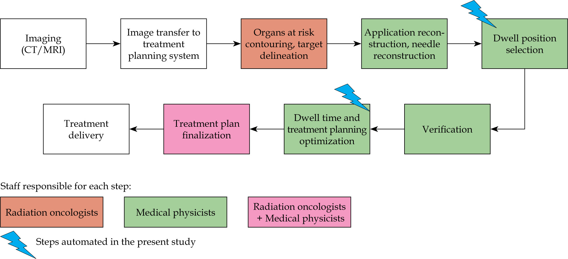
A total of 69 consecutive treatment plans were included in the analysis, where thirty plans underwent automated planning (all from Institution 2) and thirty-nine underwent manual planning (11 from Institution 1 and 28 from Institution 2). In Institution 1, four treatment plans followed the IC + IS workflow, and seven treatment plans followed the IC workflow (Figure 1). The proportionate distribution of IC and IC + IS reflected the clinical workflow in Institution 1, where up to 25-30% of patients would be treated with combined IC + IS and 70% with IC implants. Institution 2 employed combined IC + IS implants for the plans.
Median time required per step
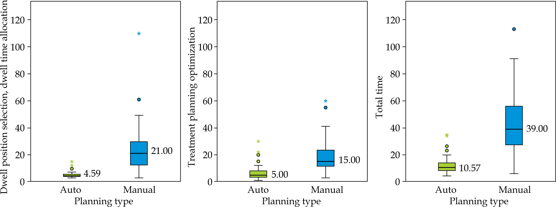
The median time (in minutes) for dwell time optimization during automated and manual planning was 4.59 minutes (IQR: 4.00-5.90) and 21.00 minutes (IQR: 12.00-30.00), respectively. For dwell time adjustments (fine-tuning), the median time for automated and manual planning was 5.00 minutes (IQR: 3.46-8.50) and 15.00 minutes (IQR: 11.00-24.00), respectively (Table 1).
Table 1
Timing between automated and manual planning
* p < 0.001, SD – standard deviation, 95% CI – 95% confidence intervals, IQR – interquartile range, 1 Mann-Whitney U test sum of ranks for treatment planning dwell time optimization: automated = 541.00, manual = 1,874.00; for dwell time adjustment (fine-tuning): automated = 679.50, manual = 1,735.50; for total time: automated = 541.50, manual = 1,873.50 Median and mean differences between automated and manual planning, with interquartile ranges and 95% confidence intervals (CI), are shown, demonstrating significantly shorter times for automated planning (Mann-Whitney U test, p < 0.001 for all components). Times exclude applicator reconstruction but include up to 3 minutes required for image export, BiCycle optimization, and re-import into treatment planning system.
For the automated workflow, the total time required for treatment plan generation, including dwell position time optimization, dwell time adjustment, and automation-specific file transfer steps, was therefore substantially shorter than for the manual workflow only, despite additional 3-4 minutes needed to export and import data to and from the automation software.
Comparative analysis of median times
An independent samples Mann-Whitney U test indicated that, for dwell time optimization, manual planning took 16.41 minutes longer (IQR: 3.22) (p < 0.001) than automated planning. Similarly, dwell time adjustments took 10.00 minutes longer (IQR: 1.50) (p < 0.001). Overall, the total time needed for manual planning was significantly greater by 28.43 minutes (IQR: 4.94) (p < 0.001) (Table 1, Figure 2) than that required for automated planning. These differences reflect the variance between group medians, while interquartile ranges for each planning method are reported separately in Table 1. These timings were averaged for the comprehensive clinical workflow of Institution 1, and were not segregated for IC and IC + IS procedures.
Sensitivity analysis
In order to address inter-institutional bias, an Institution 2-only comparison of the total time required for dwell time optimization and dwell time adjustments between manual and automated planning was performed. Among 28 manual plans and 30 automated plans within Institution 2, the median total time required was 38.00 minutes (IQR: 26.50-57.50) and 10.57 minutes (IQR: 8.57-13.87), respectively, with U = 72.5, p < 0.001 (Mann-Whitney U test).
Discussion
This study showed that integrating automation into IGABT treatment planning for cervical cancer reduced the required time by 28 minutes (p < 0.001), even after accounting for automation-specific steps, such as plan export and plan import, which required less than 4 minutes. Manual planning showed high dispersion (IQR: 27-57 min) as compared with automated planning (IQR: 8.39-13.90 min), reflecting case complexity, iterative processes, and planner experience/skill variability.
According to the International Atomic Energy Agency (IAEA) guidelines, high-volume centers can perform an average of four patient treatments per 8-hour workday [31, 32]. Whereas in the present simulated cohort, Institution 1 had 28% of patients treated with IC + IS, and it is expected that fully optimized workflows will achieve 50% or higher utilization of IC + IS implants in the future, as a reflection of anticipated adaptation of clinical practice to guideline-driven indications. Based on the present study and previous modelling [10], it is estimated that manual treatment planning for IC and IC + IS workflows require 19-38 minutes and 25-48 minutes, respectively. Automation is expected to reduce this time by 28 minutes per patient, potentially saving approximately 120-124 minutes per day for treatment planning and optimization. This extra time could allow physicians to perform an additional implant per day, therefore upscaling system capacity by 25%. These findings extend the context of previous BiCycle studies that primarily focused on dosimetric validation [29], by providing quantitative evidence of real-world workflow gains. Although earlier work demonstrated that BiCycle plans are equal or superior to manually generated plans in terms of target coverage and OAR sparing [27], the present analysis indicates that these quality standards can be maintained while substantially reducing planning time. Importantly, the decision to add an interstitial component remains fundamentally driven by tumor extent and pelvic anatomy in accordance with the EMBRACE II recommendations.
Automation is increasingly allowing efficient IGABT implementation by accelerating contouring, dose planning, and dose delivery. This is especially advantageous in low- and middle-income settings, where the complexity of delineating multiple targets and OARs must be balanced between time and resource constraints [33]. Similar time-saving effects from automation were reported in prostate cancer, where the auto-reconstruction of transperineal needles reduced the total time required by 15 minutes while maintaining the integrity of plan quality [34]. In cervical cancer, Kallis et al. reported that automation reduced dose prediction and optimization time to 3 minutes in patients treated with 0 to 3 supplemental needles [35], aligning with the present research results. Our study provides the first prospective clinical timing data for complex IC + IS implants with EMBRACE II-adherent workflows in high-volume academic centers. Building upon Kallis’ validation of the workflow algorithm, we measured complete real-world process through automation-related time reduction, including clinical review/QA. This confirms BiCycle’s practical scalability beyond technical validation to real-world practice.
Another critical step for time savings is the organ at risk and target delineation. Evidence regarding automation of organ at risk contouring is widespread [36-39], with reductions in time required to less than 1 minute [40] and a 26% of time-saving benefit for clinicians [18]. Although the present study did not assess automation of applicator reconstruction or contouring, both are likely to confer additional efficacy gains and could further enhance IGABT workflows [41, 42]. Automation has also been applied to brachytherapy quality assurance checks; Rhee et al. [43] demonstrated that a deep learning-based auto-contouring system was able to accurately distinguish between clinically acceptable and unacceptable contours.
Globally, radiotherapy demand is projected to rise substantially by 2050, particularly in Asia [9], where workforce shortages are already pronounced [9, 42]. Evidence has consistently reported that plans developed by experienced treatment planners tend to exhibit higher quality than those made by less experienced planners [44]. Automation offers a solution to address the variability in skill levels among planners, and to alleviate the existing skill shortage. Multiple studies indicated that automated planning generates plans that are equivalent or superior to manually-created plans [17, 45-47]. Consequently, automation reduces staff workload and planning time. Even modest incremental time savings in the treatment planning process can translate into significant improvements in patient management, particularly in high-volume settings. Therefore, automation in brachytherapy allows both time efficiency and standardization of the plan quality at a high level.
However, there may be some distinct challenges for integrating automated planning systems within resource-limited centers, including upfront costs and staff training. The quality of automated treatment depends on the library of previously generated plans or prior knowledge provided to machine-learning system, which may be lacking in small centers with low patient volume or skill shortage. Such variability presents challenges for the implementation of generic automation models [47]. In this case, collaborations may have to be developed with large academic centers. The BiCycle system presents such an example, where a fully automated adaptive treatment planning workflow in IGABT for LACC was recently externally validated through cross-institutional analysis [29], supporting its applicability beyond the development institution.
In HDR brachytherapy, inverse planning algorithms, such as hybrid inverse planning optimization (HIPO) and inverse planning simulated annealing (IPSA), are the most commonly available algorithms for dwell-time optimization. However, IPSA often generates heterogeneous dwell-time distributions with needle-loading spikes that may increase the risk of severe complications, whereas HIPO produces smoother dose gradients. However, semi-automated HIPO requires human intervention, and generating clinically acceptable plans typically takes 20-40 minutes [48]. Although HIPO and IPSA are known to reduce dose-optimization time [49] once the applicator reconstruction is complete, these reports often exclude the additional documentation, review, and protocol compliance steps that characterize the EMBRACE II workflows.
Despite the availability of HIPO and IPSA, inverse optimization has not been particularly successful for gynecological brachytherapy, where planning objectives require maintaining high doses centrally while ensuring full coverage of the implant periphery (100% of the prescribed dose) and limiting organ at risk doses to 60-70% of the prescription. Achieving these competing objectives necessitates prioritization of the central applicator components (tandem and ring/ovoids) over peripheral needles, but this aspect is often not sufficiently addressed by generic inverse planning systems. The AI-based BiCycle workflow directly addresses these limitations by enabling fully automated, rule-based multicriteria optimization aligned with the EMBRACE II treatment planning aims. It achieves plan generation in approximately 8-13 minutes with minimal planner input, producing plans with improved organ at risk sparing compared with IPSA and HIPO [29]. Recent time-action analyses of EMBRACE II-consistent workflows, including complex IC + IS implants and comprehensive plan evaluation, have reported substantially longer (more than 5-10 minutes) overall planning durations, particularly in academic high-volume settings [11, 29]. In contrast, the present study measured the complete planning stage preceding clinical approval, thereby providing a more comprehensive representation of routine clinical workload.
Potential limitations of this study include its small sample size, absence of patient-level paired comparisons, and non-inclusion of the applicator reconstruction step. A paired comparison of manual and automated plans for the same patients was not feasible in this study, as BiCycle was implemented prospectively in routine planning in Institution 2, and manual plans were not systematically re-generated for the same fraction. Additionally, potential determinants of time savings, such as tumor stage, intra-/inter-observer reliability, planner skill levels, or IC vs. IC + IS implants, could not be robustly analyzed in this study and remain critical for future research. Barriers, such as initial costs, staff training, and integration into clinical workflows, need to be additionally addressed. Key implementation challenges include initial capital costs for the applicators, software licensing, staff training, and integration into clinical workflows, which are particularly pronounced in resource-limited settings. These economic considerations require dedicated cost-effectiveness analyses beyond the scope of this study. Lastly, the automation-generated plans require a physician and medical physicist supervision to confirm clinical relevance and accuracy of contours, and to assess safety. Nonetheless, prospective measurement minimized recall bias, and the uniform application of institutional workflows ensured methodological consistency.
Conclusions
In conclusion, the automation of IGABT treatment planning for cervical cancer can significantly reduce the time required for the planning of advanced brachytherapy treatments. These findings indicate that automated plans can enhance treatment efficiency. This advancement not only has the potential to improve patient care, but also offers an opportunity to standardize treatment protocols for cervical cancer across various healthcare settings. Future research directions should include the implementation of automation and a comprehensive analysis of the associated cost.











