
Purpose
Cervical cancer remains a significant global health burden, particularly in low- and middle-income countries, where it is a leading cause of cancer-related mortality among women [1, 2]. For patients with locally advanced cervical cancer (LACC), the standard of care is definitive radiotherapy (RT) consisting of external beam radiotherapy (EBRT) to the pelvis, concurrent platinum-based chemotherapy, and a brachytherapy (BT) boost [3, 4]. The role of BT is paramount, as it allows delivery of a highly conformal, high-dose boost to primary tumor while maximally sparing adjacent organs at risk (OARs), such as bladder, rectum, and sigmoid [5].
With the advent of three-dimensional (3D) image-guided brachytherapy (IGBT) using computed tomography (CT) or magnetic resonance imaging (MRI), target volume coverage and OAR sparing have significantly improved, leading to enhanced local control and reduced toxicity [6-9]. Current GEC-ESTRO and ABS guidelines recommend an interstitial component (using needles or templates) when the tumor extends beyond what can be adequately covered by an intracavitary applicator alone, for example, in cases with large parametrial involvement, distal vaginal extension, or asymmetric disease [10, 11]. However, interstitial brachytherapy requires even more specialized expertise and equipment than intracavitary techniques, making it even less accessible in low-resource settings.
Despite its proven efficacy, access to BT is not universal. Many centers, especially in developing countries, lack the necessary equipment (e.g., afterloaders, applicators) and specialized training [12, 13]. Furthermore, some patients may be medically unfit for anesthesia, have anatomical distortions precluding applicator placement, or simply refuse an invasive intracavitary procedure [14]. In such scenarios, historically, patients have received EBRT alone with conventional techniques (e.g., AP/PA or 4-field box), often to limited total doses (60-66 Gy) due to OARs tolerance, resulting in inferior outcomes compared with combination therapy [15].
The evolution of highly conformal EBRT techniques, such as intensity-modulated radiotherapy (IMRT), volumetric modulated arc therapy (VMAT), and stereotactic body radiotherapy (SBRT), has re-ignited interest in using EBRT to deliver a tumor boost. These techniques, coupled with daily image-guided radiotherapy (IGRT), enable steep dose gradients and precise dose delivery, potentially mimicking dosimetric benefits of BT [16-18]. Several retrospective studies and a recent meta-analysis have explored the role of an SBRT boost as an alternative to BT. However, reported outcomes vary considerably across studies. A systematic review by Gazsi et al. (2025), encompassing 17 studies and 288 patients, observed local control rates varying from 57% to 95.5%, with a wide range reflecting heterogeneity in patient selection, dose fractionation, and follow-up duration [19]. Acute toxicity was generally low, but detailed late toxicity data remain limited. Silva et al. (2025) showed more favorable pooled outcomes in their meta-analysis (2-year LC of 94%, late grade ≥ 3 toxicity of 2%), though they acknowledged significant heterogeneity across the included studies [20]. These findings suggest that while EBRT boost techniques show promise, outcomes are not uniformly favorable and require prospective validation.
In this study, we presented a dosimetric analysis of 10 LACC patients treated with a curative-intent protocol using pelvic IMRT/VMAT and an EBRT boost (SIB or sequential) to HR-CTV, in a setting where ICBT was unavailable. We compared our dosimetric findings with published data from both BT and modern EBRT/SBRT series to evaluate the feasibility of this approach as a pragmatic alternative in resource-constrained settings.
Material and methods
Patient selection and eligibility criteria
All patients included in this study met the following inclusion criteria: 1. Histologically confirmed squamous cell carcinoma of the cervix; 2. FIGO stage IIB-IVA; 3. No evidence of para-aortic or distant metastasis on staging imaging (pelvic MRI and abdominal CT); 4. Eastern Cooperative Oncology Group (ECOG) performance status 0-2; and 5. Adequate bone marrow, renal, and hepatic functions to receive concurrent cisplatin-based chemotherapy.
Exclusion criteria were: 1. Prior pelvic radiotherapy; 2. Contraindications to curative radiotherapy (e.g., active pelvic inflammatory disease, fistulas); 3. Inability to comply with bladder/rectum preparation protocol; and 4. Refusal to participate in the study.
The reasons for brachytherapy unavailability included a lack of brachytherapy equipment/facilities at our institution during the study period (n = 6), patient refusal of invasive intracavitary procedure (n = 3), and medical unfitness for anesthesia (n = 1). No patient was excluded due to anatomical distortions precluding applicator placement, as all patients in this cohort had anatomy suitable for standard intracavitary applicators.
Patient characteristics
Between October 2015 and September 2016, 10 patients with histologically confirmed squamous cell carcinoma of the cervix were prospectively enrolled in this dosimetric study. All patients had locally advanced disease staged according to the International Federation of Gynecology and Obstetrics (FIGO) 2009 system. Staging included gynecologic examination, pelvic MRI, and abdominal CT. The patient characteristics are summarized in Table 1. This study was approved by the Institutional Review Board of the First Hospital of Shijiazhuang. All procedures followed the ethical standards of the Declaration of Helsinki.
Table 1
Patient characteristics (n = 10)
CT simulation and treatment planning
All patients underwent CT simulation with a full bladder protocol (500 ml of water drunk 30 minutes prior) and empty rectum. As per rectal preparation protocol, all patients were instructed to empty their rectum completely 30 minutes before CT simulation and before each treatment fraction. Rectal emptying was confirmed by visual check of planning CT scan (for simulation) or CBCT (for treatment fractions). If significant rectal gas or fecal material was observed (> 2 cm in diameter or causing visible displacement of the cervix/uterus), the patient was asked to attempt further emptying, and a repeat scan was acquired. Patients with chronic constipation received a mild laxative (lactulose 15-30 ml) the evening before simulation and before fractions if needed.
Patients were immobilized supine with a thermoplastic mask. Intravenous contrast (iopromide 100 ml; injection rate, 2 ml/s) was administered to all patients during CT simulation to improve delineation of pelvic vessels and nodal regions. Oral contrast (diatrizoate meglumine 500 ml) was given 60 minutes before scanning for small bowel opacification. CT images with 5 mm slice thickness were acquired from upper edge of the first lumbar vertebra to 5 cm inferior to the ischial tuberosity, and transferred to the Pinnacle3 planning system (Philips, The Netherlands). Pre-treatment pelvic MRI (T2-weighted and contrast-enhanced T1-weighted sequences, 3 mm slice thickness) was acquired for all patients and co-registered with CT planning using rigid registration in the Pinnacle3 planning system. HR-CTV was initially contoured on MRI and then transferred to CT dataset for planning. Target volumes were delineated according to ICRU reports 50/62 and RTOG consensus guidelines [21, 22]. Clinical target volume (CTV) included primary tumor, entire cervix, uterus, parametria, and a portion of the vagina. Nodal CTV involved bilateral common, external, and internal iliac, obturator, and presacral lymph nodes. Planning target volume (PTV) for elective nodes was created by adding a 5-8 mm margin to the nodal CTV. High-risk clinical target volume (HR-CTV) was defined according to GEC-ESTRO recommendations as the gross disease at diagnosis (including the entire cervix and any areas of suspected extra-cervical extension), as visualized on pre-treatment MRI and correlated with CT-based planning [23].
No additional PTV margin was applied to HR-CTV beyond CTV-PTV expansion described above. HR-CTV received simultaneous integrated boost (SIB) dose directly without a separate PTV expansion because: (i) daily IGRT with soft-tissue alignment to the cervix was performed to minimize setup uncertainty; (ii) a generous margin (5-8 mm) was already applied to CTV for elective PTV; and (iii) steep dose gradient of SIB plan was designed to ensure that HR-CTV remained within 95% isodose line even with residual motion of 2-3 mm (Figure 1). This approach is consistent with SIB techniques reported in the literature for cervical cancer [16-18].
Fig. 1
Dose distribution in four representative patients. The images illustrate the variability in tumor position (A, B) and size (C, D). The isodose lines show the conformal dose distribution achievable with VMAT-SIB. Red area – HR-CTV, red line – 100% isodose line for HR-CTV boost, green line – 100% isodose line for pelvic PTV
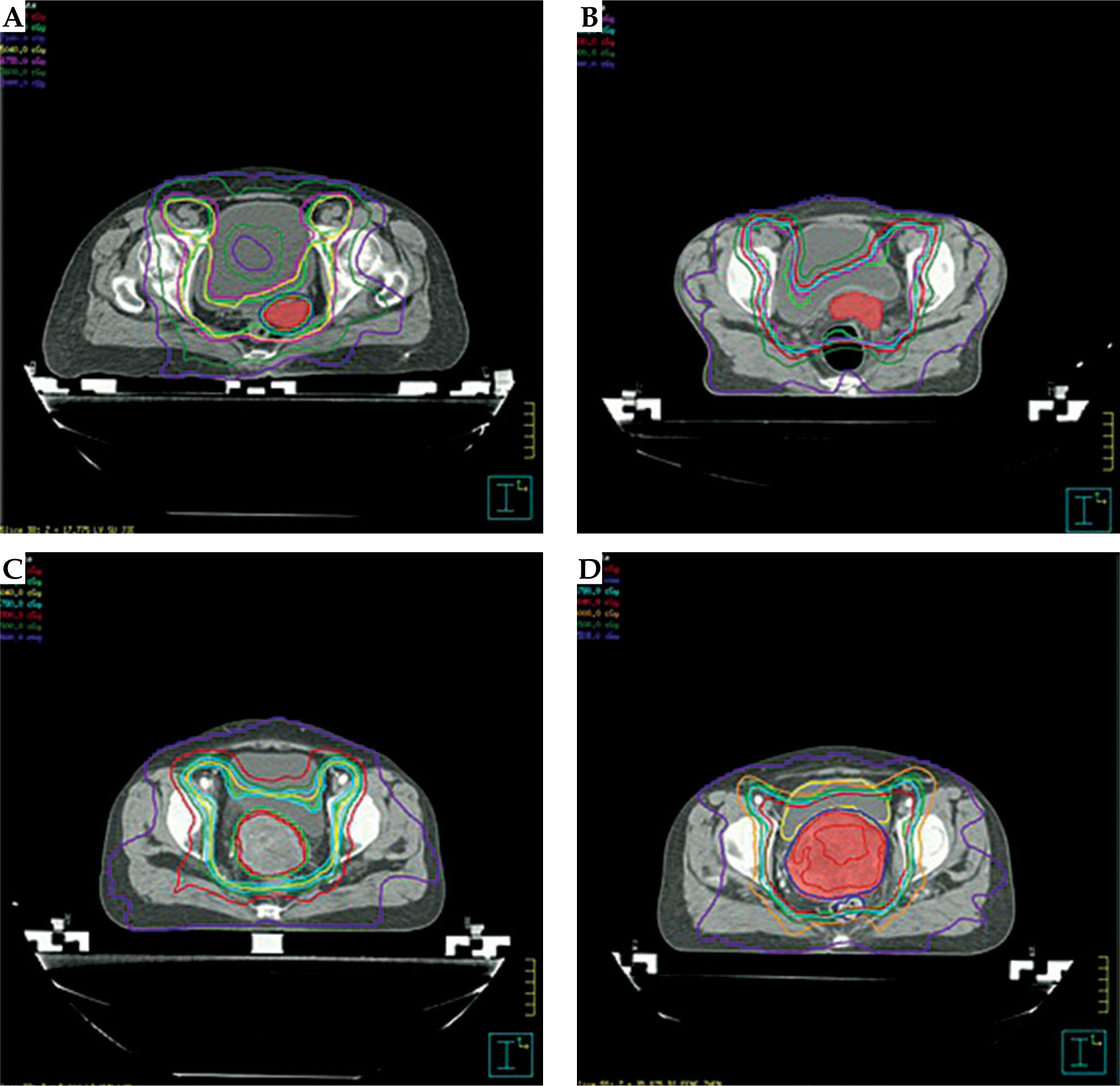
Prescribed dose was 45 Gy in 25 fractions to the pelvic PTV. A simultaneous integrated boost (SIB) was used to deliver a higher dose to HR-CTV: 2.85 Gy per fraction for 25 fractions, resulting in a total dose of 71.25 Gy. For patients treated with a sequential boost, an additional 10-15 Gy in 5 fractions was delivered after pelvic RT. All plans utilized 7-field IMRT or VMAT.
Image-guided radiotherapy and motion management
Fiducial markers were not used in this study due to unavailability at our institution during the study period. Cervical localization was performed using soft-tissue alignment on daily CBCT, with the cervix identified based on its characteristic location relative to the bladder (posterior wall), vaginal vault, and any residual gross tumor.
No online adaptive re-planning was performed, as our linac (Elekta Synergy) was not equipped with adaptive capabilities. However, if weekly CBCT showed consistent anatomical changes (e.g., significant tumor regression or change in bladder/rectum filling pattern), a new CT simulation and treatment plan were generated. This occurred in 2 of 10 patients (20%).
Daily image-guided radiotherapy with kV-CBCT was performed before each treatment fraction to verify positioning, with particular attention paid to the bladder and rectal filling.
Before each treatment fraction, CBCT was acquired and bladder volume was estimated using the planning system’s contouring tools. Target bladder volume was defined as ≥ 300 ml (approximately 70-80% of the volume achieved at simulation). If bladder volume was < 250 ml or if rectal gas/displacement was observed (> 5 mm cervical displacement from baseline position), the following protocol was followed:
mild deviation (bladder 200-250 ml, or cervical displacement 3-5 mm): Treatment was delivered after re-positioning and CBCT verification;
moderate-to-severe deviation (bladder < 200 ml, or cervical displacement > 5 mm): The patient was requested to empty and re-fill the bladder (drink 300-500 ml of water, wait for 20-30 minutes), and a new CBCT was acquired before treatment.
Using this protocol, immediate treatment was possible in 85% of fractions (with re-positioning only), while 15% required bladder re-filling. No fraction was cancelled due to persistent deviation.
Bioeffect dose calculations
Total EQD2 dose to HR-CTV was calculated using linear-quadratic model, with an α/β ratio of 10 Gy for tumor and 3 Gy for OARs [24]. EQD2 was calculated as:
EQD2 = D × [(d + α/β) / (2 + α/β)],
where D is the total dose, d is the dose per fraction, and α/β is the tissue-specific parameter. The aim was to achieve an EQD2 of > 80 Gy to HR-CTV while respecting OAR constraints.
Dosimetric analysis
Dose-volume histogram (DVH) parameters were extracted for all targets and OARs.
For target volumes (HR-CTV and PTV), we recorded D98%, D95%, D90%, D50%, D2%, Dmean, Dmax, conformity index (CI), and homogeneity index (HI). Dose to point A (left and right) was also calculated.
For organs at risk (the bladder, rectum, sigmoid, small bowel, and femoral heads), we recorded V10-V60 and Dmean. Additionally, for the bladder, rectum, and sigmoid, we recorded a minimum dose to the most exposed 1 cm3 (D1cc) and 2 cm3 (D2cc). These were converted to EQD2 (α/β = 3 Gy) for comparison with published dose constraints [10, 11, 25].
Minimum dose to 98% of HR-CTV (D98%) and the volume receiving less than 95% of the prescribed boost dose (V < 95%) were analyzed to identify potential cold spots.
Conformity index (CI) was calculated as: CI = (PTV100/PTV) × (PTV100/V100),
where PTV100 is the target volume covered by the prescribed dose, PTV is the target volume, and V100 is the volume of the prescribed dose.
Homogeneity index (HI) was calculated as: HI = D5%/D95%.
Results
Patient characteristics
All 10 patients completed the planned radiotherapy. The median patient age was 45 years (range, 35-63 years). The FIGO stage distribution was: IIB (n = 2), IIIB (n = 4), and IVA (n = 4). Seven patients had a maximum tumor diameter greater than 5 cm. All patients had histologically confirmed squamous cell carcinoma. The tumor extension details are summarized in Table 1.
Target volume dosimetry
The dosimetric parameters for target volumes are shown in Table 2. For the HR-CTV, the mean D95% was 81.6 ±0.2 Gy (EQD2), and the mean D90% was 84.9 ±0.4 Gy (EQD2). The mean D98% (approximated by Dmin) was 74.5 ±3.1 Gy (EQD2), and the mean volume receiving less than 95% of the prescribed boost dose (V < 95%) was 5.0 ±1.5%, indicating minimal cold spots, all located at the periphery of the HR-CTV adjacent to OARs. The mean HI for the HR-CTV was 1.065, indicating a homogeneous dose distribution. The mean D98% for the pelvic PTV was 50.5 ±0.7 Gy. The mean dose to point A (left and right combined) was approximately 79.8 ±7.5 Gy (EQD2).
Table 2
Target volume dosimetry (EQD2, α/β = 10 Gy)
Organs at risk dosimetry
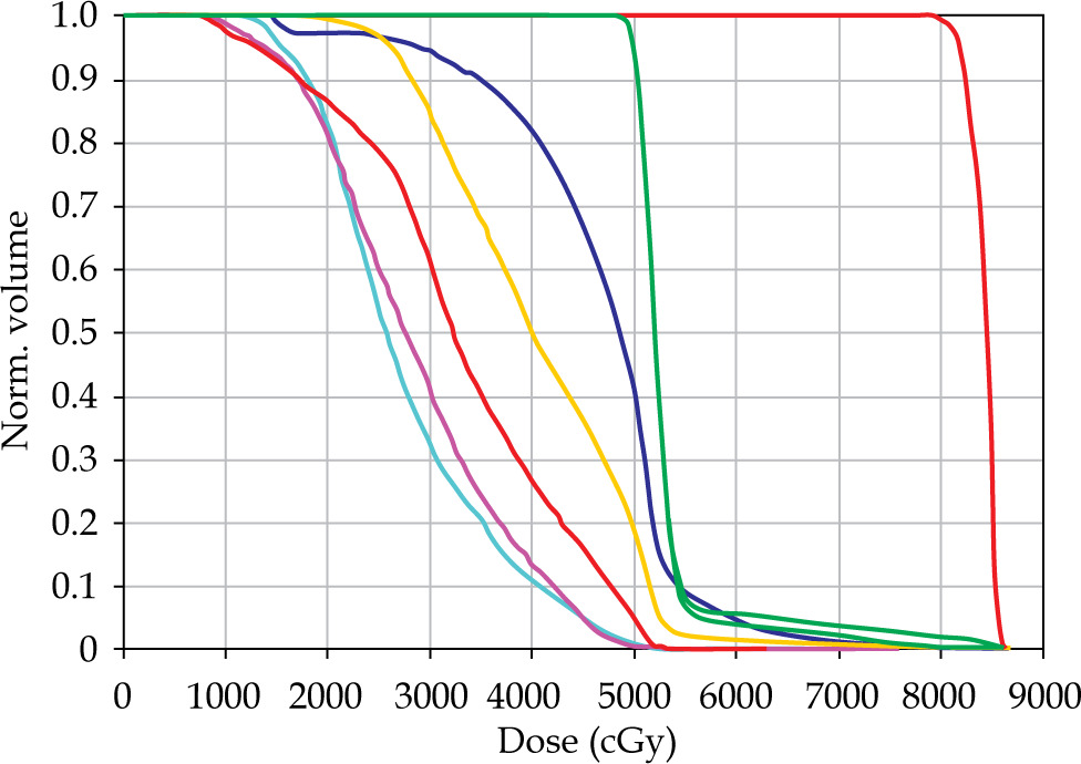
The dosimetric parameters for OARs are detailed in Table 3. The cumulative EQD2 (α/β = 3) D2cc for the rectum was 67.9 ±2.3 Gy, for the sigmoid 68.8 ±3.1 Gy, and for the bladder 74.4 ±2.7 Gy. These values were all below the commonly accepted tolerance constraints (rectum/sigmoid < 75 Gy, bladder < 90 Gy). The mean V60 for the rectum and bladder were 16.1% and 13.4%, respectively. Femoral head doses were also well within tolerance (Figure 2).
Table 3
Organs at risk dosimetry (EQD2, α/β = 3 Gy)
Discussion
The cornerstone of curative treatment for LACC is the combination of pelvic EBRT and a BT boost [3]. However, a significant proportion of patients worldwide miss the opportunity for cure due to the lack of access to BT [12, 13]. This has prompted researchers to explore whether modern, high-precision EBRT techniques can serve as a safe and effective substitute. Our dosimetric study contributes to this body of evidence by demonstrating that an IMRT/VMAT-based boost can achieve target doses approaching those of BT while respecting OAR constraints.
Historically, attempts to replace BT with conventional EBRT had resulted in poor outcomes due to limited total doses (60-66 Gy) necessitated by OARs tolerance [15]. The landscape has changed with the introduction of IMRT, VMAT, and especially SBRT, as these technologies allow dose escalation to the tumor through steep dose gradients and hypofractionation. Our study utilized a moderate hypofractionation with a SIB (2.85 Gy/fraction) to deliver a total EQD2 of over 80 Gy to HR-CTV. This aligns with the well-established dose-response relationship for local control in cervical cancer, where HR-CTV D90 EQD2 > 80-85 Gy is associated with improved outcomes [9, 26].
Our target coverage was satisfactory, with a mean HR-CTV D95 of 81.6 Gy (EQD2) and D90 of 84.9 Gy (EQD2), which – while slightly lower than what can be achieved with a state-of-the-art IGBT implant – is still within therapeutic range. Importantly, the dose homogeneity (HI = 1.065) was excellent, potentially avoiding hot spots inherent in BT. The mean V < 95% was only 5.0%, indicating that cold spots were minimal and confined to the periphery of the HR-CTV adjacent to OARs.
It is important to distinguish between intracavitary and interstitial brachytherapy. While intracavitary brachytherapy remains the most commonly performed technique worldwide, contemporary guidelines recommend adding an interstitial component when target coverage with intracavitary BT alone is inadequate [10, 11]. The EBRT boost technique described in this study may be particularly relevant for patients who would otherwise require interstitial brachytherapy but lack access to such specialized services, as interstitial techniques are available only in highly specialized centers even in developed countries [12].
Regarding OAR sparing, our results are particularly encouraging. The mean D2cc EQD2 for the rectum (67.9 Gy), sigmoid (68.8 Gy), and bladder (74.4 Gy) were all below the GEC-ESTRO/ABS recommended limits [11, 25]. These figures compare favorably with recent publications on SBRT boosts. A 2025 systematic review and meta-analysis by Silva et al., which included 13 studies on SBRT boost, reported a pooled 2-year local control rate of 94% and late grade ≥ 3 gastrointestinal toxicity rate of only 2% [20]. Another systematic review by Gazsi et al. (17 studies, 288 patients) showed local control rates ranging from 57% to 95.5% with acceptable toxicity [19]. Our dosimetric data provide a plausible explanation for these clinical findings, confirming that OAR constraints can be met with meticulous planning and daily IGRT.
It is important to acknowledge that despite the dosimetric feasibility demonstrated in this study, clinical outcomes with EBRT-based boosts have not shown equivalent to brachytherapy. Gill et al. analyzed the National Cancer Data Base, and found that the use of brachytherapy was independently associated with improved overall survival compared with EBRT alone [27]. More recently, Robin et al. reported that disparities in access to brachytherapy were associated with a significant survival decrement in patients with locally advanced cervical cancer [28]. These findings underscore that our proposed EBRT boost technique should not be viewed as a replacement for brachytherapy, but rather as a ‘second-best’ alternative in resource-constrained settings where brachytherapy is unavailable.
The issue of target motion, particularly of the cervix and uterus, remains a primary concern for EBRT boosts. Inter- and intra-fractional variations due to bladder and rectal filling can be significant [29]. In our study, we strictly adhered to a bladder-filling protocol and performed daily IGRT with CBCT, with a structured protocol for managing deviations (as described in the Methods section). For patients receiving a sequential boost, daily imaging was performed during the entire boost phase. This is, in our view, non-negotiable when delivering an EBRT boost to ensure target coverage and safe OAR sparing. Some advanced centers are now exploring online adaptive radiotherapy (oART) to further mitigate motion effects, which represents the next frontier in precision for these cases [30].
In terms of OAR margins, in conventional radiotherapy planning, margins are not typically applied to OARs because the goal is to minimize dose to these structures, not to ensure coverage. However, the reviewer raises an important point about the uncertainty in OAR position due to organ motion. In our study, we accounted for this by: (i) using daily IGRT to verify OAR position before each fraction; (ii) adhering to a strict bladder/rectum preparation protocol to minimize inter-fraction variation; and (iii) employing a planning approach that prioritize OAR sparing while maintaining target coverage. We did not add explicit ‘margins’ to OAR contours because the optimization algorithm inherently considers the entire OAR volume. If one wished to account for organ motion conservatively, OAR ‘planning organ at risk volume’ (PRV) could be contoured by adding a 3-5 mm margin, but this was not performed in our study as it would unnecessarily increase the dose to normal tissue without clear clinical benefit.
An important consideration when delivering an EBRT boost is the timing relative to tumor regression. Cervical tumors typically decrease significantly in volume during pelvic EBRT (often by 40-60% by the end of treatment). In a sequential boost approach, the boost is delivered after completion of pelvic EBRT, allowing the boost target volume to be based on a reduced tumor volume, theoretically improving OAR sparing. In a SIB approach, the boost target is defined on pre-treatment imaging and receives a higher dose from the first fraction, which may deliver unnecessary high-dose to regions that would have regressed. However, SIB offers the advantage of shorter overall treatment time and may be more effective against radio-resistant clones. In our cohort, 7 patients received SIB and 3 received sequential boost due to logistical reasons. Visual check of DVH parameters suggested no clear advantage of one technique over the other in terms of target coverage or OAR sparing, but this observation is limited by small numbers. Future studies should prospectively compare these two approaches, potentially using adaptive re-planning to account for tumor regression in a SIB arm.
Our study has several drawbacks. The small sample size (n = 10) is its most obvious weakness as well as dosimetric nature of the study without long-term clinical follow-up. We cannot draw conclusions about local control, survival, or late toxicity from these data. Our study should be viewed as a hypothesis-generating, proof-of-concept investigation that provides dosimetric rationale for larger prospective studies. Furthermore, we did not use fiducial markers for target tracking, but relying on soft-tissue alignment on CBCT, which has limitations. The use of IMRT/VMAT also inherently increases low-dose bath to normal tissues compared with BT, which in theory, could have implications for second malignancy risk, although this risk is likely small and outweighed by the benefit of curative treatment [31].
Conclusions
This dosimetric analysis demonstrates that an image-guided EBRT boost using IMRT/VMAT with daily IGRT is technically feasible for patients with LACC who cannot receive ICBT. It can deliver a target dose (HR-CTV EQD2 > 80 Gy) that is within the range associated with high local control rates, while maintaining OAR doses below standard tolerance constraints. When compared with published data on SBRT boosts, our OAR doses are comparable, suggesting this approach can be a safe and pragmatic alternative in centers without SBRT capability. We emphasize that this is not a replacement for brachytherapy, which remains the gold standard. However, in the face of a patient refusing BT or in a low-resource setting lacking BT infrastructure, a well-executed EBRT boost with high quality IGRT is a vastly superior option to EBRT alone. The dosimetric findings from this study support the design of a prospective clinical trial (in development, planned registration) to evaluate clinical outcomes, including toxicity and local control. We strongly encourage the formation of a multicenter registry or prospective trial to collect clinical outcome data and establish standardized protocols for this important patient sub-group.











