Purpose
High-dose-rate (HDR) brachytherapy enables highly conformal dose delivery with steep gradients, suitable for localized, anatomically accessible malignancies. Surface mold plesiotherapy is a non-invasive HDR technique, in which radioactive sources are positioned close to the target via an external applicator, allowing precise dose depth modulation while sparing adjacent tissues. Effective implementation relies on accurate and reproducible applicator positioning, stable source-to-surface geometry, and conformity to patient-specific anatomy.
In lip and oral cavity malignancies, radiotherapy is an important approach of organ and function preservation, particularly where surgical resection may result in unacceptable cosmetic and functional outcomes or poor quality of life [1-3]. Interstitial brachytherapy provides excellent dose conformity, high rates of local control with excellent cosmetic and functional results. Moreover, it is an essential salvage option for locally recurrent disease [4] and adjuvant treatment following non-radical surgery [5]. Although implantation may be technically challenging or contraindicated, plesiotherapy is a non-invasive alternative for superficial, well-demarcated lesions, and a combination of these two techniques has shown dosimetric advantages even for larger or irregular lesions [6-9]. Current recommendations of the Indian Brachytherapy Society (IBS) recognize surface mold technique as appropriate for small, superficial, and accessible head and neck lesions as well as those involving the lip, buccal mucosa, and hard palate in primary and salvage settings [10].
Wider implementation of surface mold plesiotherapy is limited by heterogeneity of target anatomy, immobilization challenges, and the absence of standardized design and fabrication pathways for applicators. Conventional approaches, including handcrafted molds using thermoplastic sheets or wax, dental acrylic casting, and silicone-based techniques, demonstrate common limitations of operator-dependent fabrication, geometric imprecision, introduction of workflow delays, and reliance on commercially available templates, which may not conform to patient-specific anatomy [11]. Access to customized applicators is inconsistent across institutions, and there is a lack of standardized, scalable methods for producing patient-specific surface brachytherapy devices that meet contemporary quality assurance and safety requirements.
Additive manufacturing (3D printing) enables rapid fabrication with precise geometry and reproducible construction, with reviews supporting its integration into modern brachytherapy practice [4, 12-14]. Reports have also discussed printable materials, and the influence of these choices on dosimetry and treatment requirements [13]. The current technical note showed a design of a customized HDR surface mold applicator, manufactured in collaboration with an external 3D printing provider, emphasizing practical considerations, quality assurance, and clinical implementation.
Material and methods
Patient selection
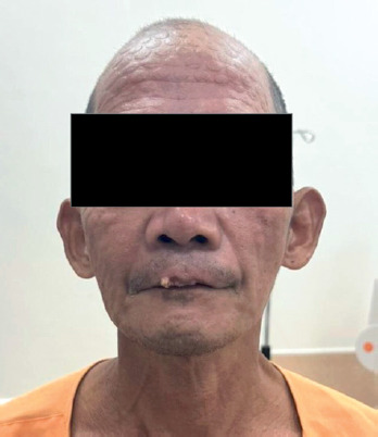
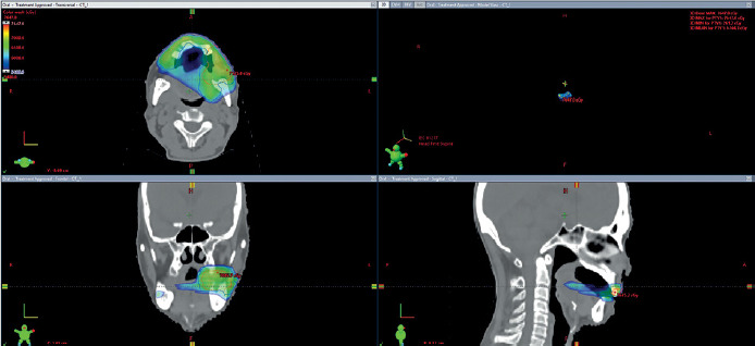
A 52-year-old male with synchronous upper lip and left buccal squamous cell carcinoma (Figure 1) underwent definitive chemoradiotherapy to a total dose of 70 Gy. Following completion of external beam radiotherapy (EBRT), there was residual disease in the upper lip with good response in the buccal region. Surface dose deficit in the upper lip sub-site was observed on dosimetric review, with the surface receiving less than 54 Gy (Figure 2). Because the residual disease was superficial, anatomically accessible, and corresponded directly to an under-dosed EBRT region, a non-invasive HDR surface mold plesiotherapy boost was selected to provide targeted dose escalation to this site.
Clinical workflow
Clinical workflow was adapted from a custom surface mold brachytherapy plan outlined in Figure 25 of AAPM-GEC-ESTRO Task Group No. 253 (TG-253) [15], AAPM Task Group No. 59 [16] for safe HDR treatment delivery, and AAPM medical physics practice guideline 13.a [17] for HDR brachytherapy program standards, along with existing institutional policies (please refer to TG-253 for the full reference workflow).
Patient was selected following clinical examination, anatomic documentation, and immobilization planning. Targets were defined, and data was used for CAD modeling and prototype fabrication by 3D printing. Verification included dimensional assessment, catheter patency evaluation, and patient fitting, with iterative prototyping performed as needed. Subsequently, CT simulation was done with the applicator in situ, followed by contouring, catheter reconstruction, dwell position activation, and plan optimization. Final plan was reviewed prior to treatment delivery. At each fraction, patient identity, applicator positioning, prescription parameters, and catheter passage were verified before HDR treatment delivery under direct physician supervision, with all steps documented according to institutional quality management standards.
Applicator design and fabrication
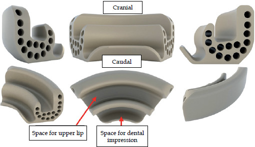
A detailed examination was performed to define targets, with anatomical measurements obtained as follow: mouth opening of approximately 2.0-2.2 cm secondary to trismus, lip width of 5.0-5.2 cm, lip thickness of 7.2 mm, inferior lip edge-to-vestibule distance of 11 mm, vestibule-to-incisor tip distance of 12.5 mm, and involved upper lip width of approximately 3.4 cm. The applicator was then digitally modeled using scaled anterior and lateral photographs of the lip with obtained anatomical measurements. Cross-sectional imaging or dedicated CT-based surface modeling were not available at the time of initial prototype design. Design parameters included an 8-12 mm inter-channel spacing constraint applied during CAD optimization to minimize overlap of high-dose regions, with final channel spacing adjusted locally across channels based on anatomical fit and patient-specific geometry. A 4-mm source-to-surface offset was planned to act as bolus. Dimensional verification was performed against CAD model, with an acceptance tolerance of ±1.0 mm for overall geometry and channel positioning, while catheter patency was confirmed by full insertion and retraction testing of clinical catheter without resistance or deformation. Channels were dimensioned to accommodate FlexiCath single-leader 6F catheters (Kalyani Radiotherapy Specialty India, Ltd.). Iterative computer-aided designs were developed in coordination with the clinical team, with five iterative prototypes made by additive manufacturing, with reference to AAPM guidelines on surface mold applicators and GEC-ESTRO working group recommendations.
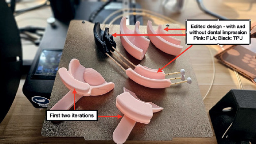
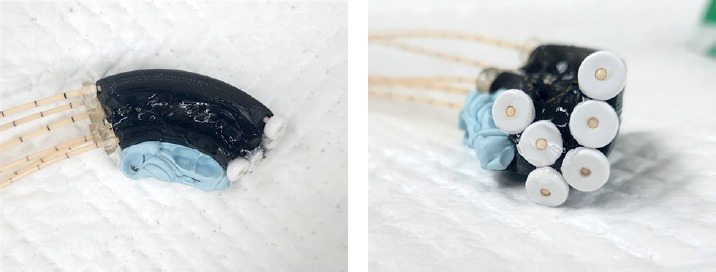
The applicator development process was completed over three days. On day 1, after clinical assessment, two initial prototypes were manufactured with additive manufacturing and delivered to radiotherapy center later that day. On day 2, the patient returned for fitting of the initial prototypes. During evaluation, the applicator material was found to be excessively rigid, and the device dimensions were too wide to accommodate the patient’s severe trismus. Following these fitting sessions, the design specifications were further refined and shared with the external manufacturer. By the end of day 2, three revised applicators incorporating the updated design were fabricated (Figure 3), one using polylactic acid (PLA) and the other made by thermoplastic polyurethane (TPU). On day 3, the patient returned for evaluation of the revised applicators. TPU applicator demonstrated improved flexibility, conformity to the upper lip contour, and more favorable adjustment into the patient’s limited oral aperture while maintaining stable catheter geometry. Based on these findings, TPU applicator was selected for clinical use (Figures 4 and 5). Following satisfactory fitting, acceptance testing was performed, including dimensional verification, catheter spacing assessment, catheter patency testing, and technical review prior to CT simulation and treatment planning.
Simulation, treatment planning, and delivery
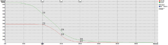
CT simulation was performed with the finalized applicator in situ secured by dental putty, followed by insertion of radiopaque CT markers. A sterile water-based gel lubricant was introduced into the cavity around the anterior lip as tissue equivalent bolus material and to remove air gaps. Planning CT was acquired with 1.25-mm slice thickness using Oncentra BT planning system (Nucletron BV, Veenendaal, The Netherlands). High-risk clinical target volume (HR-CTV) was contoured to encompass residual disease, and intermediate-risk clinical target volume (IR-CTV) was defined as the remaining upper lip with a 3-5-mm margin around HR-CTV. Volumes were compared with the original EBRT plan (Figure 2) to ensure coverage of previously under-dosed areas of the upper lip. Prescription doses were 3.5 Gy to HR-CTV corresponding to residual gross tumor to 5-mm depth from the mucosa, and 3.0 Gy to IR-CTV, to be delivered twice daily at least 6 hours apart in 6 fractions. Planning parameters were: HR-CTV D90 > 100%, HR-CTV V100 > 90%, HR-CTV V200 < 20%, dose non-uniformity ratio (DNR): V150/V100 < 0.36, and dose homogeneity index (DHI): [V100-V150] / V100 > 0.70, as per GEC-ESTRO guidelines. Dose calculations used TG-43 formalism according to GEC-ESTRO recommendations [4].
For planning, catheters were reconstructed from the tip’s end, and paths of the needles were reconstructed in the treatment planning system. Dwell positions were manually selected based on the length of the target, and points were activated every 3 mm. Doses were initially normalized to points along the contoured target, and then optimized graphically. Upon plan review and approval, treatment was implemented using Elekta Flexitron (Nucletron BV, Veenendaal, The Netherlands) with Flexisource Ir-192.
Across six fractions, HR-CTV D90 received 3.95 Gy in 6 fractions and IR-CTV D95 received 2.26 Gy in 6 fractions; fractions 1 and 6 were delivered once daily, and fractions 2 to 4 were administered in a twice-daily schedule with 6-hour interval. In EQD2 calculations for the tumor, no tumor repair was assumed as per LQ framework for short interfraction intervals [18]. Cumulative tumor EQD2 (α/β = 10) was 84.6 Gy for HR-CTV and 67.4 Gy for IR-CTV, including prior EBRT of 54 Gy in 35 fractions (EQD2: 51.8 Gy) combined with brachytherapy of 3.95 Gy in 2 once-daily fractions, 7.9 Gy in 2 daily fractions, 2.26 Gy in 2 fractions, and 4.52 Gy in 2 fractions. For normal tissue dose calculations, interfraction repair was modeled for twice-daily schedules based on incomplete repair after 6 hours using α/β = 3 and a 4.4-hour repair half-time for subcutaneous fibrosis endpoints [19]. Assuming complete overnight repair, cumulative EQD2 estimates yielded 86.9 Gy (HR-CTV-adjacent normal tissue) and 65 Gy (IR-CTV-adjacent normal tissue). Dosimetric evaluation confirmed that coverage, dose homogeneity, and high-dose constraints were within acceptable ranges of GEC-ESTRO recommendations and institutional plan review.
The applicator was cleaned between fractions using 70% isopropyl alcohol as part of routine infection control. No surface degradation, deformation, or mechanical failure were observed. Repositioning was guided by external skin markings and verified with radiographs at each fraction. Although formal robustness analysis was not performed, daily physician-supervised setup with verification imaging demonstrated minimal interfraction variation and acceptable geometric reproducibility across fractions.
Results
Applicator design and fabrication
Multiple design iterations were required to address challenges related to the patient fitting and severe trismus as well as positional reproducibility and conformity to the upper lip contour. Early prototypes contained an intra-oral component based on a design of commercial dental tray, but could not be accommodated within the patient limited oral aperture. Instead, the design was modified to minimize intra-oral extension, and the patient dentition was utilized as an anchoring point to stabilize the applicator during treatment. Catheter channel spacing and layout were refined to enable dose distribution dorsal and ventral to the lip surface and along the vermilion border. The applicator was additionally designed to physically space the upper lip from the lower lip and the mandible, thus reducing mucosal contact and limiting dose to adjacent structures. The number of catheters insertable at any given time was physically constrained by fixator buttons; hence, optimal catheter positions were determined based on the proximity to target volume and the requirement to maintain hyperdose sleeve (200% isodose) outside the lip surface. The presence of an air-tissue interface between the applicator and anterior lip surface was addressed by incorporating water-based gel as a tissue-equivalent spacer and bolus material to minimize dose inhomogeneity.
The final applicator was fabricated with TPU, providing more flexibility over initial PLA prototypes, and facilitating catheter insertion and patient comfort while maintaining geometric stability during treatment delivery. From a radiological perspective, TPU demonstrates near water-equivalent dosimetric behavior in megavoltage photon beams, with percentage depth-dose differences within approximately 2% relative to a Solid Water reference, supporting its suitability as a tissue-equivalent radiotherapy material [20].
Post-processing was performed to remove minor surface irregularities and filament artifacts along the mucosa-contacting edges and channel entry points, with trimming of excess material and smoothing of contact surfaces using a small blade. Following these iterative refinements, the final applicator achieved adequate conformity to the upper lip, stable anchoring to dentition for reproducible positioning, and appropriate catheter geometry for dose modulation across the lip margin.
Catheter setup and CT simulation
During CT simulation, multiple catheter configurations were possible due to the modular channel design of the surface mold applicator. Six catheter channels were selected based on anatomic positioning, proximity to clinical target volume, and adequate inter-catheter spacing to optimize target coverage and dose modulation along the curved upper lip surface. Channel selection was guided by clinical judgment to ensure coverage of the lesion margins while maintaining appropriate spacing to avoid excessive dose heterogeneity.
After selection of the catheter configuration, the patient setup for CT simulation demonstrated good applicator fit and positional stability (Figure 6). Catheters were clearly visualized on CT, with adequate contrast between the patient lip tissue and the thermoplastic polyurethane (TPU) applicator material. No clinically relevant imaging artifacts were observed, and catheter trajectories were reconstructed without ambiguity.
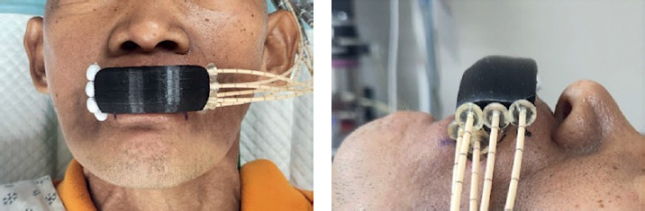
A slice thickness of 1.25 mm was selected given the small treatment volume and the narrow catheter diameter, ensuring reliable visualization of all catheters and accurate reconstruction in treatment planning. This slice thickness provided sufficient spatial resolution to delineate clinical target volumes and evaluate source-to-surface geometry.
Treatment planning
Final catheter configuration, with channels positioned both ventral and dorsal to the lip surface and spaced to provide circumferential anterior coverage, enabled adequate coverage of the delineated target volumes. On iterative planning, only four of the six catheters positioned within the applicator needed to be activated to provide adequate dose coverage. The selected dwell stop positions provided complete longitudinal coverage of HR-CTV and IR-CTV while maintaining appropriate inter-catheter spacing to support dose homogeneity across the curved lip surface (Figure 7).
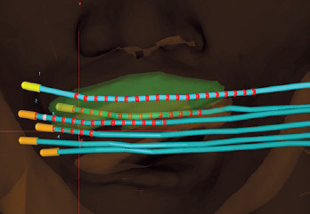
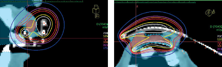
Tissue-equivalent spacer gel served as separation between the high-dose region surrounding the HDR source channels and the lip surface. This mitigated excessive surface dose from the hyperdose sleeve, limiting surface dose receiving 200% of the prescription volume. The 100% isodose line encompassed the delineated target volumes, confirming adequate target coverage while limiting excessive dose to adjacent uninvolved mucosa and underlying structures, which had already received dose from prior external beam radiotherapy (Figures 8 and 9).
Discussion
This case demonstrates the integration of 3D printing into the design-to-treatment workflow of a surface mold brachytherapy applicator for a patient with upper lip squamous cell carcinoma. The applicator was prepared in 3 days, allowing prompt initiation of treatment. This is of clinical value in the salvage or boost setting, where it is critical to avoid prolonged treatment breaks. In this patient, previous chemoradiation has been completed in June 2025, with residual tumor identified two weeks after treatment. Following referral from the outside institution, the first plesiotherapy fraction was delivered four weeks after completion of chemoradiation.
The applicator designed in this paper have site-specific limitations; it was optimized for lesions at the vermilion border of the upper lip based on circumferential spacing and placement of modular catheter channels, while lesions extending significantly cranial to this region may not be adequately covered. Lateral target extent was additionally constrained by applicator width and the patient mouth opening, limiting the coverage of more laterally extensive disease. Careful positioning and verification of full applicator contact are essential, whereas complete filling of the cavity with tissue-equivalent gel spacer have to be ensured for each fraction to minimize air gaps and dose inhomogeneity.
Although formal comparison with conventional handcrafted molds was beyond the scope of this single-case technical report, additive manufacturing offer several practical technical advantages in this setting, informed also by prior institutional experience with conventional mold fabrication using native or commercial materials [21, 22]. The use of computer-aided design (CAD) enabled precise geometric accuracy in channel placement, adhering to planning requirements for catheter spacing, source-to-surface offset, and target coverage. Moreover, 3D printing allowed to iteratively modify the applicator according to patient-specific anatomic constraints while retaining manufacturing consistency and geometric accuracy. In the present workflow, CAD modeling was performed using manual anatomical measurements and clinical photographs, as cross-sectional imaging was not available during prototype development due to workflow limitations. Incorporation of CT surface data or other 3D imaging methods may further improve geometric reliability and reproducibility in future implementations. These observations are consistent with emerging trends in head and neck brachytherapy, where 3D-printed templates and applicators are increasingly applied across multiple sub-sites to improve workflow efficiency, reproducibility, and treatment precision [23].
While additive manufacturing has demonstrated growing uptake in superficial brachytherapy, current evidence is largely composed of single-institution case reports, proof-of-concept studies, dosimetric evaluations, and design process assessments, with limited prospective clinical outcome data. As highlighted by a contemporary review of Bellis et al., studies on 3D-printed applicators have increased substantially in recent years, reflecting growing clinical interest driven by advances in efficiency, geometric consistency, and workflow standardization; however, higher level evidence is still needed to define best practices and quantify clinical benefit. Furthermore, its implementation into clinical practice requires institutional access to 3D printing infrastructure, biocompatible and dosimetrically validated materials as well as expertise in CAD-based device design and quality assurance. Workflow optimization is particularly important for higher volume centers, whereas variability in available hardware, materials, and technical expertise across institutions may limit generalizability and scalability of this approach at present [11].
As a single-case experience, this report has technical and operational limitations and the workflow described may not be directly applicable to other anatomic sites or clinical indications for plesiotherapy. In this case, the absence of pre-existing applicator design libraries necessitated de novo engineering based on patient-specific anatomy, which was resource- and time-intensive, requiring dedicated clinical time for anatomical measurements as well as iterative in-clinic applicator testing and preparation. These involved multidisciplinary brachytherapy team consisting of a radiation oncologist, radiation therapist, brachytherapy nurse, and medical physicist, with additional participation of an external 3D-printing service responsible for applicator modeling and fabrication. Based on the complexity index of interventional radiotherapy implants (COMIRI) framework described by Fionda et al., this procedure would likely meet criteria for a high complexity interventional radiotherapy procedure given the multicatheter implant configuration, CT-based image guidance, multidisciplinary procedural workflow, and customized applicator fabrication [24]. Alternatively, surface mold brachytherapy for intra-oral and skin sub-sites has also been described as a low-complexity procedure in terms of interdisciplinary requirements, considering its comparatively limited need for sedation, airway protection, or surgical collaboration relative to other head and neck sub-sites [25]. Cost implications were not formally quantified, but the overall resource utilization was likely comparable with other image-guided HDR brachytherapy procedures, with variability mainly driven by external printing and material expenses. While adding to initial workflow demands, the CAD-based design and 3D printing may ultimately provide more precise, reproducible, and efficient fabrication of applicators, compared with improvisation from native or commercial materials. Also, it can reduce complexity over time as institutional experience and design libraries can be established.
Conclusions
This paper demonstrates the feasibility of a structured, collaborative workflow for designing, fabricating, and implementing a patient-specific additive manufactured (3D-printed) surface mold applicator for HDR plesiotherapy in upper lip squamous cell carcinoma. In the presented patient, this process was implemented in collaboration with an external printing service provider and allowed rapid design for treatment delivery within 72 hours, yielding an applicator with good anatomic conformity and positional reproducibility without undue technical or clinical complications. The circumferential design and catheter channel configuration enabled stable source positioning along the curved vermilion border, providing effective target coverage with clinically acceptable dosimetry. Iterative prototyping and material selection while adhering to recognized quality assurance standards supported safe implementation.
This experience demonstrates technical feasibility of integrating additive manufacturing into a clinical interventional radiotherapy setting, particularly for surface brachytherapy in anatomically complex and mobile regions. Further development of processes, including parametric design libraries, standardized materials, and streamlined quality assurance policies, will be crucial for establishing additive manufacturing as a practical and reliable component of modern brachytherapy practice.