
Purpose
Synchronous bladder carcinoma and prostate adenocarcinoma is extremely rare. In most cases, prostate cancer is discovered incidentally following radical cystoprostatectomy performed for bladder cancer [1]. Large cystoprostatectomy series reported incidental detection of prostate cancer in 23-54% of specimens, though the majority were considered clinically insignificant and of limited prognostic impact [2, 3]. Truly synchronous presentations, where both malignancies are clinically suspected, investigated, and confirmed at diagnosis, are far less common and largely described in case reports [4, 5]. Management of dual primary malignancies is particularly challenging, given overlapping pelvic treatment fields, differing disease biology, and the need to balance curative intent with toxicity. These complexities underscore the importance of a methodical, multidisciplinary approach to treatment planning [6, 7]. This report described a patient with T2 high-grade bladder carcinoma and NCCN very high-risk prostate adenocarcinoma (stage IIIC), who was successfully managed with concurrent chemoradiation and high-dose-rate (HDR) brachytherapy. To our knowledge, this represents one of the first reported cases of bladder-prostate synchronous malignancy treated with this combined modality approach.
Case presentation
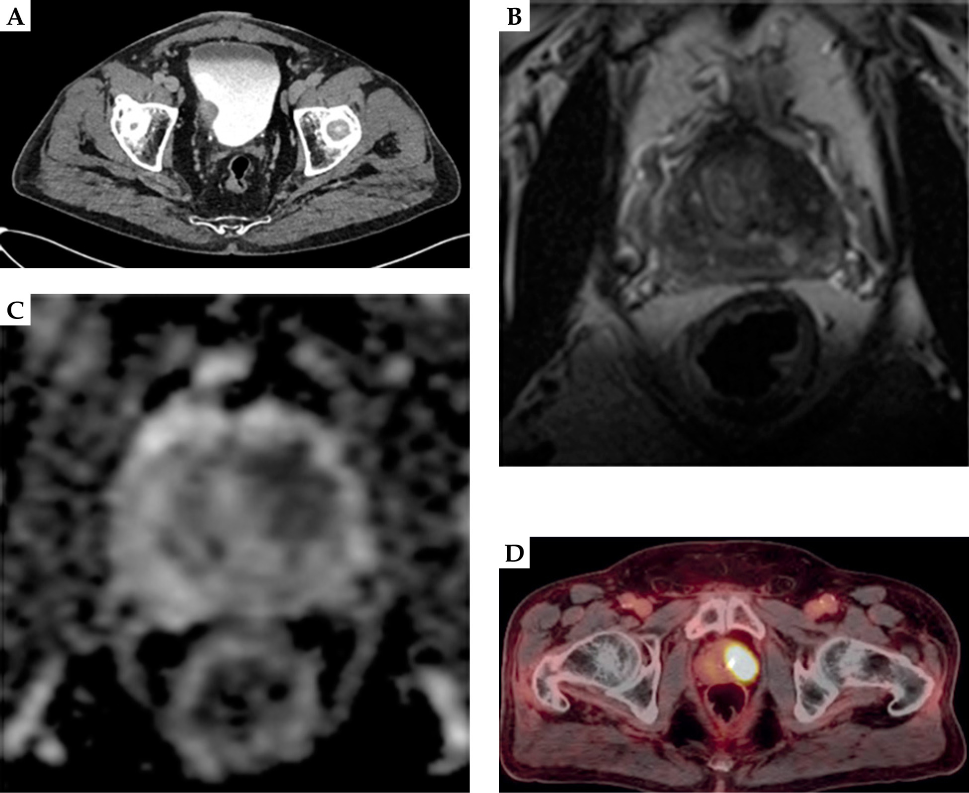
A 70-year-old man with a medical history of emphysema (36-pack-year smoking history), hypertension, and polycythemia, presented to his primary care physician in April 2023 with gross hematuria. He denied dysuria, frequency, or incontinence. Laboratory evaluation revealed a prostate-specific antigen (PSA) of 13.59 ng/ml in April 2023 that increased from 3.57 ng/ml in October 2020. A CT urogram in April 2023 demonstrated a 5 cm right lateral bladder wall mass without associated hydronephrosis (Figure 1A). The patient was referred to urology department, and underwent transurethral resection of bladder tumor (TURBT) with concurrent prostate biopsy. Digital rectal exam was performed with no nodules or masses found. Pathology of the prostate revealed acinar adenocarcinoma involving 6 of 12 cores, Gleason score 5 + 4 = 9 (ISUP grade group 5). MRI of the prostate in August 2023 showed a normal-sized gland with a suspicious PIRADS 4 lesion in the left superior/base transition zone, characterized by decreased T2 signal (Figure 1B) and reduced apparent diffusion coefficient (ADC) (Figure 1C) signal, without significant diffusion restriction or enhancement, and no evidence of extra prostatic extension nor seminal vesicle invasion. Additionally, MRI re-demonstrated a mass along the posterior lateral inferior right bladder wall with associated focal bladder wall thickening. Bladder specimen revealed high-grade, poorly differentiated invasive urothelial carcinoma with papillary and solid features, indicating invasion into the lamina propria and staged as T1 high-grade. Subsequent staging with prostate-specific membrane antigen positron emission tomography (PSMA PET) in June 2023, showed left-sided prostate tumor with no evidence of nodal or distant metastatic disease (Figure 1C). Re-staging CT urogram in October of 2023 prior to initiating treatment, demonstrated subtle asymmetric thickening of right lateral urinary bladder wall, with evidence of mild bilateral hydroureteronephrosis and no sign of metastatic disease within the abdomen or pelvis. After work-up, he was staged with AJCC 8th ed. stage II, cT2 cN0 cM0 urothelial carcinoma of the bladder, and AJCC 8th ed. stage IIIC, cT1c cN0 cM0, NCCN very high-risk prostate adenocarcinoma.
Fig. 1
A) CT urogram from April 2023 demonstrating a 4 cm right lateral bladder wall mass. B) MRI prostate from August 2023 with T2 sequence and C) ADC sequence revealing PIRADS 4 left superior base/transition zone lesion, with no extraprostatic extension. D) PSMA scan showing the prostate tumor without evidence of metastatic disease
Treatment
The patient underwent three transurethral resections of the bladder tumor (TURBT). The initial resection in April 2023 revealed high-grade T1 urothelial carcinoma, while the second TURBT in June 2023 showed no residual disease or muscle invasion. However, the third TURBT in September 2023 confirmed progression to muscle-invasive disease (T2). In parallel, the patient received a six-week induction course of intravesical bacillus Calmette-Guérin (BCG) immunotherapy beginning in July 2023. For prostate adenocarcinoma, androgen deprivation therapy (ADT) with goserelin (Zoladex®) and abiraterone (Zytiga®) was initiated in August 2023. ADT was continued with repeat goserelin injections administered every three months through April 2025, for a total duration of 20 months. Given progression of bladder cancer to muscle-invasive disease, the patient was offered cystoprostatectomy, which he declined. In an effort to preserve his bladder, he selected to proceed with definitive combined modality therapy.
Prior to initiating treatment, three fiducial markers in the prostate were placed. One fiducial marker was intentionally positioned as close as possible to the bladder neck in the right prostate base. From November 6, 2023 to December 27, 2023, he completed external beam chemoradiation, receiving 45 Gy to the pelvis in 25 fractions, with a sequential boost of 64.8 Gy in 36 fractions to the bladder tumor bed with intensity modulated radiation therapy, delivered concurrently with weekly cisplatin 40 mg/m2 over the course of approximately 7 weeks with no treatment breaks. External beam radiation treatment fields were created, so that the inferior aspect of the 64.8 Gy boost field did not extend inferiorly beyond the inferior border of the bladder, and did not extend beyond the fiducial marker placed at the prostate base/bladder neck.
The pelvic clinical target volume (CTV) treated to 45 Gy included the prostate, bladder, and pelvic lymph nodes. Pelvic lymph nodes encompassed the common iliac nodes up to the aortic bifurcation, presacral nodes, external and internal iliac nodes, and obturator nodes, representing the elective pelvic nodal regions for both high-risk prostate cancer and bladder cancer. A 5 mm isotropic expansion was used to generate the pelvic planning target volume (PTV), while the whole bladder CTV included in the 45 Gy volume was treated with an empty bladder to minimize the treated volume. An isotropic 1.2 cm expansion was employed to generate the bladder PTV. The prostate and seminal vesicle (SV) CTV included the entire prostate and full seminal vesicles. The PTV expansion for the prostate and SVs was 7 mm isotropically, except posteriorly where a 5 mm margin was applied. Cone-beam CT was used daily for image guidance. For bladder tumor boost, gross tumor and tumor bed were contoured based on the original location of the tumor prior to TURBT fusing the original CT urogram. CT-SIM was performed when intravenous contrast reached the bladder, so the contrast in the bladder was easy to visualize. The inferior border of boost volume was at the bladder prostatic interface, and we ensured that the contour encompassed the spot where the contrast in the urine stopped and was at least 5 mm superior to the prostate base fiducial. PTV expansion of the bladder tumor boost was 1.2 cm isotropically and 5 mm inferiorly to minimize overlap with the anticipated brachytherapy boost field to the prostate and seminal vesicles. The external beam radiation treatment fields and sequential bladder boost volumes are illustrated in Figure 2A. To address higher curative dose requirement of very high-risk prostate cancer, 6 days after completing external beam chemoradiation, an HDR brachytherapy boost (15 Gy in a single fraction) was delivered to the prostate on January 3, 2024. CTV treated to 15 Gy using HDR brachytherapy included the prostate and proximal seminal vesicles. CTV of the distal seminal vesicles (SVs) was treated with 11 Gy to achieve an equivalent dose in 2 Gy fractions (EQD2) greater than 70 Gy while respecting bladder constraints. No PTV expansion was performed for brachytherapy part of treatment. When designing brachytherapy field, we ensured coverage of the CTV without exceeding the D2cc bladder constraint of 90 Gy, assuming 64.8 Gy from external beam radiation treatment (Table 1). In addition, we sought to further minimize urethral dose by applying stricter constraints than published guidelines, keeping urethral volume receiving 125 Gy (V125) < 0.1 cc (Table 1). The HDR brachytherapy implant and dose distribution to the prostate and seminal vesicles are shown in Figure 2B. ADT was continued throughout this period, and completed in April 2025.
Fig. 2
A) External beam radiation therapy treatment fields demonstrating 45 Gy pelvic isodose line (dark blue) with sequential boost to gross bladder tumor of 64.8 Gy (yellow). A 45 Gy pelvis treatment in 25 fractions encompassed the prostate, whole bladder, and pelvic lymph nodes (common iliac lymph nodes to the aortic bifurcation, pre-sacral, external iliac, internal iliac, and obturator lymph nodes). A 64.8 Gy bladder boost for additional 11 fractions (total 36 fractions) included gross bladder tumor, resection bed, with isotropic PTV expansion of 1.2 cm and inferior expansion of 0.5 cm to limit overlapping with anticipated HDR brachytherapy treatment volume. B) High-dose-rate brachytherapy plan delivering 15 Gy in a single fraction to the prostate and seminal vesicles
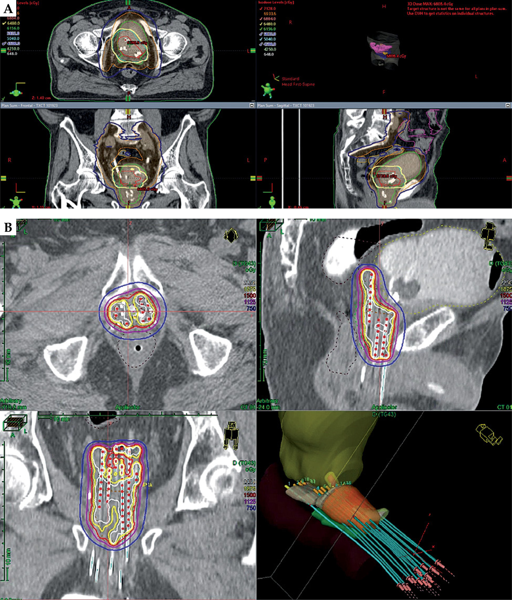
Table 1
Dosimetric goals and constraints taking into account external beam radiation treatment and high-dose-rate brachytherapy doses
[i] Prox. SVs – proximal seminal vesicles, EQD2 – equivalent dose in 2 Gy fractions, D2cc – dose to the hottest 2 cubic centimeters of an organ, V15 Gy – volume receiving 15 Gy, V75 – volume receiving 75% of the prescription dose, V125 – volume receiving 125% of the prescription dose, D10 – dose to 10% of the organ
Post-treatment follow-up
Cystoscopy performed two months after completion of radiotherapy demonstrated no evidence of disease (NED). PSA levels declined to 0.09 ng/ml shortly thereafter and continued to decrease, reaching < 0.01 ng/ml by one year following treatment. A CT urogram in April 2024 revealed linear calcification along the bladder suture line, consistent with post-treatment changes. Surveillance cystoscopy, last in October 2025, almost two years post-treatment, again confirmed NED. Repeat CT urogram and chest imaging, most recently performed in January 2026, showed no evidence of recurrence or metastatic disease. At recent follow-up (January 2026), the patient remains disease-free with undetectable PSA (< 0.01 ng/ml) and preserved functional status. He has minimal urinary symptoms requiring tamsulosin 0.4 mg nightly, and does not report any bowel issues. His testosterone is slowly recovering. Late effects include stable bladder wall calcification and right-sided hydronephrosis, both managed conservatively. These findings suggest excellent early disease control using the above described multimodality treatment approach.
Discussion
Studies reporting synchronous bladder and prostate malignancy are limited. Most prostate cancers in this setting are discovered incidentally at cystoprostatectomy, and are often low-grade [3]. Reports of clinically significant synchronous presentations remain scarce, largely confined to case reports [4, 5]. Our patient represents an unusual scenario, with high-risk disease in both organs requiring definitive local and systemic therapy. Management of synchronous pelvic malignancies poses unique challenges, because each cancer has distinct therapeutic dose requirements and treatment paradigms, often necessitating individualized treatment strategies developed through multidisciplinary collaboration.
Published reports describing synchronous genitourinary malignancies most commonly involve incidental prostate cancer identified at the time of radical cystoprostatectomy for bladder cancer. Conversely, cases in which both tumors are clinically significant and require definitive treatment, are far less frequently described. When both malignancies warrant curative therapy, clinicians must balance the need for adequate oncologic dosing for each cancer with the cumulative radiation tolerance of surrounding pelvic organs. As a result, treatment decisions often require adaptation of established disease-specific protocols to safely address both malignancies concurrently.
Bladder preservation with chemoradiation is an accepted alternative to radical cystectomy for selected patients with T2 disease [6]. For very high-risk prostate cancer, combined modality therapy with ADT, dose-escalated external beam radiotherapy, and brachytherapy boost, is associated with improved biochemical control [7]. Importantly, prostate adenocarcinoma generally requires a higher effective dose for disease control compared with urothelial bladder cancer. In our case, pelvic chemoradiation was delivered to a dose appropriate for bladder cancer (64.8 Gy), and therefore HDR brachytherapy was incorporated to escalate the dose to the prostate to curative range. HDR brachytherapy allowed dose escalation to the prostate and seminal vesicles while minimizing additional dose to the bladder and rectum, an important consideration in the setting of prior pelvic chemoradiation.
This strategy is consistent with other reports describing the use of HDR brachytherapy as a method of safely escalating dose to the prostate when treating synchronous pelvic malignancies. For example, Konat-Baska et al. reported a case of synchronous prostate and rectal cancer treated with pelvic chemoradiation, followed by an HDR brachytherapy boost to the prostate. The authors demonstrated that brachytherapy can effectively deliver ablative prostate doses while maintaining acceptable toxicity profiles [8]. Their report highlighted that combining external beam radiotherapy with an HDR boost enabled curative-intent treatment of both malignancies without compromising surrounding organs at risk.
Additional reports have described similar strategies for managing synchronous prostate and rectal malignancies. A recent 2024 single-center retrospective review evaluated 16 patients with locally advanced synchronous rectal and prostate cancers. In this cohort, the investigators reported the use of multimodality treatment approaches incorporating pelvic radiation and systemic therapy. Prostate brachytherapy was the most frequently utilized modality, allowing many patients to avoid prostatectomy as well as additional external beam radiation to the rectum, highlighting the treatment’s potential role in facilitating definitive treatment of both malignancies while limiting overlapping pelvic radiation exposure [9]. Another report evaluating management strategies for synchronous rectal and prostate cancers emphasized the feasibility of treating both malignancies with curative intent using tailored multimodality therapy, stressing the importance of multidisciplinary planning when standard treatment pathways for each disease overlap [10]. Similarly, a case published in Medicine (Baltimore) described the management of locally advanced synchronous prostate and rectal cancer using coordinated chemoradiation and systemic therapy, followed by a HDR prostate brachytherapy boost and subsequent anterior resection. This report further demonstrated how individualized treatment sequencing can facilitate definitive management of both malignancies while maintaining acceptable organ at risk dose constraints [11].
Although these reports involved rectal rather than bladder malignancies, the underlying treatment principle is comparable: external beam radiation can address regional disease and the second pelvic malignancy, whereas HDR brachytherapy provides the necessary dose escalation for prostate cancer control. In scenarios where pelvic radiation is already required for another malignancy, HDR brachytherapy represents a practical strategy to achieve prostate dose intensification without exceeding tolerance limits of adjacent organs. To our knowledge, this is the first report of chemoradiation followed by HDR brachytherapy for treatment of synchronous prostate and bladder malignancies.
Despite the need for prostate dose escalation, bladder and rectal dose constraints were respected, with cumulative EQD2 values remaining within accepted tolerance limits (Table 1). Careful treatment planning was particularly important in this case, given the prior delivery of definitive-dose pelvic radiation for bladder cancer, as excessive cumulative dose to organs at risk could increase the risk of late genitourinary or gastrointestinal toxicity. This dual-targeted approach allowed organ-preservation for the bladder while ensuring definitive management of the very high-risk prostate malignancy. Our patient’s plan integrated these evidence-based strategies concurrently.
The patient tolerated combined therapy without unexpected toxicity, and remains without evidence of disease at 24 months of follow-up. However, given the natural history of high-risk prostate cancer, longer follow-up is necessary to determine durability of disease control and to fully assess late treatment-related toxicity. Although further long-term outcomes remain to be seen, this report emphasizes the importance of multidisciplinary coordination and individualized treatment planning in rare presentations of synchronous bladder and prostate cancer. A limitation of this report is the relatively short duration of follow-up, which precludes definitive conclusions regarding long-term oncologic outcomes. Longer follow-up is needed to evaluate outcomes and to understand potential long-term toxicities associated with this treatment approach. Considering the limited number of published cases addressing synchronous genitourinary malignancies requiring definitive radiation therapy, reports such as this one contribute to the growing body of literature supporting tailored radiation strategies, which integrate external beam therapy with brachytherapy dose escalation.
Conclusions
This case demonstrates that concurrent chemoradiation and HDR brachytherapy boost can be a safe and effective treatment approach for synchronous bladder and prostate cancer. Because prostate adenocarcinoma requires a higher dose for curative control than urothelial bladder carcinoma, pelvic radiation was delivered at a dose appropriate for bladder preservation, while an HDR brachytherapy boost was incorporated to escalate the prostate dose to curative levels. This tailored approach achieved excellent disease control with acceptable toxicity. Multidisciplinary coordination remains essential in such complex dual primary scenarios, and ongoing follow-up will be critical to assess long-term efficacy and late effects.











