
Purpose
High-dose-rate (HDR) brachytherapy is an essential component in the management of locally advanced gynecologic cancers [1-4]. The standard of care for many locally advanced cervical cancers includes an initial external beam radiation therapy, followed by intra-cavitary or interstitial brachytherapy [4]. Brachytherapy allows the delivery of a highly conformal dose to the target with a rapid dose fall-off that spares healthy tissues and nearby organs at risk (OARs) [5]. The steep dose gradient allows the escalation of dose to the target while minimizing toxicity to OARs, including the bladder, rectum, sigmoid, and small bowel [6]. However, it also necessitates precise OAR contouring to ensure accurate dosimetric evaluation, plan optimization, and toxicity minimization [4].
In cervical cancer brachytherapy, a tandem and ovoids (T&O) applicator system is commonly used in conjunction with needles (T&O + N) when necessary. Since the applicators and needles (if used) must be inserted before each treatment fraction, a new set of planning images, contours, and treatment plans have to be generated for every fraction. This adaptive planning process leads to a repetitive and time-consuming workflow, particularly for OAR contouring, which can be a challenge in busy clinics treating several T&O cases daily.
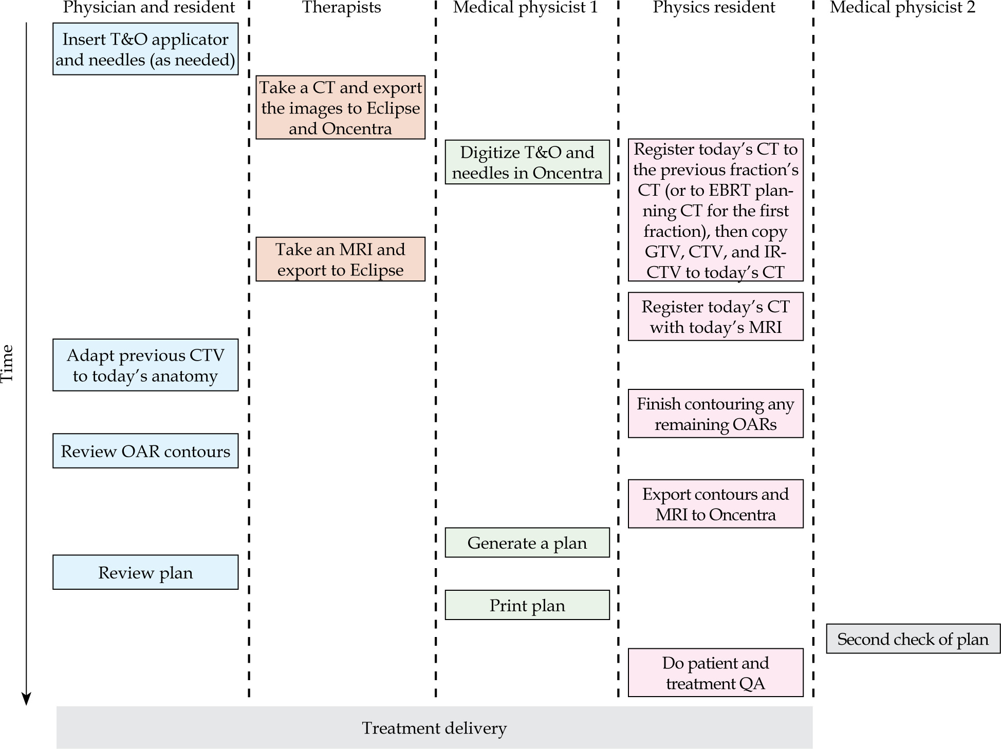
To illustrate the complexity of this workflow, Figure 1 outlines the step-by-step process required for each T&O fraction for a single patient in our clinic. Multiple team members are involved in applicator insertion, computed tomography (CT) and magnetic resonance imaging (MRI) acquisition, image registration, contouring, treatment planning, plan review, and patient-specific quality assurance (QA). The repeated need for full OAR contouring across all fractions, contributes significantly to clinical workload and planning time, particularly in high-volume settings.
Fig. 1
Clinical workflow for each fraction of tandem and ovoid (T&O) treatment. The diagram shows multi-disciplinary coordination and step-by-step process required in adaptive planning for a single patient
To reduce the burden of contouring OARs fully without compromising on dosimetric accuracy, patient-specific adaptive rings around the target volume have been investigated in the setting of online adaptive external beam radiation therapy as a method to identify where OAR contouring is necessary [7]. By limiting OAR delineation to these clinically relevant regions, this approach has the potential to significantly reduce contouring and, as a result, planning time while improving clinical efficiency. In this study, we investigated the feasibility and effectiveness of using a patient-specific adaptive ring structure to guide OAR contouring in T&O cases for cervical cancer. Also, we evaluated the time savings from this approach, quantified the dosimetric accuracy compared with full OAR contours, and determined appropriate adaptive ring margins, based on anatomic and dosimetric analysis across a cohort of previously treated patients.
Material and methods
For this study, a total of 50 previously treated HDR brachytherapy fractions were investigated across 25 patients. Of these, 29 fractions were delivered with T&O alone, while the remining 21 fractions with T&O + N. Most cases (n = 48) were treated with a prescription dose of 5.5 Gy × 5 fractions, whereas the remaining two patients received 5.25 Gy × 5 fractions. For all cases, contouring was performed in the Varian Eclipse treatment planning system (TPS) (Palo Alto, CA, USA) contouring workspace, and planning was completed in the Varian BrachyVision TPS (Palo Alto, CA, USA). All clinical reference contours were the contours drawn by the physicist at the time of actual HDR treatment.
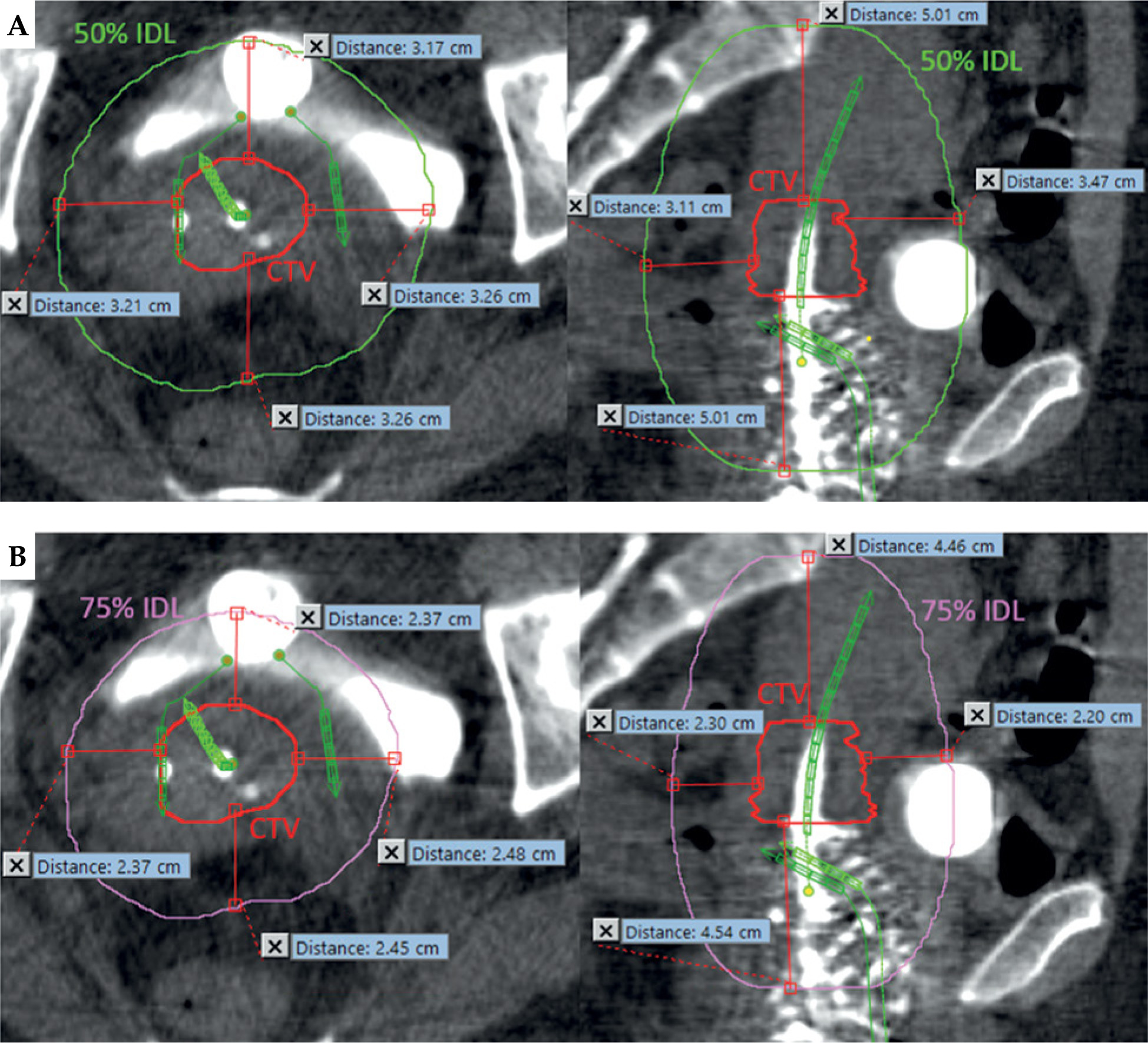
In this study, we evaluated two types of adaptive rings, generated as anisotropic expansions in the Eclipse TPS, for contouring: one defined by the margin between clinical target volume (CTV) and 75% isodose line (IDL), and another defined by the margin between CTV and 50% IDL. Henceforth, these will be referred to as the 75% IDL adaptive ring and the 50% IDL adaptive ring, respectively. The margins for the 75% and 50% IDL adaptive rings were determined by measuring the distance from the CTV to the corresponding IDLs in three orthogonal directions: anterior-posterior (A-P), right-left (R-L), and superior-inferior (S-I), as illustrated in Figure 2. Then, 95% confidence intervals (CIs) were calculated as mean ± 2 × standard deviation, and were based on the measured distances for each orthogonal direction. These became the anisotropic expansions used to generate the adaptive rings. A leave-one-out validation was performed to assess the robustness of the calculated CIs. For each iteration, one case was randomly excluded, and the CIs were re-calculated using the remaining data. The excluded data was then evaluated against the recalculated CIs. This process was repeated 10 times.
Fig. 2
Measurement of distances from clinical target volume (CTV; shown by the red contour) to A) the 50% isodose line (IDL; shown by the green contour) and B) 75% IDL (shown by the pink contour) in three orthogonal directions (anterior-posterior [A-P], right-left [R-L], superior-inferior [S-I])
For dosimetric evaluation, contours for the bladder, rectum, sigmoid, and small bowel were modified to include only portions inside the adaptive rings. D2cc values for the ring-bound OARs were compared with D2cc values from the original, full OAR contours. The D2cc was the dose delivered to the hottest 2 cc of a structure. This comparison was done to assess whether limiting contouring to the region inside the rings introduced clinically significant dosimetric differences.
To assess potential time savings, OAR contouring times were recorded for 10 fractions using three different strategies: full OAR contouring, contouring within the 50% IDL adaptive ring, and contouring within the 75% IDL adaptive ring. All the contouring for this study was done by one physics resident to minimize inter-user variability and ensure consistency across all cases.
Results
Adaptive ring dimensions
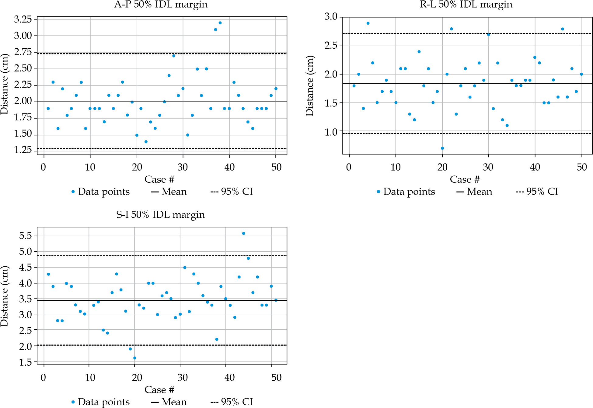
The distances from the CTV to the 75% and 50% IDLs were calculated for each case to determine the final margins for the adaptive rings using 95% CI. Figures 3 and 4 show the distribution of these distances for all cases along the A-P, R-L, and S-I directions for both the 75% and 50% IDLs, respectively. From Figure 3, the mean distances (cm) for the 75% IDL were: A-P = 1.20 (95% CI: 0.44-1.96), R-L = 1.02 (95% CI: 0.25-1.79), and S-I = 2.90 (95% CI: 1.41-4.38). From Figure 4, the mean distances (cm) for the 50% IDL were: A-P = 2.01 (95% CI: 1.27-2.74), R-L = 1.84 (95% CI: 0.94-2.73), and S-I = 3.46 (95% CI: 1.99-4.92).
Fig. 3
Distribution of distances from the clinical target volume (CTV) to the 75% isodose line (IDL) in anterior-posterior (A-P), right-left (R-L), and superior-inferior (S-I) directions across 50 high-dose-rate (HDR) brachytherapy fractions
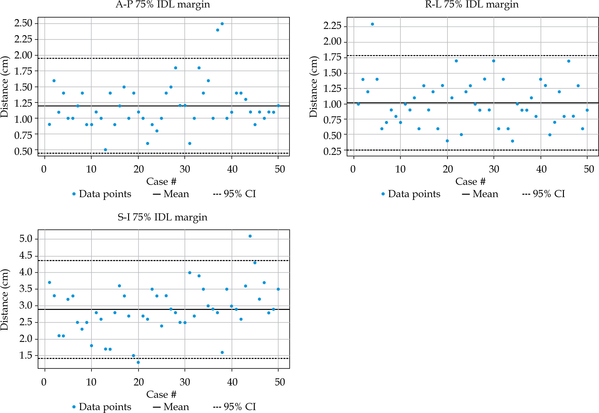
Fig. 4
Distribution of distances from the clinical target volume (CTV) to the 50% isodose line (IDL) in anterior-posterior (A-P), right-left (R-L), and superior-inferior (S-I) directions across 50 high-dose-rate (HDR) brachytherapy fractions
A Shapiro-Wilk normality test was performed on these distances, and the resulting p-values were: A-P 75% IDL (p = 1.68 × 10⁻⁴), R-L 75% IDL (p = 0.03), S-I 75% IDL (p = 0.46), A-P 50% IDL (p = 7.71 × 10⁻⁴), R-L 50% IDL (p = 0.37), and S-I 50% IDL (p = 0.68). Although the Shapiro-Wilk test indicated some non-normality in certain datasets, visual inspection of Q-Q plots suggested that the data was approximately normally distributed, with only minor deviations at the tails. Therefore, normality was assumed.
Substantial variability was observed across all cases in all directions, with the widest CIs occurring in the S-I direction. This variability was due to the differences in the CTV shape, applicator geometry, patient anatomy, and tandem loading. To assess whether there were significant variabilities between T&O and T&O + N patients, an independent t-test was performed. The results from the t-test are summarized in Table 1. No statistically significant differences were noted between the two groups for any of the six distance metrics (p > 0.05). Due to non-significant differences from this sub-group analysis, the T&O and T&O + N groups were combined for calculating 95% CIs.
95% confidence interval (CI) calculations
The 95% CIs were calculated as the mean ± 2 × standard deviation. To assess the robustness of calculated CIs, a leave-one-out validation was performed, where one case was randomly removed from the dataset, and the CIs were calculated from the remaining 49 cases. The excluded data was evaluated against the calculated CIs to determine whether the data was within the CIs. This was repeated for 10 times. Across all ten iterations, 100% (10/10) excluded data points fell consistently within the CI ranges, indicating the stability of the derived margins. Therefore, the full dataset was used to derive the final confidence intervals.
The upper limits of these 95% CIs were employed to create conservative margins for the adaptive rings. These limits were rounded to simplify the creation of margins around the CTV. The margins used to generate the 75% IDL adaptive ring were: A-P = 2.0 cm, R-L = 2.0 cm, and S-I = 4.5 cm. The margins used to generate the 50% IDL adaptive ring were: A-P = 2.75 cm, R-L = 2.75 cm, and S-I = 5.0 cm.
Dosimetric impact of adaptive rings on OAR D2cc
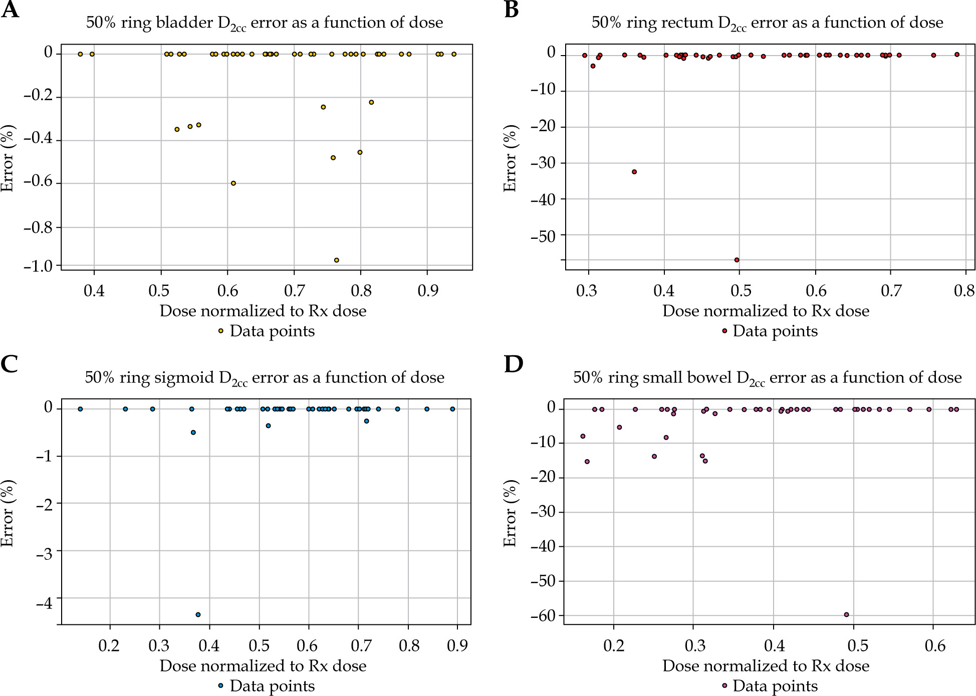
Figures 5 and 6 show the percent error in D2cc as a function of dose normalized to the prescription dose for the (A) bladder, (B) rectum, (C) sigmoid, and (D) small bowel with the 75% and 50% IDL adaptive rings, respectively. A normalized threshold of 0.6 was chosen to define clinically relevant D2cc values for the rectum, small bowel, and sigmoid, and 0.7 was chosen for the bladder. These thresholds were selected based on typical OAR constraints in HDR brachytherapy, which generally range between 60-90% of the prescription dose, depending on the OAR and fractionation scheme. The selected thresholds corresponded to a range of acceptable D2cc values from 65-75 Gy EQD2, assuming 45 Gy in 25 fractions of external beam radiation therapy followed by 27.5 Gy in 5 fractions of brachytherapy. Below this threshold, the D2cc was well within tolerance, so errors at this dose level were less likely to impact dosimetry. For doses above the 0.6 or 0.7 threshold, percent errors did not exceed 3% for all OARs contoured using both the 75% and 50% IDL adaptive rings. The 3% difference was not relative to prescription dose, but relative to clinical dose the OAR received. The maximum absolute dose per-fraction deviation in D2cc between the full and 50% IDL ring-based contouring was observed in the small bowel (1.61 Gy), corresponding to 2.70 Gy for the full contouring and 1.09 Gy for the ring-based contouring; the clinical reference contour received a D2cc of 2.69 Gy. Similarly, the maximum absolute deviation for the 75% IDL ring-based contouring was observed in the rectum (1.98 Gy), corresponding 3.25 Gy and 1.27 Gy for the full and ring-based contours, respectively, with a clinical D2cc of 1.98 Gy.
Fig. 5
Percent error in (A) bladder, (B) rectum, (C) sigmoid, and (D) small bowel as a function of dose normalized to prescription dose for organ at risk (OAR) contours within the 75% isodose line (IDL) adaptive ring
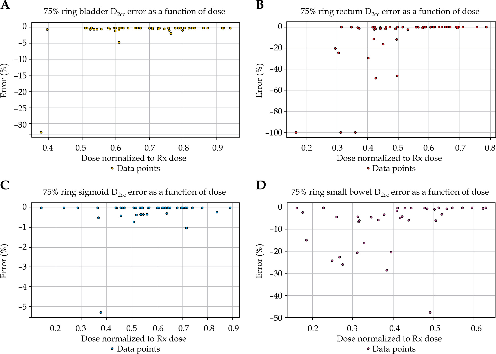
Fig. 6
Percent error in A) bladder, B) rectum, C) sigmoid, and D) small bowel as a function of dose normalized to prescription dose for organ at risk (OAR) contours within the 50% isodose line (IDL) adaptive ring
To further evaluate the clinical impact of these per-fraction differences, EQD2 analysis was performed. The median D2cc EQD2 values were similar across all contouring methods for all OARs. For the bladder, the values were 67.78 Gy (53.77-85.44 Gy), 68.62 Gy (54.02-85.31 Gy), and 68.30 Gy (49.36-85.41 Gy) for the full contouring, 50% IDL ring, and 75% IDL ring methods, respectively. Corresponding values for the rectum were 58.93 Gy (48.61-74.94 Gy), 58.93 Gy (48.13-74.97 Gy), and 58.59 Gy (48.29-74.94 Gy); for the sigmoid, they were 63.13 Gy (46.10-81.78 Gy), 62.98 Gy (46.23-81.70 Gy), and 62.98 Gy (46.25-81.69 Gy); and for the small bowel, they were 52.17 Gy (45.66-65.55 Gy), 51.71 Gy (46.15-65.55 Gy), and 50.75 Gy (46.57-65.55 Gy). The bladder, rectum, and small bowel D2cc EQD2 values were within EMBRACE II constraints, but the sigmoid D2cc EQD2 exceeded the constraint in one case. This estimate assumed that the sigmoid received the same D2cc across all HDR fractions. In practice, the position of the sigmoid relative to the CTV can vary between fractions, and the highest dose region may not necessarily be reproduced. Therefore, EQD2 values derived from a single fraction may overestimate the true cumulative dose. When reviewing this patient’s full course of treatment, including external beam radiotherapy and brachytherapy, the sigmoid dose remained within the constraint. Therefore, this supports the interpretation that the apparent dose increase reflects this limitation of the EQD2 calculation rather than a true dose constraint violation.
Contouring time savings using adaptive rings
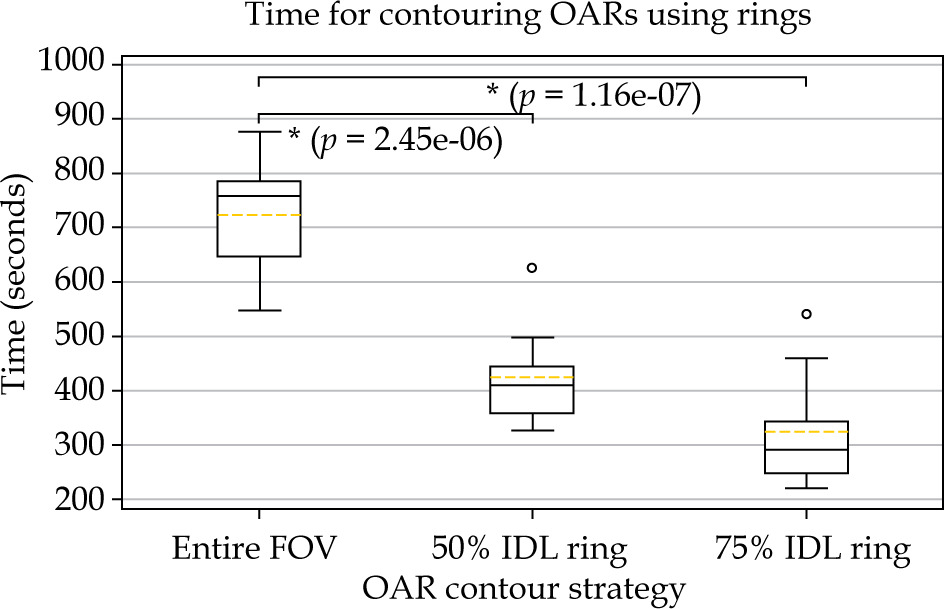
To evaluate potential time savings from this approach, OAR contouring times were recorded for 10 fractions using three strategies: (1) full OAR contouring, (2) contouring within the 75% IDL ring, and (3) contouring within the 50% IDL ring. By only contouring the OARs within the 50% IDL adaptive ring, the mean contouring time decreased from approximately 700 sec to 400 sec (p = 2.45 × 10⁻⁶), while only contouring within the 75% IDL adaptive ring decreased the contouring time further to approximately 300 sec (p = 1.16 × 10⁻⁷), as shown in Figure 7. These times only included the contouring times and not the time to create the rings. Since the rings were created based on Boolean expansion, an additional of 10-15 sec can be added to these contouring times, assuming only one ring is created. Both methods reduced the contouring time by half compared with full OAR contouring, showing a significant improvement in clinical workflow and efficiency. An additional observer independently contoured a sub-set of three cases to evaluate inter-observer variability in contouring time savings. A t-test comparing contouring times for full OAR contouring and contouring within the 50% IDL ring, demonstrated a significant difference (p = 6.62 × 10⁻⁴), and a similar comparison with the 75% IDL ring also showed a significant difference (p = 6.96 × 10⁻⁵). However, no significant difference was observed between contouring within the 50% and 75% IDL rings (p = 0.08).
Intra- and inter-observer contour reproducibility
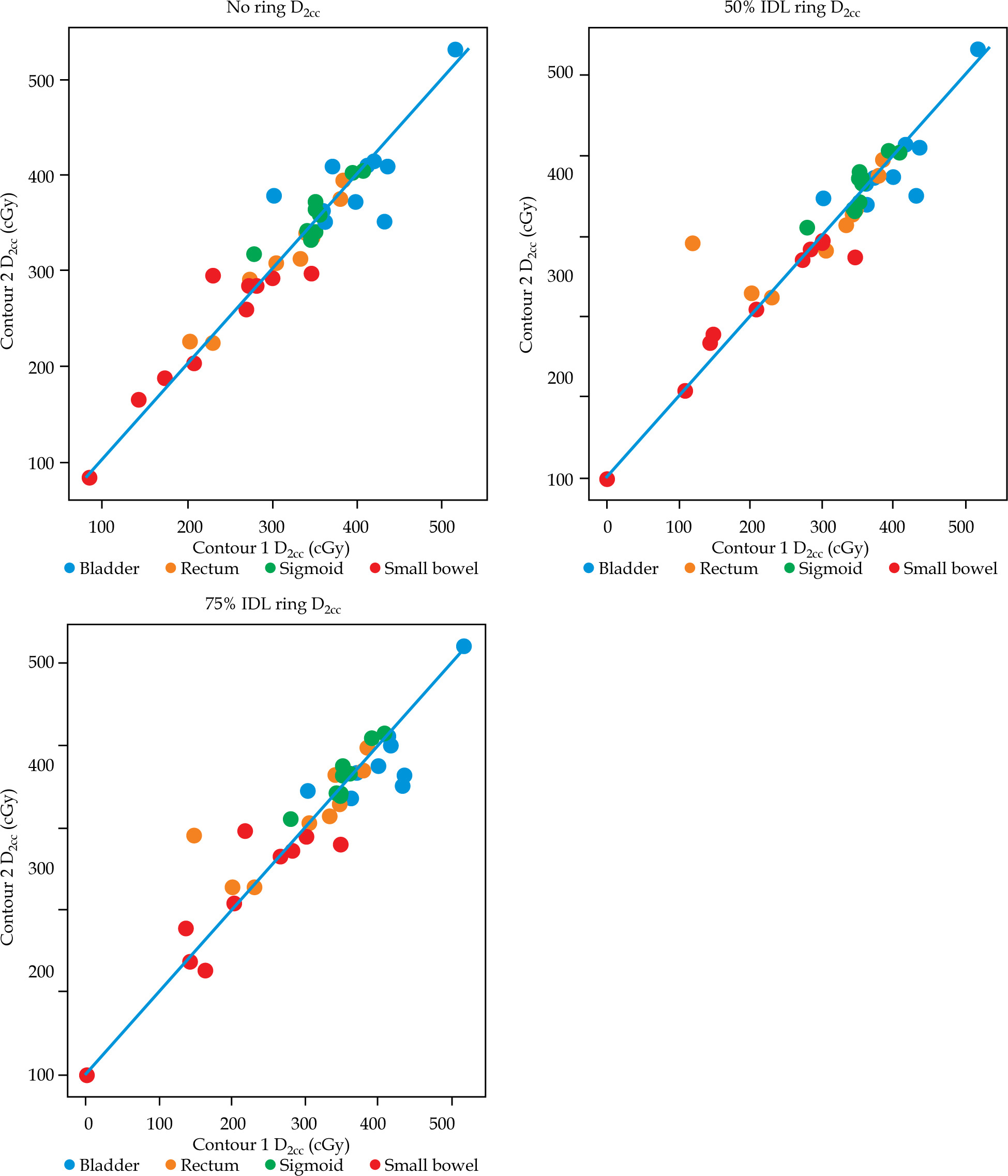
To evaluate intra-observer contour reproducibility, the ten fractions used for assessing OAR contouring times were re-analyzed. The same resident re-contoured the bladder, rectum, sigmoid, and small bowel for a second time using the three strategies: (1) full OAR contouring, (2) contouring within the 50% IDL ring, and (3) contouring within the 75% IDL ring. D2cc values for the four OARs were compared between the two iterations. Figure 8 displays a strong agreement between the two iterations, with most of the data points closely distributed along the straight line across all dose levels, indicating good reproducibility. Quantitative analysis revealed no statistically significant differences for the bladder, rectum, sigmoid, and small bowel (p > 0.05) across the contouring methods. The maximum absolute per-fraction difference in D2cc between the two contouring iterations across all three contouring strategies was 0.81 Gy, corresponding to 4.32 Gy and 3.52 Gy for the first and second iterations, respectively.
Fig. 8
Comparison of D2cc values between two contouring iterations for each organ at risk (OAR). Each point represents an individual case, and diagonal line indicates perfect agreement
To evaluate inter-observer contour reproducibility, a sub-set of three cases was independently contoured by a second observer, who contoured all four OARs using the same three strategies. The D2cc values were compared with those obtained from the resident’s contours, and a quantitative analysis demonstrated no statistically significant differences for the bladder, rectum, sigmoid, and small bowel (p > 0.05) across the contouring methods. The maximum absolute difference in D2cc between contours created by the resident and the second observer was 1.15 Gy (3.22 Gy vs. 4.37 Gy) for full contouring, 0.62 Gy (3.11 Gy vs. 3.73 Gy) for the 50% IDL ring-based contours, and 1.00 Gy (2.56 Gy vs. 3.56 Gy) for the 75% ring-based contours.
Discussion
This study demonstrates that adaptive rings can streamline OAR contouring in HDR gynecologic brachytherapy without compromising dosimetric accuracy. For clinically relevant D2cc values, the percentage errors between full OAR contouring and contouring within the rings were less than 3% for all OARs, confirming that contours confined to the adaptive rings accurately capture metrics for high-dose regions. Additionally, contouring time decreased by a factor of two, highlighting a significant potential for improving clinical efficiency.
A time-consuming part of HDR T&O planning is OAR contouring during each fraction to account for changes in anatomy and applicator positioning within the patient. Previous works have shown that contouring is a labor-intensive task that is susceptible to inter-observer variability, especially in brachytherapy [8, 9]. Therefore studies have investigated auto-segmentation methods to address this limitation and reduce clinical workload [10, 11]. Though the results from the studies were promising, with time savings of about 30-50%, editing was still required to improve contour accuracy [10, 12]. The reported contouring times in this study did not include the time to generate the rings. Since the rings were created manually, an additional 10-15 seconds can be added to the contouring times, assuming only one ring is created. Compared with time savings achieved by the auto-segmentation methods, our adaptive ring methodology achieved approximately 60% reduction in time while maintaining dosimetric accuracy. This suggests that the adaptive ring approach is a feasible alternative compared with auto-contouring methods, and is achievable without requiring a technical background in automation.
Intra- and inter-observer analyses demonstrated good reproducibility of OAR D2cc values across all contouring approaches, with no statistically significant differences observed between contouring iterations or between observers. These results suggest that adaptive rings produce consistent dosimetric outcomes when applied by the same and different observers. While MRI provides superior soft tissue contrast for target and OAR delineation, inter-observer variability in contouring remains an important consideration [13]. Adaptive rings may help reduce this variability by focusing contouring on clinically relevant high-dose regions, particularly in MRI-based workflows where contouring complexity is greater.
While this study demonstrates significant time savings, there are some limitations of the current approach. Firstly, only 50 fractions from 25 patients were analyzed, which may limit the study’s generalizability of findings. In order to combat this drawback, the margins for each adaptive ring were purposefully increased compared with the true 95% CI values. A leave-one-out validation was performed to assess the robustness of the CIs. Secondly, the impact of these rings on physician contour and plan review was not evaluated as part of this study. However, since the adaptive rings limit contouring to relevant high-dose regions, it reduces the extent of contour review and editing by the physician, which may reduce the physician workload during contour and plan review. Although, inter-observer variability was evaluated and demonstrated minimal variations, it was performed on a limited sub-set of three cases. A larger multi-observer analysis may be needed to evaluate inter-observer variability more comprehensively. However, we believe the significant finding of saving time by drawing less would hold true for any user. Finally, prospective validation of this workflow in a true clinical setting to evaluate time savings was not performed in the current study. Contouring is only one part of the planning and treatment workflow, and if the bottleneck exists in a different process, overall planning and treatment time may not be reduced. However, the advantage of this scenario is it would still free time for the clinical team to focus on other tasks. Future studies will investigate the use of the 75% and 50% IDL adaptive rings prospectively to evaluate time savings in the overall process.
Conclusions
Adaptive rings provide an efficient solution for OAR contouring in HDR gynecologic brachytherapy. This approach reduces contouring time by 50-60% while keeping D2cc accuracy within clinically acceptable limits. This method is independent of TPS and imaging modality, meaning this method can be implemented into other clinical workflows. However, clinics should determine the margins for the adaptive rings based on their clinical data. These adaptive rings offer a solution to streamline HDR brachytherapy workflows without compromising on plan quality and patient safety, and without requiring advanced, automated methods.











