
INTRODUCTION
The purpose of this case study is to present factors influencing therapeutic decisions and patterns of language recovery in a bilingual post-stroke patient, focusing on the impact of single-language therapy on both the treated and untreated language.
Over half of the world’s population communicates in more than one language [1], with about 43% being daily bilinguals [2]. The first language (L1) is usually acquired through early exposure, whereas the second (L2) is often learned later, with varying proficiency levels. Types of bilingualism are distinguished mainly by the age and manner of L2 acquisition [e.g., early vs. late (after age 6-7), additive, subtractive, passive] [2].
Due to the high incidence of stroke [3], bilingual individuals are at risk of developing post-stroke aphasia – a disabling disorder of language comprehension, expression, reading, and writing, typically resulting from damage to language-related brain regions around the left Sylvian fissure, supplied by the left middle cerebral artery. Aphasia varies in symptom patterns, ranging from word-finding difficulties (anomic aphasia) to impairments across all language modalities (global/mixed aphasia), and in severity from mild to severe [4, 5]. In bilingual aphasia, all known languages are generally affected, though not equally, producing unique profiles sometimes marked by pathological code-mixing, language switching, and translation difficulties [4, 6].
Post-stroke language recovery typically relies on spontaneous recovery – natural reorganization triggered by sudden injury, most intensive in the first months and largely completed within the first year after onset – and on clinically guided therapy, consisting of structured interventions aimed at language improvement, tailored to the patient’s abilities and communication needs [7, 8]. In bilingual patients, recovery is shaped not only by factors identified in monolingual patients (e.g., lesion size, time since injury, aphasia severity), but also by premorbid bilingualism-related variables (e.g., age of acquisition, proficiency, use patterns) and sociocultural context (e.g., language preferences, dominant language in the patient’s environment) [4, 6, 9, 10]. According to Paradis [11], bilingual language outcomes may follow synergistic patterns, where both languages improve to some extent – either in parallel (similar deficits and progress across both languages), or differentially (different deficits and reco-very rates). Alternatively, antagonistic patterns may occur, with improvement in one language accompanied by deterioration in the other. Other patterns, such as successive (one language returns first), selective (only one language improves), or mixed (interference between languages), are also observed [6, 11].
The effects of bilingual aphasia rehabilitation remain poorly understood. Since therapy aims to improve every-day communication, treatment should ideally address both languages. However, this is often unavailable or challenging due to the patient’s condition or limited resources, making language choice crucial. Some recommend therapy in the mother tongue, others in the less impaired language, or the one most useful for the patient’s daily life, often the majority language of the country [12-14]. A key unresolved issue is cross-linguistic transfer to the untreated language, which is particularly relevant in common bilingual profiles given their high prevalence. English-Polish bilingualism is a pertinent example: English is the most widely taught foreign language in Poland [15], while Polish is the second most spoken language in the UK [16].
Case description
This case concerns a 68-year-old right-handed English-Polish bilingual male who underwent four years of language therapy for severe post-stroke aphasia; the results are presented here. Written informed consent for publication was obtained.
Acquisition and use of two languages
The patient was born in Scotland to a Polish father and a Scottish mother. According to an interview with his sister, conducted using the Polish version of the Bilingual Aphasia Test (BAT) [17], English was his L1 and Polish his L2. L1 was the home language from birth, and all formal education – from primary school through university and postgraduate studies – was conducted in L1. More intensive exposure to L2 began around age 10, during visits to his father’s family in Poland and through extracurricular classes. Around the age of 30, he moved to Poland for work and has resided there since. By profession, he was an accountant and auditor in a managerial position, regularly using both languages in spoken and written communication depending on context. Spoken L1 predominated in professional settings and in interactions with colleagues and many friends (~60% of speaking time), whereas L2 was more common in everyday life and at home (e.g., conversations with friends, shopping, watching TV; ~40%). He read (e.g., books, newspapers, and work documents) and wrote in both languages, with only occasional minor errors in Polish. As a result, prior to the brain injury, the patient was a balanced late bilingual, with native-like proficiency in English and high proficiency in Polish. He actively used both languages across modalities and contexts, with a slight dominance in L1. The two languages are unrelated: English belongs to the Germanic family, while Polish is Slavic [18].
Brain damage
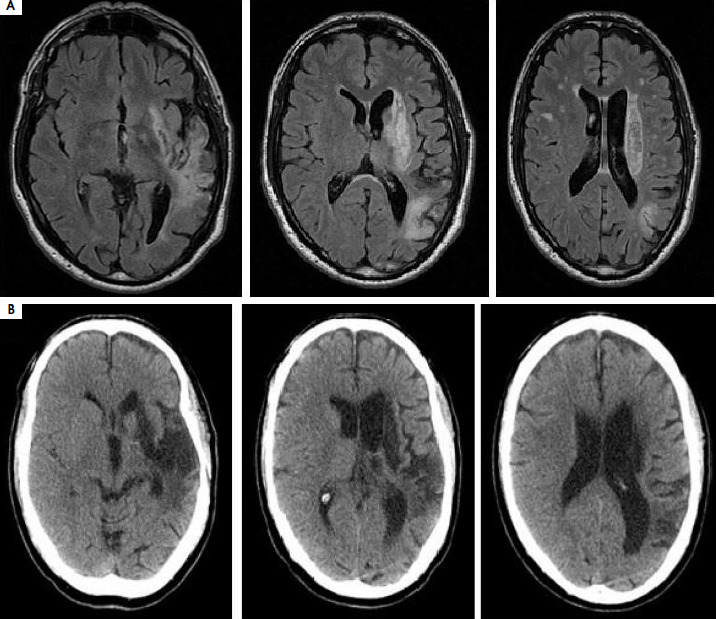
Within two months, the patient experienced two consecutive ischemic strokes affecting peri- and extrasylvian regions of the left hemisphere. Post-second-stroke MRI revealed acute ischemic lesions in the left thalamus and extensive prior damage in the left insula, temporal and occipital lobes, deep structures, and multiple small chronic infarcts. A follow-up CT scan two years later showed a large post-stroke scar in the left temporal and parietal lobes, insula, and basal ganglia, with secondary dilatation of the left lateral ventricle. Neuroimaging from both time points is shown in Figure I.
Figure I
Axial MRI images from the acute phase of the second stroke (A), and axial CT images from the chronic phase of the illness, obtained two years after the second stroke (B). A) Acute ischemic lesions were identified in the left thalamus, coexisting with chronic ischemic damage in the left hemisphere involving the insular cortex, temporal and occipital lobes, deep brain structures, and multiple small chronic infarct foci in both hemispheres. B) An extensive encephalomalacic area of post-ischemic origin involving the left insular cortex, temporal and parietal lobes, and basal ganglia, with secondary dilatation of the left lateral ventricle
Bilingual aphasia assessment and therapy
In line with Fabbro’s recommendation [12], the patient’s aphasia was assessed at three post-stroke stages: acute (1 week after the second stroke), lesion (4 months), and late phase (2 and 4 years). The first two assessments took place during the spontaneous recovery period, when therapy effects overlap with natural improvement. The latter two occurred after this period, with therapy as the primary source of functional gains [7, 19]. L1 and L2 were assessed by an experienced clinical neuropsychologist (A. Zielińska-Michalak) on separate days, with test order varied to minimize fatigue and order effects. Depending on communication ability, assessments included language tasks and formal testing with the English [20] and Polish [21] short versions of the Boston Diagnostic Aphasia Examination (BDAE), which evaluates auditory comprehension, oral expression, repetition, reading, writing, and overall communicative functionality. In this study, individual language functions were monitored using BDAE scores, while overall speech functionality was rated on the 6-point (0-5) Aphasia Severity Rating Scale (ASRS), indicating: very severe aphasia (0; non-functional speech and/or comprehension), severe (1-2; fragmented simple expression or limited conversation on familiar topics), moderate (3; able to discuss various topics despite limitations), or mild (4-5; reduced fluency or subjective difficulties).
Therapy language was selected based on the patient’s linguistic profile, clinical condition, and psychosocial context. A Polish-English bilingual specialist provided individual training within a restitutive–compensatory framework, including impairment-based therapy (progressive repetitive exercises targeting disrupted language functions using paper-based materials and computer-assisted programs) and functional communication practice, supported by gestures and visual aids when necessary. The program incorporated widely used techniques, including phonological and semantic cueing (providing initial sounds or contextual hints to facilitate word retrieval), semantic feature analysis (activating semantic attributes associated with the target word), script training (rehearsing personalized everyday dialogues), and guided conversational practice (structured interpersonal interactions) [22].
Language changes during therapy
One week after the second stroke, the patient presented with very severe aphasia in both languages. Although he appeared to grasp basic conversational phrases, single- word comprehension was poor, indicating primarily contextual understanding. Speech was nonfluent and effortful. He was unable to name objects, repeat words, or read and write in either language, though he occasionally produced spontaneous L1 words, even when prompted in L2.
Given that L1 had been the dominant, native language that was more frequently used pre-stroke and better preserved early post-stroke, therapy was initiated in L1 to support natural recovery and cross-linguistic generali-zation. This choice was reinforced by the fact that family and friends also spoke English. The initial phase (16 weeks, ~80 hours) focused on spoken language, targeting auditory comprehension (yes/no questions, word–picture matching, following commands) and oral expression (word repetition, automatic series, retrieval of high-frequency words with cueing, simple question responses).
At four months post-stroke, improved communication enabled BDAE assessment in both languages. Performance was severely impaired in each, with abilities better preserved in L1. In L1, auditory comprehension was impaired at the single-word level; expressive output was limited to simple phrases, occasional object naming, and word repetition. Reading showed difficulty with letter and word recognition, frequent perseverations, and poor interpretation. In writing, the patient attempted only his name and simple sentence copying, with errors in letter selection and formation. Although score differences between L1 and L2 were small, spontaneous expression clearly favored L1. In the picture description task, the patient produced short L1 phrases despite anomia and paraphasias (ASRS = 1). In contrast, in L2 he produced no utterance, switched involuntarily to L1, and could not return to L2 even with prompts (ASRS = 0).
Following this assessment, L1 therapy was continued, with a 4-month interruption during which L2 therapy was introduced due to the patient’s temporary transfer to another facility (16 weeks, 40 hours). Upon return, L1 therapy was resumed and continued for over 1.5 years, with breaks (~65 weeks, ~290 hours). The program targeted comprehension of complex sentences (matching sentences to pictures, following multi-step instructions, interpreting embedded clauses); vocabulary expansion and longer, more varied utterances (facilitated naming with cueing, sentence completion, answering open questions, generating short stories from picture sequences); basic conversational skills (role-play of contextually relevant interactions); reading comprehension (word-to-picture matching, answering questions about short paragraphs); and word-level writing (single-word copying, dictation, completing sentences with missing words). L2 exposure was limited to interactions with a Polish-speaking caregiver, medical staff, and television.
Two years post-stroke, BDAE scores continued to indicate severe aphasia, with better performance in L1. In L1, improvements occurred across domains, particularly in reading and simple writing, with modest gains in expression. Communicative functionality also improved: despite non-fluent speech, imprecise articulation, and paraphasias, the patient could engage in basic conversation on familiar topics with support (ASRS = 2). In L2, progress was most evident in reading, while expressive abilities remained markedly impaired. A notable feature was the substitution of dictated Polish words with morphologically similar but semantically incorrect English words [e.g., “last” for lata (Polish for “it flies”)]. Spontaneous speech remained the weakest domain: in open-ended tasks (e.g., Cookie Theft description), the patient produced no utterances and failed to name scene elements, even with prompts (ASRS = 0).
Following this assessment, L1 therapy continued for another two years, in accordance with the patient’s and family’s preferences, with periodic breaks (~80 weeks, ~360 hours). Sessions targeted spoken and written language, supplemented by extended thematic conversations (personal questions, scaffolded dialogues, picture-based description, simulated everyday situations).
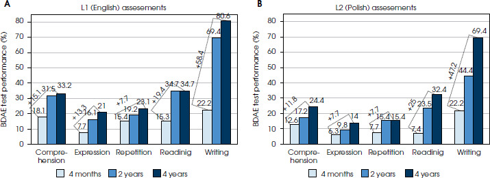
Four years post-stroke, a final evaluation of both languages was conducted. Compared with earlier assessments, BDAE scores showed less pronounced improvement. The greatest gains were observed in written language – writing in L1 and both reading and writing in L2. However, the gap in communicative functionality between the languages widened: the patient could engage in supported conversation in L1 (ASRS = 2), while communication in L2 remained absent (ASRS = 0). Table 1 presents BDAE scores, and Figure II shows the assessment timeline across therapy stages.
Table 1
Test scores obtained from successive assessments conducted in both languages

Changes in both languages: a comparison
Over four years of language therapy, the patient demonstrated differential patterns of improvement across L1 and L2, with BDAE score increases were greater when spontaneous recovery coincided with therapy than in the later therapy-driven phase, and overall progress remained more pronounced in L1. The most substantial improvements were noted in reading and writing (reading: ~60% in L1, ~50% in L2; writing: ~20% in L1, ~25% in L2), while changes in expressive language remained limited (speech repetition: ~8% in both; oral expression: 13% in L1, 8% in L2) (Figure III). Communicative functionality evolved unevenly: L1 recovered gradually – from single words to thematic conversation – whereas L2 remained largely nonfunctional and prone to involuntary switching into L1, with no capacity for intentional return to L2.
Commentary
This case study describes a patient with severe post-stroke bilingual aphasia in English (L1) and Polish (L2) who underwent four years of therapy to restore verbal communication. Clinical symptoms were analyzed in relation to therapy decisions and outcomes in both languages.
The primary therapeutic challenge was selecting the appropriate language for treatment. As no standardized guidelines exist for language choice in bilingual aphasia therapy, decisions should be based on a comprehensive evaluation of the patient’s language history, aphasia profile, communicative environment, and emotional– motivational factors [23]. In this case, L1 (native English) was chosen, as it was expected to promote faster and broader improvement with potential cross-linguistic benefits (a brief trial of therapy in L2 was also attempted, but its lack of effect led to resuming L1). Although the patient had lived in Poland for nearly 30 years, English was prioritized due to earlier acquisition, higher proficiency, and broader use across communicative contexts and modalities. These features align with classical Ribot’s rule (more recently acquired memories, including language, are more vulnerable to loss) and Pitres’ rule (better recovery in the language most familiar before brain injury) [6, 9].
Evidence from bilingual aphasia research supports this approach, showing that early acquisition and pre-stroke dominance significantly affect therapy outcomes [9]. Recent meta-analyses also confirm greater therapy effectiveness in languages acquired in early childhood compared with those learned later (after age 6) [4]. An additional rationale was that L1 remained consistently better preserved and was the primary language used with the patient’s relatives and friends.
To evaluate whether L1 therapy improved that language and positively influenced untreated L2, the patient was monitored over four years – a duration well beyond the expected window of spontaneous recovery, which is most pronounced during the first year post-stroke, especially in the initial months [7, 19]. During this time, positive but asymmetric changes were observed, with greater gains in the treated L1. In this language, the patient’s performance improved across all domains (ranging from ~8% in repetition to ~60% in writing), as well as in overall communication, progressing from producing isolated, error-prone words to participating in simple conversations on various topics. Beyond the previously mentioned factors supporting the potential for greater improvement in L1, it is a well-established finding that stronger outcomes are typically observed in the trained language [4].
Another explanation may lie in the specificity of the brain lesion. In severe aphasia, extensive damage often forces neuroplastic reorganization beyond the classical perisylvian network (covering Broca’s and Wernicke’s regions), engaging perilesional areas in the left hemisphere as well as contralateral homologues [24]. While such compensatory recruitment may support partial recovery, it may remain inefficient for sustaining parallel improvement across both languages and may even involve maladaptive processing that interferes with residual left-hemisphere function.
Given the focus on a single language (L1), possible generalization effects to the untreated language (L2) were also analyzed. According to Green’s neural convergence hypo-thesis, as bilinguals gain proficiency in a new language, its neural representation converges with that of the native language. Neuroimaging evidence shows that bilinguals engage a widespread language network, with substantial overlap of L1 and L2 representations in the left perisylvian areas, particularly when L2 is acquired early and to a high level of proficiency [6]. These intermixed neural systems may enable therapy in one language to co-activate overlapping representations, facilitating cross-linguistic transfer. Meta-analyses indicate it occurs most often following therapy in an early-acquired or immersion language [4, 23], with effects reported primarily in the lexical– semantic domain [25, 26].
In this patient, potential cross-linguistic transfer from L1 to L2 varied by domain. L2 improvements paralleled L1 gains in specific functions, with the largest effects in reading and writing, smaller in comprehension, and mini-mal in spoken expression. The greatest discrepancy between the languages was in functional communication, which was virtually absent in L2. Enhanced reading and auditory comprehension may reflect activation of a shared semantic system, particularly in late L2 learners whose L2 semantics are mediated by L1. Most prior studies have examined L1–L2 transfer in naming therapy targeting lexical retrieval [25-27]. In our case, it remains unclear whether comprehension gains stemmed from therapy or from ongoing Polish exposure in daily life, consistent with evidence on the role of the language environment in the-rapy outcomes [4, 28]. The limited improvement in oral expression likely reflects differences in sound structure, as English and Polish share minimal phonological overlap. Their morphosyntactic systems also diverge considerably: Polish relies on rich inflection and flexible word order, whereas English is largely analytic with fixed word order, further constraining cross-language transfer [29]. Cross-linguistic generalization is strongest between similar or cognate languages [30]; thus, greater transfer would be expected in closer pairs (e.g., Polish-Ukrainian, English-German) than in Polish-English bilinguals. Beyond language-specific factors, in this case limited transfer also likely reflected aphasia severity. In milder forms, better-preserved networks can support shared activation and transfer, whereas in severe aphasia – after extensive stroke – network disruption strongly constrains cross- linguistic effects [10].
Another crucial aspect of bilingual aphasia therapy is the role of cognitive control in verbal communication, a component of executive functions commonly associated with the left dorsolateral prefrontal cortex [23, 31, 35]. Successful communication in bilinguals requires selecting the target language while suppressing the non-target one [6], a process supported by a complex cortico-subcortical network (prefrontal cortex, dorsal anterior cingulate cortex, presupplementary motor area, inferior parietal cortex, left basal ganglia, thalamus, right cerebellum) [33]. In this patient, as expressive abilities improved, his pathological tendency to switch from L2 to L1 hindered the initiation and maintenance of speech in L2, reflecting impaired inhibitory cognitive control. Although post-stroke recovery has been linked to strengthened connectivity between language systems and cognitive-control areas [34], impaired regulation of language selection may exacerbate aphasia [35], underscoring the importance of providing therapy in the patient’s spontaneously used language.
Clinical implications
With bilingual aphasia becoming increasingly common among stroke patients, there is a growing need to better understand its clinical features to improve both diagnosis and treatment.
In single-language therapy, choosing the language for intervention is crucial and should take into account not only aphasia severity but also the patient’s language history, personal needs, and sociocultural background.
Recovery in bilingual aphasia is often uneven across languages, both in specific linguistic functions and in overall communication.
Limited transfer to the untreated language is likely multifactorial, involving aphasia severity, language similarity, and the efficiency of language control me-chanisms.








