Purpose
Breast-conserving surgery followed by adjuvant radiotherapy is the standard of care for early-stage breast cancer. Although whole-breast irradiation (WBI) effectively reduces ipsilateral recurrence, most local relapses occur near the original tumor bed, providing the rationale for accelerated partial breast irradiation (APBI). Randomized GEC-ESTRO phase III trials have demonstrated that interstitial multicatheter brachytherapy (MIBT) achieves local control and cosmetic outcomes similar to WBI in appropriately selected patients [1, 2].
Among APBI techniques, MIBT has the longest clinical track record and is regarded as a reference method due to its highly conformal dose distribution and steep dose gradients, enabling effective sparing of the skin, lung, ribs, and, in left-sided cases, the heart. These advantages are reflected in the GEC-ESTRO and ESTRO-ACROP recommendations on patient eligibility, implant geometry, and treatment planning [3-5].
Advanced external beam radiotherapy (EBRT) methods, such as intensity modulated radiation therapy (IMRT) and volumetric modulated arc therapy (VMAT), have been increasingly incorporated into APBI protocols, offering improved dose shaping and planning efficiency. Several dosimetric studies have indicated that EBRT-based APBI can provide satisfactory target coverage, yet may expose larger breast volumes and nearby organs to higher low- and intermediate-dose regions [6-10].
Because APBI techniques differ fundamentally in their dose fall-off characteristics, a detailed comparison of organ at risk (OAR) exposure is clinically relevant, particularly in terms of relative doses to the skin and ipsilateral non-target breast. While previous studies have compared MIBT with general external beam techniques, few have specifically evaluated different VMAT arc arrangements against the brachytherapy gold standard.
The objective of this study was therefore to perform a focused dosimetric comparison of multicatheter interstitial high-dose-rate (HDR) brachytherapy and two VMAT-based (tangential and continuous arcs) APBI techniques, using identical patient imaging and standardized contouring. The analysis focused on OAR doses to clarify the relative radiobiological and clinical implications of each modality.
Material and methods
Patient selection
Fifty consecutive female patients with early-stage breast cancer treated between 2022 and 2024 were included in this comparative analysis. Patient eligibility for APBI followed the GEC-ESTRO Breast Cancer Working Group recommendations, including age ≥ 50 years, unifocal invasive carcinoma or ductal carcinoma in situ (DCIS), tumor size ≤ 30 mm, negative margins, node-negative disease (pN0), and absence of lymphovascular invasion [4].
The baseline patient and tumor characteristics are presented in Table 1. Study cohort consisted of women aged 52-82 years (median, 65 years), and tumors were located in the left breast (n = 32, 64%) and the right breast (n = 18, 36%). In the left breasts, lesions were distributed as follows: 11 in upper inner quadrant (UIQ), 12 in upper outer quadrant (UOQ), 5 in lower outer quadrant (LOQ), 2 in lower inner quadrant (LIQ), and 2 in central region. In the right breasts, lesions were located in UOQ (n = 10), UIQ (n = 4), LOQ (n = 3), and central region (n = 1). Most patients had pT1 disease (94% classified as pT1b or pT1c), with pTis in 4% and pT2 tumors in 2%. All patients were pN0 on final pathology. Median invasive tumor size was 12 mm (range, 5-23 mm), while minimal surgical margin width ranged from 2.5 to 19 mm.
Table 1
Baseline patient and tumor characteristics (n = 50)
All breast cancers were of the luminal sub-type, with estrogen receptor (ER) expression positive in all patients, and progesterone receptor (PR) expression positive in 88%. Histologic grading demonstrated a distribution of G1 in 52% and G2 in 48% of all tumors (Table 1).
Simulation and contour delineation
Computed tomography (CT) simulation was performed using dedicated CT scanners (SOMATOM Definition AS until 2023, and SOMATOM go.Open PRO thereafter; Siemens Medical Solutions, Germany), with a slice thickness of 3 mm. Patients were positioned supine on an indexed breast board, with the ipsilateral arm elevated above the head, according to standard APBI imaging practices. All CT simulations were performed during free breathing. For brachytherapy, CT imaging was performed after implantation of multicatheter applicators, with flexible plastic catheters secured to the skin to prevent displacement, following technical recommendations described in the ESTRO-ACROP guideline [3]. For better visualization, radiopaque markers were inserted into the catheters before imaging. Despite the presence of radiopaque markers, no significant CT artifacts were observed.
Target delineation followed the principles outlined in the GEC-ESTRO recommendations for APBI patient selection [4] and the anatomical guidance defined in the GEC-ESTRO Working Group II consensus [5]. Clinical target volume (CTV) was generated by expanding the lumpectomy cavity by 20 mm after subtraction of the surgical margin in all directions, limited to 5 mm from the skin surface, pectoral muscles, and chest wall, in accordance with the GEC-ESTRO Working Group II [5]. In brachytherapy planning, CTV was considered equivalent to planning target volume (PTV), in line with fixed-geometry nature of catheter implants described in the GEC-ESTRO Working Group II target delineation recommendations [5]. For VMAT planning, PTV was generated by adding a 5 mm isotropic margin to CTV, and subsequently cropped 5 mm from the body contour to exclude the build-up region. This was done as per EBRT-based APBI planning strategies described by Major et al. [6].
Organs at risk were delineated according to contemporary breast radiotherapy contouring standards. The following structures were contoured: ipsilateral and contralateral breasts, ipsilateral lung, heart, ribs adjacent to the implant region, and skin. The skin was defined as a 5 mm superficial structure generated by inward contraction of the body contour, consistent with published recommendations on skin dosimetry in breast brachytherapy by Hilts et al. [10].
The ipsilateral non-target breast was defined by subtracting CTV (in brachytherapy) or PTV (in VMAT planning) from the ipsilateral breast volume, enabling analysis of dose to uninvolved breast tissue [6].
For EBRT planning, CT images and all contours were transferred from Oncentra Brachy treatment planning system (TPS) to Monaco TPS v. 6.1.4 (Elekta, Sweden) to generate VMAT plans. This ensured that CTV and OAR contours were identical across modalities, allowing unbiased comparison between multicatheter HDR brachytherapy and VMAT-based APBI [6].
Multicatheter interstitial brachytherapy
All patients underwent multicatheter interstitial HDR brachytherapy delivered as APBI, using an iridium-192 stepping source on the Flexitron afterloading platform (Elekta, Sweden). Catheter implantation, target definition, dose prescription, and treatment planning were performed as per the updated ESTRO-ACROP guidelines for interstitial multicatheter brachytherapy, which provide contemporary consensus recommendations on APBI technique, applicator geometry, image-guided CTV delineation, and acceptable dose fractionation schedules [3]. The procedure was carried out under sterile conditions using either template-based technique or free-hand approach, depending on tumor bed location and patient anatomy. In the template-based technique, a dedicated breast template with a pre-defined grid was positioned over the tumor bed region, ensuring stable geometry and adequate spacing between insertion channels. Metallic guiding needles were first introduced through the template in two to four parallel planes, typically spaced 12-16 mm apart, to achieve a uniform implant geometry. Needles were subsequently exchanged for flexible plastic catheters, which served as channels for the stepping iridium-192 source.
Following catheter implantation, a planning CT scan was obtained with all catheters in situ, and the lumpectomy cavity, CTV, and OARs were delineated. The reconstructed catheters were then used to generate HDR treatment plan in the Oncentra Brachy TPS, in which the AAPM TG-43 formalism is implemented for dose calculation. All plans were created using geometrical and graphical optimization. The number of applicators ranged from 6 to 14, with the source stepping through the catheters at intervals of 3 mm. The prescribed dose was 32 Gy delivered in 8 fractions administered twice daily, with a minimum inter-fraction interval of 6 hours, resulting in an overall treatment time of 4-5 days [1, 2]. Treatment plans were made according to the GEC-ESTRO recommendations, ensuring target coverage of V100 ≥ 90% and meeting OAR dose-volume constraints [3].
External beam radiotherapy planning
In routine clinical practice at our institution, APBI delivered with EBRT is prescribed as 30 Gy in 5 fractions of 6 Gy each [11]. However, to enable a direct dosimetric comparison across all modalities in this planning study, a uniform schedule of 8 × 4 Gy was applied to VMAT plans, matching HDR brachytherapy dose regimen, in line with methodologies used in previous comparative APBI studies [6].
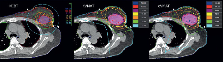
For each patient, two VMAT plans were generated with 6 MV flattened photon beams in the Monaco v. 6.1.4 TPS (Elekta, Sweden), using Monte Carlo dose calculation algorithm: (a) Continuous VMAT (cVMAT) treatment plan with one dual arc (range, 160°-180°); (b) Tangential VMAT (tVMAT) with two tangential dual arcs (range, 50°-70°). In all comparative VMAT plans, the same isocenter, gantry rotation range, and cost functions were employed to ensure that at least 95% of the PTV received at least 95% of the prescribed dose (V95 ≥ 95%). All VMAT plans were prepared for the Versa HD (Elekta, Sweden) linear accelerator (Figure 1).
Plan analysis
To analyze dose distribution in target volume and OARs, dose-volume histograms (DVHs) were used. The analysis included relative volume parameters, specifically low-dose exposure (V5%, V10%) for the lungs and heart, and V25% and V50% for the ipsilateral non-target breast. To evaluate high-dose regions in OARs (skin, ribs, contralateral breast, and heart), near-maximum absolute doses were reported as D0.1cc and D1cc. Additionally, mean dose (Dmean) was recorded for the lungs, heart, non-target breast, and contralateral breast. To evaluate conformality and homogeneity of dose distribution in PTV for VMAT plans, homogeneity index (HI) and conformity index (PCI) were calculated according to the International Commission on Radiological Units and Measurements (ICRU) Report No. 83 [12], as follow:
where D2%, D98%, and D50% are the minimum doses received by 2%, 98%, and 50% of the PTV, respectively. HI equal to 0 corresponds to an ideal homogeneous dose distribution in the volume of the PTV.
where TVRx is the volume of the PTV covered by the prescription dose, TV is the volume of the PTV, and VRx is the total volume of the prescribed dose. PCI equal to 1 corresponds to a perfect coverage of and fit to volume of the PTV.
There are no international recommendations on acceptable values for the HI (Eq. 1) and PCI (Eq. 2). However, the published studies on VMAT plans for APBI have reported PCI values between 0.60 and 0.90, and HI values between 0.05 to 0.28 [6-9, 13].
To evaluate the impact of the tumor bed location on cardiac sparing in left-sided breast cancer cases, patients were categorized into sub-group pairs based on anatomical quadrants: Inner (UIQ + LIQ, n = 13) vs. outer (UOQ + LOQ, n = 17), and upper (UIQ + UOQ, n = 23) vs. lower (LIQ + LOQ, n = 7), while centrally located tumors (n = 2) were excluded from quadrant sub-groups.
Statistical analysis
Results were statistically analyzed. The median values with interquartile range (IQR: 25th-75th percentile) are presented for each parameter in Tables 2, 3, and 4. Normality of data distribution was assessed using Shapiro-Wilk test. Since most of the parameters were not normally distributed, non-parametric paired Wilcoxon test was employed. For all tests, a significance level was set to 5% (p < 0.05).
Table 2
Dosimetric parameters of target volumes (CTV, PTV) for MIBT, tVMAT, and cVMAT plans –
Table 3
Dosimetric parameters of organs at risk for MIBT, tVMAT, and cVMAT plans
Table 4
Cardiac dosimetric parameters for left-sided breast lesions (n = 30) according to tumor bed location
Results
Target volumes
The median CTV in MIBT was 37.12 cc (range, 18.23-121.09 cc). For the VMAT-based APBI plans, the addition of safety margins resulted in a significantly larger target volume (PTV), with a median of 85.05 cc (range, 45.20-229.63 cc), representing a median volume increase of approximately 129% when using external beam techniques compared with the interstitial approach. Differences in CTV V100% among all three techniques were highly significant (p < 0.0001). VMAT plans provided better coverage than MIBT, while cVMAT plans achieved the highest coverage. cVMAT plans also provided higher V95% coverage of the PTV (p = 0.0003) as well as lower HI (p < 0.05) and higher PCI (p < 0.0001) indices compared with tVMAT plans. The representative dose distributions for a multicatheter interstitial brachytherapy and external beam intensity-modulated radiotherapy in a transversal CT slice are shown in Figure 1 (Table 2).
Non-target breast
The analysis demonstrated that MIBT provided the most effective sparing of healthy ipsilateral breast tissue surrounding the target volume. The volume of the breast receiving 50% of the prescribed dose (V50%) was significantly lower in MIBT (15.84%) compared with external beam techniques (p < 0.0001). Among the VMAT approaches, cVMAT resulted in lower V50% values (23.91%) than tVMAT (27.66%, p < 0.05). V25% followed a similar distribution (34.05%, 38.25%, and 43.17% for MIBT, cVMAT, and tVMAT, respectively). MIBT was associated with a significantly lower mean dose than tVMAT (p < 0.05), while no significant difference was observed between MIBT and cVMAT.
Contralateral breast
All techniques provided excellent sparing of the contralateral breast. MIBT achieved the lowest D1cc (0.00 Gy), which was significantly lower than that observed for tVMAT and cVMAT (0.85 Gy and 0.86 Gy, respectively, p < 0.05). No significant differences were found among the techniques for Dmean and D0.1cc.
Heart
For left-sided lesions, EBRT techniques significantly reduced cardiac exposure compared with MIBT. The median mean heart dose was 1.54 Gy in MIBT vs. 0.38 Gy and 0.62 Gy in tVMAT and cVMAT, respectively (p < 0.0001). V10% showed a comparable distribution (9.08%, 0.09%, and 1.80%, respectively). Within the VMAT cohort, tVMAT yielded significantly lower V10%, V5%, and Dmean than cVMAT (p < 0.05). A similar trend was observed for right-sided lesions, where MIBT resulted in higher doses than VMAT. V10% was 3.54% in MIBT, while it was negligible (0.00%) in both VMAT techniques (p = 0.0007). The mean dose was also significantly higher in MIBT (0.48 Gy) compared with tVMAT (0.24 Gy, p < 0.05). Comparing external beam plans, tVMAT achieved significantly lower doses than cVMAT for V10%, V5%, and Dmean (p < 0.05).
Sub-group analysis by tumor quadrant revealed that the cardiac sparing advantage of VMAT was strongly location-dependent (Table 4). For lesions in the upper quadrants (UIQ + UOQ, n = 23) and outer quadrants (UOQ + LOQ, n = 17), both VMAT techniques achieved significantly lower Dmean than MIBT (p < 0.005), with tVMAT providing the lowest cardiac exposure. For tumors in the inner quadrants (UIQ + LIQ, n = 13), tVMAT significantly reduced mean dose (p = 0.0030) and low-dose volumes (V5%, V10%) compared with MIBT, whereas high-dose metrics (D1cc, D0.1cc) were similar (p = 0.1520). However, for lesions in the lower quadrants (LIQ + LOQ, n = 7), EBRT lost its advantage, showing no significant differences compared with MIBT for Dmean, D1cc, D0.1cc, and V5%.
Ipsilateral lung
There was a major difference in low-dose exposure. cVMAT delivered a low-dose exposure to a large part of the lung, with a V5% of 52.60%. This was significantly higher than tVMAT (19.26%) and MIBT (15.48%) (p < 0.0001). The mean dose to the lung was also highest in cVMAT (2.50 Gy). MIBT and tVMAT resulted in much lower mean doses (1.61 Gy and 1.21 Gy, respectively), with no statistically significant difference between them.
Skin and ribs
MIBT provided superior sparing of both the skin and ribs. The maximum skin dose (D1cc) was 14.75 Gy in MIBT, significantly lower than the 24-26 Gy range observed in VMAT techniques (p < 0.0001). Similarly, rib D1cc values were significantly lower in MIBT (13.73 Gy) compared with tVMAT (17.36 Gy) and cVMAT (18.07 Gy) (Tables 3 and 4).
Discussion
To our knowledge, this is one of the few studies focused on a dosimetric comparison of MIBT vs. VMAT (two different arc arrangements), where treatment plans were based on identical CT datasets. While all techniques are valid options for APBI, our results show they deliver dose to OARs very differently. Our findings confirm that MIBT is generally the best method for protecting the skin, ribs, and healthy breast tissue. However, VMAT plans, especially tVMAT, spare the heart better than MIBT, particularly in left-sided tumors [14].
MIBT provided the best sparing of non-target breast tissue: V50% was significantly lower (15.84%) than in VMAT techniques (over 23%). These results are in line with the finding of Major et al. [6], who reported a V50% of 13.7% in MIBT vs. 25.5% in IMRT. This difference stems from the absence of CTV-to-PTV margins in brachytherapy, targeting a much smaller breast volume. Furthermore, cVMAT spared the non-target breast slightly better than tVMAT due to a wider range of beam angles, which more effectively increased the dose fall-off gradient outside the target volume. In tVMAT, these angles are intentionally restricted to protect the lung and heart, which consequently reduces the potential to spare the non-target breast tissue. Regarding the ipsilateral lung, MIBT proved to be a superior technique, delivering the lowest mean and low-dose volumes. Our observation is consistent with Major et al. [6], who reported that MIBT spares the lung better than IMRT, with significantly lower volumes irradiated by V5%, 32.9% vs. 41.7%, respectively. However, a key finding of our study is the significant dosimetric advantage of tVMAT over cVMAT. By restricting the arc angles, tVMAT reduced the V5% to approximately 19% compared with over 52% in cVMAT. This demonstrates that if VMAT is chosen, tangential arcs can effectively limit the low-dose bath, achieving lung sparing results much closer to those of brachytherapy. This observation is consistent with other recent dosimetric studies, which confirm that restricting gantry rotation to tangential arcs significantly reduces the low-dose spread to ipsilateral lung than continuous arc arrangements [15-19]. Because low-dose lung exposure (particularly V5%) contributes to pulmonary toxicity, the reduction of V5% remains clinically relevant [20, 21].
Generally, brachytherapy is considered superior for sparing all OARs. However, our study demonstrates that for left-sided lesions, tVMAT results in a significantly lower mean dose to the heart (0.38 Gy) compared with MIBT (1.54 Gy). This supports Major et al. results [6], showing higher mean heart doses in MIBT (~1.35 Gy) compared with IMRT (~0.60 Gy). This difference stems from the distinct geometric capabilities of each technique. While VMAT optimization can effectively modulate beam shape and intensity to spare the heart, dose distribution around interstitial sources is isotropic and cannot be “shaped” away from the heart if the implant is in close proximity. The ability of tangential VMAT to effectively minimize heart exposure has also been demonstrated in other dosimetric comparative studies [15, 18-22]. Given the established linear relationship between mean heart dose and long-term risk of ischemic heart disease, with an estimated nearly 7% of relative increase in risk per 1 Gy, even small absolute reductions in cardiac exposure may translate into clinically meaningful benefits [23]. Furthermore, our sub-group analysis showed that the heart-sparing advantage of VMAT over brachytherapy depends heavily on lesion location. For tumors in the upper (UIQ + UOQ) and outer (UOQ + LOQ) quadrants, tangential tVMAT achieved significantly lower mean heart doses (0.34 Gy and 0.41 Gy) compared with MIBT (1.54 Gy and 1.50 Gy) (p < 0.005). However, this cardiac advantage depends on the quadrant. In inner quadrant tumors, tVMAT effectively reduces mean and low heart doses, but high doses remain similar to MIBT. For lower quadrant tumors, the cardiac benefit of VMAT disappears completely, with no significant differences compared with MIBT. These findings align with Major et al. [6], who stated that assuming VMAT as universally superior for left-sided lesions is a generalization, and technique selection must be tailored to tumor location. Incorporating deep inspiration breath hold (DIBH) into VMAT workflows would further increase the distance between the heart and target volume in left-sided cases, potentially reducing cardiac exposure even beyond free-breathing VMAT levels observed in our study [15, 16, 24, 25].
With respect to the skin and ribs, MIBT demonstrated a consistent dosimetric advantage. Both maximum skin dose (D1cc) and rib D1cc were significantly lower in MIBT compared with VMAT techniques. These findings are in line with Major et al. [6], who similarly reported reduced high-dose exposure to the skin and ribs in interstitial brachytherapy. The higher skin and rib doses observed with VMAT are largely driven by the required PTV margins, which physically expand the high-dose region closer to both the skin and underlying chest wall. Additionally, photon beams in external beam delivery must necessarily traverse these structures to reach the target, contributing to a greater incidence of radiation-induced skin toxicity. In contrast, in MIBT, the radioactive sources are positioned within the target volume, allowing for a steep dose fall-off towards the skin and chest wall structures [26]. Future prospective studies integrating clinical outcomes with detailed dosimetric analyses are warranted to better define optimal technique selection in APBI.
In our cohort, the median CTV volume was 37.12 cc, which is smaller than the median target volumes reported in major APBI trials, such as the GEC-ESTRO phase III trial (81 cc) [1] and the planning study by Major et al. (60 cc) [6]. Despite these differences in absolute target size, the observed dosimetric relationships and relative OAR dose differences between MIBT and external beam techniques are in line with published literature [6, 13]. These findings suggest that the distinct physical and geometric characteristics of MIBT and EBRT have a greater impact on determining OAR sparing than variations in target volume.
A major methodological difference between the techniques studied is the dose calculation algorithm utilized. Model-based dose calculation algorithms (MBDCAs) demonstrated that TG-43 systematically overestimated OAR doses. Sinnatamby et al. [27] found that using MBDCA instead of TG-43 reduced calculated low-dose cardiac parameters (V5% and V10%) by 22% (p = 0.001) and 26% (p = 0.055), respectively. While their study examined metal catheters, Roussakis et al. [28] reported that plastic catheters also produce similar scatter deficits at tissue boundaries, reducing real doses to the chest wall (D0.1cc), skin (D0.1cc), and lung (V5 Gy). Therefore, the actual doses delivered to the heart, lungs, and chest wall during brachytherapy in our patients are likely lower than our TG-43 calculations indicate. This means that while tVMAT shows a statistically significant reduction in cardiac dose compared with brachytherapy for left-sided upper quadrant tumors, the true clinical advantage of VMAT over brachytherapy may be smaller in practice than TG-43 planning suggests.
Strengths and limitations
This study has several strengths, including the direct comparison of three contemporary techniques using identical CT datasets and a uniform fractionation schedule, enabling meaningful dosimetric evaluation. However, several limitations should be acknowledged. This was a planning study without clinical outcome correlation. The sample size was limited, while advanced motion management strategies, such as deep inspiration breath hold were not included. All VMAT plans were generated on post-implant CT scans, where the breast was compressed by the fixation buttons of the catheter applicators. Although the use of identical CT datasets minimized anatomical variability and allowed direct dosimetric comparisons by ensuring identical target and OAR contours, the localized compression altered the natural breast contour relative to standard external beam positioning. Consequently, VMAT plans generated on uncompressed breasts during routine EBRT simulation may result in different OAR doses. Therefore, the clinical implications of the observed dosimetric differences should be interpreted with caution. A limitation of the dosimetric analysis is the difference in dose calculation methods, i.e., the Monte Carlo algorithm was used in VMAT, whereas the TG-43 formalism in MIBT. Since TG-43 may overestimate doses to OARs, the actual OAR doses during MIBT may be lower than those reported in the analysis.
Conclusions
APBI technique selection must be strictly individualized based on patient anatomy and tumor bed location. MIBT delivers optimal target conformality and superior sparing of healthy breast tissue, skin, and chest wall structures. tVMAT effectively minimizes low-dose lung exposure compared with cVMAT plans, providing superior cardiac sparing for left-sided tumors located in upper and outer quadrants. For inner quadrant lesions, tVMAT retains an advantage in reducing mean and low heart doses, though protection against high-dose hot spots is comparable with MIBT. However, for lower quadrant lesions, the cardiac advantage of VMAT is entirely absent, where MIBT offers comparable or superior overall OAR protection. Furthermore, clinical dose interpretation should consider TG-43 calculation limitations, as MBDCA algorithms indicate that real-world cardiac and tissue doses in brachytherapy may be even lower than estimated. Therefore, VMAT cannot be universally regarded as superior for all left-sided lesions without accounting for specific tumor bed quadrant location.