
Purpose
Tissue repair occurs through regeneration or scarring, depending on the extent and site of tissue damage [1]. Skin injuries may heal by scar formation. When the repair process is disrupted, complications, such as insufficient healing, excessive tissue deposition, and contracture formation, can occur. Increased responsiveness to transforming growth factor (TGF)-β1 and 2, along with excessive fibroblast activity and collagen production, can lead to the formation of hypertrophic scars and keloids [1]. Keloids are characterized by an increased ratio of type 1 to type 3 collagen deposition in a haphazard pattern, whereas hypertrophic scars show parallelly organized excessive type 3 collagen fibers [2]. One of the characteristic of clinical differences between the two is that keloids tend to grow beyond the scar boundaries, unlike hypertrophic scars.
Keloids frequently develop following skin injuries, including piercings, surgical incisions, burns, infections, or even minor traumas, such as vaccinations. They tend to appear in areas with high skin tension or mechanical stress, e.g., the ear lobes, chest, sternum, and shoulders. The risk of keloids’ formation is strongly associated with darker skin tone, puberty, pregnancy, female sex, and a positive personal or family history suggesting genetic predisposition [3]. Although keloids are benign and frequently untreated, an increasing number of patients pursue treatment because of cosmetic concerns and pain. Treatment options vary widely, ranging from medical to surgical approaches, along with pressure therapy, application of topical agents, such as 5% imiquimod, intra-lesional injections of corticosteroids, or 5-fluorouracil laser therapy (erbium-doped yttrium aluminum garnet Er:YAG, or carbon dioxide laser) and cryotherapy [4]. Despite the variety of available options, treatment of these lesions is often challenging due to high chances of recurrence. One of the most commonly used modalities is surgery, which has a high recurrence rate, ranging from 45% to 100% with excision alone [4].
Post-operative radiation therapy is an effective treatment to prevent keloids’ re-growth. While it is commonly offered for cases refractory to non-surgical interventions, such as corticosteroid injections, laser therapy, or cryotherapy, there is evidence supporting its use even after the first surgical excision to reduce recurrence [5]. Radiation prevents recurrence by inducing DNA damage in proliferating fibroblasts and downregulating TGF-β1 to suppress collagen synthesis [6].
The use of combined surgical excision and radiotherapy for treating keloids was first reported by De Beurmann and Gougerot in 1906 [7]. Post-operative radiation can be administered externally using superficial X-rays and electron beams, or internally with brachytherapy techniques. When external beam therapy is employed, dose to normal skin or surrounding structures, which are distant from the scar, is unavoidable. This drawback can be overcome by brachytherapy, as first described by Malaker et al. in 1976, who used a polyethene tube inserted intra-operatively and later loaded with iridium-192 (192Ir) source [8]. The physical advantage of interstitial brachytherapy over external beam modalities (electrons or superficial X-rays) lies in its superior dose conformality due to the inverse square law. By delivering radiation directly within surgical bed, brachytherapy achieves rapid dose fall-off, providing high-dose to target fibroblasts while sparing surrounding healthy tissues.
Currently, among the available techniques, high-dose-rate (HDR) brachytherapy is the most widely used, involving temporary placement of a highly radioactive source, typically for 5-10 minutes, delivering dose at a rate ≥ 12 Gy per hour. A biologically effective dose (BED) in the range of 15 to 22.5 Gy has been shown to achieve favorable control rates and cosmetic outcomes [9]. Supported by existing literature on the efficacy of immediate post-operative brachytherapy in keloids’ management, our study aimed to evaluate treatment outcomes in patients who underwent surgical excision and peri-operative brachytherapy at our institution. Additionally, we assessed whether any correlations existed between patient-related factors and clinical outcomes.
Material and methods
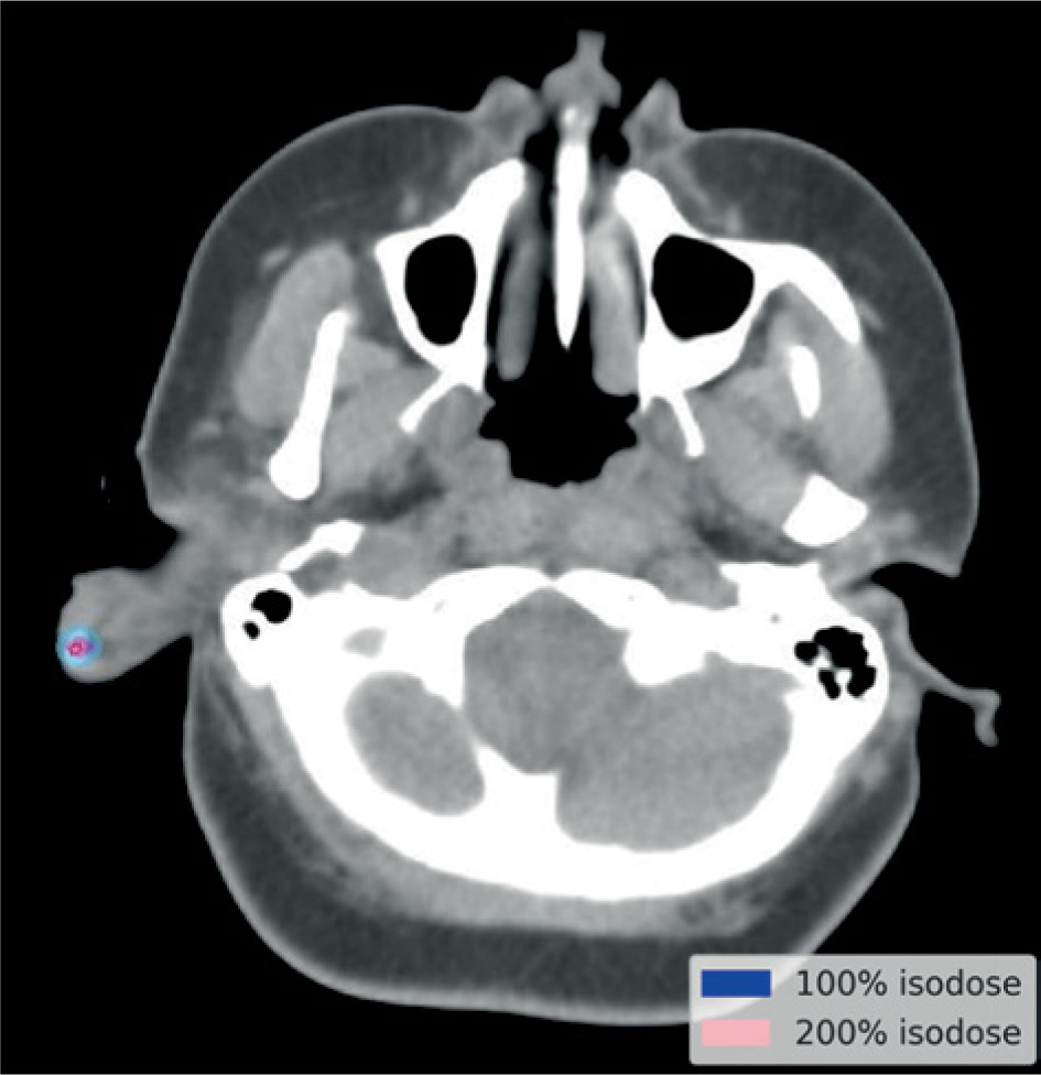
In this retrospective study, after obtaining ethics committee approval, a total of 32 patients treated at our institute from 2019 to 2025 were identified. All patients were examined by both a surgeon and radiation oncologist before undergoing surgery, refer Figure 1A. Under local anesthesia, complete extra-lesional excision of the keloid was performed up to healthy skin margins. The wound was closed in layers using absorbable sutures for subcutaneous tissue. One hollow, flexible catheter was then placed in the surgical bed, after which the skin was closed over the catheter using monofilament, non-absorbable nylon sutures, preventing damage to the catheter. The catheters were secured on both edges of the surgical site, as shown in Figure 1B and Figure 2 and the surgical area was then dressed under sterile conditions. Patients were taken for computed tomography (CT) simulation scans after post-operative recovery period of an hour . During simulation scan, under strict aseptic precautions, radio-opaque markers were inserted through the flexible catheter for better visualization of the post-operative bed on imaging. The simulation images were then exported to a planning system. The catheter was digitized and dwell positions were optimized to deliver the prescription dose along the length of catheter, which was inside the post-operative bed, while limiting V200% on the skin as shown in Figure 3. After obtaining informed consent, radiation was delivered using a cobalt-60 radioisotope from inside the lesion, targeting only the desired area. Treatment was initiated within 24 hours of surgery, and a prescribed dose of 15 Gy in 5 Gy per fraction was delivered with a minimum six-hour interval; treatment was completed within 48 hours. Post-treatment, the catheter was removed and the tumor bed was tightly bandaged.
Fig. 1
Clinical images showing A) lesion at presentation, B) intra-operative view after excision and catheter insertion, and C) post-operative status after 4 weeks of recovery
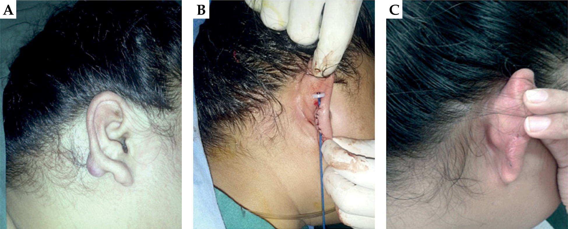
Fig. 2
A) Catheter placed in post-operative bed and fixed with buttons. B) 3D reconstructed image showing the catheter with surrounding 100% isodose
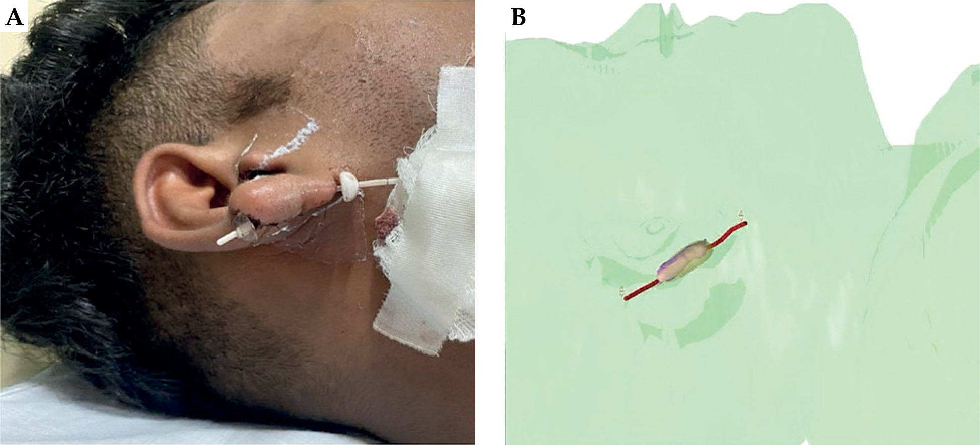
Fig. 3
Axial section of planning CT showing 100% (blue line) and 200% (pink line) isodose curves around the intracavitary catheter. The 100% isodose line represents the prescription dose coverage, while the 200% isodose line highlights the region of dose concentration close to the applicator
All patients were followed up with telephonic interviews to evaluate treatment outcomes. The assessment focused on three primary aspects, i.e., recurrence, acute toxicities, and late toxicities. Recurrence was defined as re-appearance of a keloid or mass within all or part of the treated area and growing beyond the boundaries of original wound [10, 11]. Acute and late toxicities were assessed and graded according to CTCAE version 5.0 [12]. Acute toxicities included radiation-induced dermatitis and delayed wound healing beyond 4 weeks post-treatment, while late toxicities were pigmentary changes, skin sclerosis, and other cosmetic effects.
From the 32 patients, CT simulation scans could be retrieved for 24 patients only. In the acquired scans, high-risk clinical target volume (HR-CTV) was contoured 3 mm around the catheter along its entire length, according to Anderson et al. [13]. Treatment length and volume, with V100%, V150%, and V200% were recorded for these patients.
Statistical software SPSS v. 19.0 was used for data analysis. Descriptive statistics were employed to summarize patients’ demographics and treatment-related variables. Continuous variables, such as age, lesion length (cm), and volume (cm3), were presented as mean and standard deviation (SD), while categorical variables, including sex, pre-treatment history, and possible cause, were expressed as absolute frequencies and percentages. Recurrence rate was calculated, whereas associations between patient, treatment characteristics, and clinical outcomes were evaluated using chi-square test for categorical variables and Spearman’s correlation for continuous variables. An independent sample t-test was employed to compare variables between patients with recurrence and those without.
Results
This retrospective study included 32 patients with recurrent keloids, who were treated with surgical excision followed by immediate post-operative brachytherapy at our institute. Patient accrual was completed over a period of six years. All patients had a prior history of keloid being treated with either intra-lesional steroid injections (68.8%) or surgery alone (31.2%). The study cohort included 17 males (53.1%) and 15 females (46.9%), with age ranging from 21 to 65 years (median age, 33 years). Keloids of the ear pinna were most frequent (50%), followed by the anterior chest and extremities, as shown in Table 1. Ear lobe keloids were most commonly observed in females aged 24-35 years, with majority (53.1%) having ear piercing as a preceding cause. None of the patients reported family history of predisposition to keloids.
Table 1
Patient characteristics in terms of frequency and percentages
The median length of treated volume was 3.75 cm (range, 1.5-21 cm), and the median volume treated was 1.7 cm3 (range, 0.1-9.9 cm3), with V200% isodose curve covering an average of 25.8% of the treated volume. The V100% coverage ranged from 86.42% to 96.16%, as presented in Table 2. The interval between surgery and initiation of the first radiation fraction was 3-4 hours for all patients, including those with and without recurrence, indicating no delay in treatment initiation. The total radiation treatment time ranged from 30 to 36 hours for all patients. The me- dian follow-up was 25 months (range, 7-82 months) after completion of brachytherapy. No acute toxicities were reported. Late toxicity was observed in only one patient treated for a keloid of the right ear, who experienced grade 1 paraesthesia. No other adverse effects, such as delayed wound healing or skin pigmentation, were reported.
Table 2
Summary of patient characteristics and dosimetric parameters
| Parameter | Mean | Median | SD |
|---|---|---|---|
| Age (years) | 37.6 | 33 | 14.40 |
| Length (cm) | 5.68 | 3.75 | 4.68 |
| Volume (cm3) | 3.22 | 1.7 | 3.26 |
| V100% (%) | 91.15 | 91.55 | 4.87 |
| V200% (%) | 26.71 | 27.00 | 7.99 |
Four out of 32 (12.5%) patients developed recurrence at the same site. The median time to relapse after completion of peri-operative brachytherapy was 2.5 months, with a range of 1-6 months post-treatment. The V100% among the four patients who had recurrence was 92.5%, 88.3%, 92.5%, and 85.8%. The length of treated volume and volume of treatment among these patients ranged from 1.5 cm to 15 cm and 0.1 cm3 to 9.9 cm3, respectively. Volume receiving 200% dose (V200%) for the one patient who reported paraesthesia was 28.6%, which was not skewed from the rest of the study subjects.
Moreover, there was no statistically significant association observed between sex and recurrence (χ2 = 0.87, p = 0.34). Recurrence by anatomical site occurred in 2 of 12 chest lesions (16.6%) and 2 of 16 ear lesions (12.5%), with no recurrences of lesions involving the extremities or scapula; this association was not statistically significant (χ2 = 7.6, p = 0.11). Similarly, recurrence did not significantly differ by etiological factors, occurring in 1 of 12 burn-related cases (8.3%), 2 of 17 piercing-related lesions (11.8%), and 1 of 3 trauma-related keloids (33.3%) (χ2 = 1.38, p = 0.49). In addition, prior treatment history showed no significant association with recurrence, as no recurrences were observed among patients previously treated with surgery alone, while 4 of 22 patients who had received intra-lesional steroids developed recurrences (χ2 = 2.07, p = 0.14).
The Spearman’s correlation analysis revealed no statistically significant association between recurrence and age (p = 0.41), treatment length (p = 0.94), lesion volume (p = 0.42), or V100% (p = 0.54). Patients with recurrence had slightly greater treatment length and volume, and slightly lower V100% compared with those without recurrence; however, none of these differences were statistically significant.
Discussion
Radiation therapy, though traditionally associated with the treatment of malignant neoplasms, has also been used in the management of selected benign conditions, such as arteriovenous malformations, heterotopic ossification, trigeminal neuralgia, acoustic neuroma, and keloid scars, among others. The use of radiation therapy for benign conditions is gaining significance in the current era due to a better understanding of the biological mechanism of radiation and the availability of advanced techniques.
In the immediate post-operative period, unstable collagen fibers and naïve fibroblasts predominate the wound environment, a phase during which the tissue is more responsive to ionizing radiation [6]. According to Casarett’s classification, fibroblasts are categorized as reverting post-mitotic cells with moderate radiosensitivity due to their limited but inducible proliferating potential [14]. In their colony-forming assay studies, Ma et al. demonstrated that fibroblasts derived from keloid tissue have greater radiosensitivity compared with normal dermal fibroblasts, as indicated by lower D0 (dose that reduces the surviving fraction of cells to 37%) and D10 (dose that reduces the surviving fraction to 10%) [15]. This highlights the biological susceptibility of keloid fibroblasts to ionizing radiation, and suggests that targeting these highly proliferative cells early can inhibit fibroblast multiplication and collagen synthesis, thereby reducing the risk of recurrence. The law of Bergonié and Tribondeau states that cells are more radiosensitive when rapidly dividing, and radiation should ideally be administered during the early proliferative phase [16]. At our institution, brachytherapy is routinely commenced within 24 hours of surgical excision and completed within 30 to 36 hours post-procedure, with most patients transferred to the radiotherapy department within an hour after surgery, allowing treatment to begin 3-4 hours post-excision. This approach aligns with evidence that early initiation of radiotherapy is crucial for minimizing recurrence. Multiple studies have explored different surgery to RT time schedules; for instance, Lee et al. assessed factors affecting recurrence, showing that recurrence rates were higher when treatment was initiated more than 72 hours after surgical excision [17]. Fernandes et al. reported a recurrence rate of just 18.8% when treatment was initiated within 24 hours [18]. Furthermore, a meta-analysis by Peng et al. highlighted the clinical importance of early initiation of radiotherapy, within 2 hours [19]. The low recurrence rate observed in our cohort may partly be attributed to strict adherence to the early post-operative radiotherapy interval of 3-4 hours, consistent with the GEC-ESTRO ACROP recommendations for skin brachytherapy, emphasizing starting brachytherapy as soon as possible after keloid excision [20].
One of the earliest studies demonstrating the benefit of adjuvant radiation was conducted by Escarmant et al. in 1993, where 192Ir interstitial brachytherapy was administered post-operatively, resulting in a significantly reduced recurrence rate of 21% [21]. In a meta-analysis by Mankowski et al., various radiation modalities were compared in the management of recurrent keloids. Their findings showed that brachytherapy had the lowest recurrence rate (15%), outperforming both electron beam and X-ray therapies (each with a recurrence rate of 23%), as it is a highly conformal technique, where a hollow catheter is placed peri-operatively after excision of the scar, through which a radioactive source is directed [22]. In line with these findings, our study showed a recurrence rate of 12.5%, further supporting the efficacy of peri-operative brachytherapy in keloid management.
In the past decades, HDR brachytherapy has replaced low-dose-rate (LDR) brachytherapy in many centers, but its outcomes are comparable to that of HDR brachytherapy, as demonstrated by De Cicco et al., who reported no difference in recurrence rates of patients treated with LDR and HDR brachytherapy (30.4% vs. 38%, p = 0.52); however, significantly higher symptom improvement was noted in HDR- than LDR-treated patients (92% vs. 68%, p = 0.03) [23]. This was also highlighted by studies where HDR brachytherapy showed low recurrence rates, such as 4.9% in a study conducted by Barragán et al., who assessed 61 keloids [24].
High-dose-rate brachytherapy dose protocol in keloid management is not typical, but a few of the regimens reported across the literature include 12 Gy in 3-4 fractions [11], 15 Gy in 3 fractions [25, 26], 18 Gy in 2-3 fractions [27], and a single fraction ranging from 8 to 13 Gy [28]. However, it is important to mention that even with these variations in doses, the recurrence rates post-brachytherapy are low. The recurrence rates in the above-mentioned research ranged from 4.7% to 24%, with 24% being in the study using 13 Gy delivered in a single fraction. With a 5 Gy dose per fraction, BED of 22.5 Gy, and 30-36 hours of overall radiation time, we report high local control of 86%.
A study by Anderson et al. assessed the dosimetric characteristics of keloids treated by peri-operative brachytherapy, showing the mean HR-CTV volume of 3.9 cm3 (SD ±3.1), mean V100% of 56.5% (SD ±26.4), and mean V200% of 25.1% (SD ±14.4), with no reported recurrences [13]. In our study, the mean volume was 3.2 cm3 (SD ±3.2) and the mean V200% was 26.71% (SD ±7.99), similar to the previously-mentioned study, whereas the mean V100% coverage in our study was higher, i.e., 91.15% (SD ±4.87).
In the current study using HDR brachytherapy, a recurrence rate of 12.5% was reported, which is in line with a recent study by Garg et al., who demonstrated a recurrence rate of 12% with surgery followed by HDR brachytherapy, with the same dose prescription (15 Gy in 3 fractions) and a median follow-up of 26 months [25].
Moreover, Gonzalez-Alaña et al. described substantially higher toxicity rates after immediate post-operative 192Ir HDR brachytherapy, with minor adverse events occurring in 68% of patients, including wound dehiscence, infection, and mild radiodermatitis, showing a 25% recurrence rate [29]. In contrast, our cohort experienced minimal toxicity: only one patient (3.1%) developed transient paresthesia, demonstrating a lower recurrence rate of 12.5%.
Previous literature reported that recurrence rates were typically higher in the sternal and chest regions compared with other sites, likely due to skin tension, while earlobe keloids showed favorable healing with lower recurrence rates [23]. In our cohort, chest wall recurrences (16.6%) were not significantly higher than ear recurrences (12.5%), with two recurrences observed at each site.
With a control rate of 87.5%, this study emphasizes the role of peri-operative brachytherapy in the prevention of keloids’ recurrence. With practical advantages, such as shorter overall treatment time, minimal side effects, and good patient compliance, brachytherapy stands out as a potent tool in keloids’ management.
Secondary malignancies are a major concern, and patients often hesitate accepting radiation therapy. However, multiple studies have demonstrated that the overall estimated risk of developing a secondary malignancy is less than 0.1% [24, 30, 31]. Importantly, radiation-induced malignancies have latency periods of 5-7 years for leukemia and ≥ 10 years for solid tumors [32]. Although none of our participants reported a history suggestive of secondary malignancy, the median follow-up duration of 25 months is substantially shorter than these latency periods; therefore, late carcinogenic effects cannot be excluded.
A study by Nangole et al. analyzed the histopathological factors that influence recurrence after surgical excision and radiotherapy. They found a correlation between the absolute count of more than 50 per HPF of lymphocytes, fibroblasts, and macrophages with recurrence of keloid (p = 0.000, 0.000, and 0.007, respectively) [33]. Future research directions should include further understanding of histopathological features, which influence recurrence and guide treatment decision accordingly, especially with wide variety of options available. Research should focus on the establishment of uniform recommendations, and reporting the impact on quality of life and cosmetic outcomes.
The main strengths of the present study include a standardized treatment protocol and detailed dosimetric analysis (V100%, V200%, treated length, and volume) as well as alignment of outcomes with existing HDR keloid literature, supporting external validity. Limitations include its retrospective design, telephone-based outcome assessment without clinical follow-up, and the absence of a comparison arm (e.g., surgery alone or external beam radiotherapy).
Conclusions
Surgical excision and immediate peri-operative brachytherapy provide acceptable local control with minimal side effects in the management of keloids. Therefore, it can be considered in the first occurrence of keloid, while attention should be paid to the interval between surgery and RT starting as well as to the dosimetric optimization of RT treatment.











