Eosinophilic inflammation
Eosinophils (acidophils) play an important role in the immune response. Interest in these cells has grown significantly over the past 30 years and has contributed to a deeper understanding of their importance in both health and disease. Eosinophils have an extremely wide range of functions within the human body [1–3]. Due to their cytotoxicity, they are involved in protecting the host from viral, bacterial, and parasitic infections. Eosinophils also participate in processes related to tumor immunology and are involved in immunoregulation by modulating the function of lymphocytes, dendritic cells, neutrophils, mast cells, and NK cells. In addition, they can act as antigen-presenting cells. Moreover, eosinophils are involved in the homeostasis of individual tissues and play an important role in wound healing and tissue remodeling [1–4]. On the other hand, their cytotoxicity is responsible for their involvement in the pathogenesis of allergic and autoimmune diseases. Elevated eosinophil counts and eosinophilic-like inflammation are central to the etiopathogenesis of allergic and non-allergic diseases such as asthma, allergic rhinitis, chronic rhinosinusitis with nasal polyps, atopic dermatitis, or eosinophilic diseases of the gastrointestinal tract [4–6]. In these diseases, eosinophilic inflammation is local in character/nature, mainly involving a single organ.
Another group of diseases is hypereosinophilic diseases presenting with generalized inflammation, such as eosinophilic granulomatosis with polyangiitis (EGPA) or hypereosinophilic syndrome (HES) [1–6]. The main factor in the pathogenesis of allergic diseases presenting with eosinophilic inflammation is the predominance of Th2 lymphocyte-mediated immune response. The mechanism begins with the antigen being presented to T cells by antigen-presenting cells, which include dendritic cells, monocytes, macrophages, and B cells. In the presence of high prostaglandin E2 concentration, IL-12 is reduced to result in the production of clones of type 2 T-helper cells and the initiation of type 2 inflammation. The predominance of type 2 response is associated mainly with parasitic allergens and antigens [7]. The leading role in this mechanism is played by type 2 T-helper cells, which can be compared to a conductor for an effector trio of B cells, mast cells, and eosinophils [8]. This regulation is mediated by the production of cytokines, mainly IL-4, IL-5, IL-13, and chemokines that affect the synthesis of IgE within B cells as well as the production, growth, and maturation of mast cells and eosinophils. Interleukin 4 is responsible for the maturation of Th2 lymphocytes, activation of B lymphocytes, and promotion of IgE synthesis in a class switching process. This cytokine also enhances the expression of vascular endothelial adhesion molecules and increases the production of eotaxin, which contributes to eosinophil chemotaxis. IL-5 is a cytokine closely related to eosinophils, as it is responsible for enhancing their production, maturation, and inhibition of apoptosis. Interleukin 13 increases the proliferation of plasma cells and influences antibody class switching to enhance the production of IgE. Once released from the plasma cell, IgE binds to high-affinity receptors on the surface of mast cells and basophils. Upon re-exposure to the allergen, mast cell degranulation occurs, and numerous immediate reaction mediators are released. Thus, the early phase of the immediate reaction is started, with the main mediators preformed at this stage including histamine, tryptase, chymase, and carboxypeptidase A as well as the generated cysteinyl leukotrienes, prostaglandins, and platelet-activating factor (PAF). These substances are responsible for increasing the permeability and dilatation of blood vessels as well as the contraction of smooth muscles and excessive mucus production. Mast cells also release numerous cytokines and growth factors (IL-4, IL-5, IL-6, IL-8, IL-13, TNF-α, GM-CSF). Thus, mast cells are responsible for the initiation of the late phase of the allergic reaction and the development of the chronic inflammatory process [7–10]. Mediators released from mast cells present with strong chemotactic effects and contribute to eosinophilic, neutrophilic, as well as CD4+ and CD8+ lymphocytic infiltration of tissues. Tryptase contributes to the spread of inflammatory factors and cellular infiltration in tissues. In turn, TNF-α, IL-4, IL-5, IL-13, and GM-CSF enhance the expression of adhesion molecules. The number of eosinophils in the blood and tissues increases in allergic diseases. This is due to a variety of chemotactic and activating factors, including GM-CSF, IL-2, IL-3, IL-4, IL-16, TGF-α, and, most importantly, IL-5. IL-5 is particularly important as it contributes to the development of eosinophils, enhances their function, and prolongs their viability [11]. A characteristic feature of eosinophils consists in bilobed nucleus as well as numerous primary and secondary granules. The circulation period of eosinophils is short, the cells being deposited into the tissues after 3–8 h. Large numbers of these cells are found especially in tissues affected by allergic inflammation and parasitic infections. Eosinophils circulating in the blood can reside in three states: non-activated, pre-activated (primed), and fully activated [1, 2, 8–10]. Non-activated eosinophils are characterized by low surface expression of CD69, which is the marker of activation thereof. Pre-activated eosinophils are “primed” by IL-3, IL-5, and GM-CSF and are highly sensitive to stimulants (eotaxins, histamine, IL-3, IL-4, IL-5, IL-13, adenosine, and leukotrienes) released by immune cells such as fibroblasts, epithelial, and endothelial cells. Activation of eosinophils results in their transmigration into the tissues along with the modulation and activation of their function [11–13]. The survival of eosinophils within the tissues depends on the effect of the cytokines and may be as long as 2 weeks. The chemotaxis of eosinophils to the inflammation site is driven mainly by eotaxin (1, 2, 3), macrophage chemotactic factor (MCP-3, MCP-4), and RANTES (CCL5). Of these, CCL11 (eotaxin 1), CCL24 (eotaxin 2), and CCL26 (eotaxin 3) present with the greatest chemotactic selectivity towards eosinophils [1, 2, 10, 13]. Upon long-term allergen exposure, eosinophils become the dominant cells in the tissue destruction and subsequent remodeling processes [14]. Factors released by these cells are responsible for the damage to the tissues of the affected organ. The mediators produced by eosinophils can be divided into granular mediators and mediators generated upon activation. The granular mediators include the major basic protein (MBP), eosinophil peroxidase (EPO), eosinophil cationic protein (ECP), and eosinophil-derived neurotoxin (EDN). MBP is a protein with strong cytotoxic properties. It is located within the core parts of the granules. Eosinophil peroxidase is an enzyme found in the granular matrix of these cells which initiates the formation of reactive oxygen species capable of eliminating viruses, bacteria, and cancer cells. ECP is a protein with ribonuclease activity that exhibits potent cytotoxicity, procoagulant properties, and antiviral activity while also stimulating the production of mucus and inducing the secretion of histamine from basophils. EDN is a ribonuclease-like protein that exhibits toxic effects on the nerve tissue. In addition, activated eosinophils produce leukotrienes (LTC4) and platelet-activating factor (PAF) [1, 2, 8, 10, 14]. A very unique phenomenon regarding these cells has also been discovered, namely, that related to their ability to form extracellular structures (eosinophil extracellular traps, EETs) upon activation. The EETs consist of granular proteins embedded within a scaffold of mitochondrial DNA (mtDNA). The likely role of EETs consists in defending the host against microorganisms such as bacteria, worms, and fungi, as well as participating in mechanisms of allergic inflammation and autoimmune processes [1, 10, 14]. The release of mtDNA from eosinophils was observed to proceed rapidly in a catapult-like fashion, even after cell death. Eosinophil mediators can cause exfoliation of single cells or fragments of the entire epithelium of the organ affected by inflammation. On the other hand, cytokines such as TGF-β and platelet-derived growth factor (PDGF) are involved in the process of tissue remodeling [1, 14]. In addition, eosinophils were shown to be capable of either releasing the granules immediately or releasing the mediators in a slow, portioned manner under activating stimuli. It was also shown that even the single granules can release the contents in a controlled manner. Given the aforementioned functions of eosinophils, these cells rightly deserve the status of a “terminator” within the immune system. For safety reasons, regulatory mechanisms keep the number of eosinophils low under physiological conditions, while very aggressive eosinophilic inflammation develops once the regulatory mechanism is disturbed [14]. The chronic process of eosinophilic inflammation contributes to significant pathomorphological changes in the affected organ. These include proliferation, hypertrophy, and thickening of the epithelium and subepithelial layer, and eventually consolidation of the lesions, fibrosis, and tissue remodeling [1, 2, 10, 11, 12, 14]. An answer continues to be sought regarding the fundamental question of whether it is safe to therapeutically remove eosinophils and what consequences this might have for the human system due to their immune effector role and their being responsible for fighting parasitic and some bacterial infections. In 2023, Jackson and Pavord published a paper suggesting, on the basis of eosinophil knockout mice models and observations of humans undergoing anti-eosinophil therapy, that the absence of eosinophils results in no harmful effects. Based on the analysis of a vast body of data, the authors hypothesized that the evolution of the human species has probably produced sufficient immune compensation, allowing humans to be healthy and strong without eosinophils [15]. The mechanism of eosinophilic inflammation is common to various “eosinophilic” disorders, while the clinical manifestations resulting therefrom vary depending on the affected organ.
Eosinophilic esophagitis (EoE)
Eosinophilic esophagitis (EoE) is a chronic disease manifesting clinically with symptoms of esophageal dysfunction and histologically with eosinophilic infiltration of the esophageal epithelium [16, 17]. In 1989 Attwood et al. in their paper titled “Oesophageal asthma – an episodic dysphagia with eosinophilic infiltration” singled out 15 out of 100 adults with gastroesophageal reflux disease (GERD) who had presented with a different course of the disease. In these patients, presenting with dysphagia, the pH-metry results were normal, and a significantly greater eosinophilic infiltration was observed within the esophageal epithelium compared to the remaining subjects [18, 19]. Further research into this topic resulted in the 1993 publication titled “Esophageal eosinophilia with dysphagia. A distinct clinicopathological syndrome”, in which the same authors defined eosinophilic esophagitis as a new disease entity, which has been going by that name ever since. The authors presented the first description of the clinicohistological profile of the condition, including dysphagia, normal pH, and significant eosinophilic infiltration within the esophagus [18, 19]. Tellingly, Attwood et al. had called EoE “esophageal asthma” due to the remarkably similar pathomechanisms of the two diseases [18, 19]. Over the ensuing years, the number of publications and studies on eosinophilic esophagitis has increased dramatically. Today, eosinophilic esophagitis occupies an important place among upper gastrointestinal diseases and is a significant public health problem worldwide. This is probably related, on the one hand, to the physicians’ growing awareness of EoE and, on the other hand, to the rapid development and greater availability of diagnostic examinations, particularly endoscopic techniques [20, 21].
The pathomechanism of EoE is beyond doubt multifactorial and not fully understood [22]. Numerous animal model studies, genetic studies, comorbidities, and the effectiveness of elimination diets suggest an atopic basis for the condition [23]. Most patients with EoE are found to present with food and inhalant allergies. An important role in the pathogenesis of EoE is attributed to a Th2-dependent mechanism and eosinophilic inflammation [24]. Migrating into the tissues, eosinophils preferentially populate the lamina propria of gastrointestinal mucosa. It is primarily related to the constitutive local production of eotaxin 1. Chemokines with strong eosinophil chemotaxis and activation properties include eotaxin 1 (CCL 11), eotaxin 2 (CCL24), and eotaxin 3 (CCL26). Under the influence of these factors, eosinophils populate the esophageal mucosa to exert cytotoxic effects by means of the mediators produced thereby [25, 26]. These mediators are responsible for tissue damage, causing exfoliation of single cells or whole sections of the esophageal epithelium, which contributes to esophageal dysfunction [1, 2, 25, 26]. Eosinophils also produce TGF-β and platelet-derived growth factor (PDGF), which are responsible for fibrosis and angiogenesis and the consequent remodeling of esophageal tissues, leading to esophageal stricture [25]. Damage to the integrity of the esophageal epithelial barrier appears to be an important element in the development of EoE.
According to the so-called “two-hit hypothesis”, damage to the esophageal epithelium resulting from microinjury, infection, or the action of hydrochloric acid facilitates contact with food and inhalant allergens to trigger a Th2-dependent response in EoE patients [26, 27]. On the other hand, the ongoing eosinophilic inflammation and the active substances produced by eosinophils secondarily damage the esophageal epithelium. Another hypothesis is that some patients with EoE present with an inherent deficit of proteins responsible for the integrity and barrier function of the esophageal epithelium, such as filaggrin, desmoglein, and calpain [28–30]. IL-13 has been shown to play an important role in epithelial barrier dysfunction as it reduces the filaggrin and desmoglein levels while increasing calpain activity [30–33].
Many studies emphasize the role of genetic disorders in the development of EoE. The main immunogenetic determinants associated with an increased risk of developing eosinophilic esophagitis include the variants of genes encoding the Th2 signaling pathway, such as those encoding CRLF2 (cytokine receptor-like factor 2), CCL26 (eosinophil chemoattractant eotaxin 3, C-C motif chemokine ligand 26), FLG (filaggrin), DSG-1 (desmoglein-1), STAT6 (signal transducer and activator of transcription 6), and TSLP (thymic stromal lymphopoietin). Genome-wide association studies (GWAS) revealed changes within the 5q22 region for the TLSP and WDR36 (WD repeat-containing protein 36) genes, as well as within the CAPN14 (calpain 14) gene on chromosome 2p23 and the EMSY (EMPSY, BRCA2 interacting transcriptional repressor) gene on chromosome 11q13.5 [1, 2, 32, 33].
The environmental factors predisposing patients to the development of EoE include preterm birth, delivery by cesarean section, formula feeding, intrauterine infections, early use of antibiotics, and a diet high in animal fats (the so-called Western diet), which lead to dysbiosis [32, 33].
How to approach a patient with EoE?
According to the current guidelines, EoE should be treated as a disease with an immunological background. This is because the diagnostic criteria and the diagnosis of the disease are based equally on clinical and histopathological findings [1, 2, 34]. The diagnostic criteria for EoE are as follows:
Esophageal dysfunction symptoms.
The minimum threshold of 15 eosinophils per HPF at ×400 magnification upon histopathological examination of a biopsy specimen.
Exclusion of other causes of esophageal eosinophilia.
The clinical presentation of EoE depends on the patient’s age. In adults, dysphagia and episodes of esophageal foreign body entrapment are among the most common symptoms, while feeding difficulties and abdominal pain predominate in pediatric patients [1, 2, 34–36].
According to the ACG Clinical Guideline: Diagnosis and Management of Eosinophilic Esophagitis, patients should be asked some important questions to help diagnose EoE [34]. The questions, referred to using the IMPACT acronym, are aimed at identifying behaviors indicative of underlying swallowing problems. IMPACT: I – Imbibe fluids with meals – taking meals with large amounts of fluids; M – Modify foods – cutting foods into small pieces, blending or choosing more easily ingestible textures; P – Prolong meal times – prolonged chewing/eating times, with patients eating very slowly or being “last at the table”, as slow chewing and swallowing reduce discomfort; A – Avoid hard texture foods (e.g. meats, crusty bread, sticky foods) which are more likely to cause bite retention during meals; C – Chew excessively – chewing very thoroughly and slowly to grind the food into as fine a mass as possible to swallow, T – Turn away tablets/pills – reluctance to take tablets and difficulty swallowing tablets [34].
The diagnosis of the disease is based on endoscopic examination and mucosal biopsy [1, 2, 34–39]. Notably, there is no endoscopic imaging presentation that is pathognomonic for the disease, as some patients present with unremarkable endoscopic results. Of course, the allergy diagnostics, including skin prick tests (SPT), determination of serum sIgE levels, and molecular diagnostics, constitute an integral part of the management.
As of today, no uniform and universal protocol is available for the treatment of eosinophilic esophagitis [34–40]. The goal of the treatment is to achieve not only clinical, but also histopathological improvement [36–39]. Treatment includes dietary therapy (hypoallergenic diet, targeted diet, elemental diet), pharmacological treatment, and endoscopic treatment (endoscopic esophageal dilatation) [34–40]. Current guidelines emphasize that eosinophilic esophagitis is predominantly a non-IgE-mediated disease; therefore, dietary management strategies are not guided by IgE-based allergy testing but rather rely on empiric elimination approaches. However, a substantial proportion of patients – particularly children – present with concomitant IgE-mediated food allergy, which requires separate consideration due to the risk of immediate hypersensitivity reactions. Consequently, clinical decision-making should clearly distinguish between IgE-mediated and non-IgE-mediated mechanisms, as these pathways have different implications for dietary management, risk assessment, and patient counseling [34, 40]. Dietary therapy plays an important role in the management of eosinophilic esophagitis (EoE), as the condition is largely driven by food allergens in most patients. Although elemental diets are highly effective, their strict nature, high cost, and challenges with adherence limit their routine use. Empiric elimination diets, particularly the six-food elimination diet (6FED), are more commonly applied and can achieve histologic remission in approximately 70% of patients by removing the most frequent food triggers. A step-up elimination strategy, starting with fewer food restrictions and gradually expanding if needed, may reduce the number of endoscopic procedures and improve efficiency in identifying specific triggers. The choice of dietary approach should be individualized and ideally supported by a multidisciplinary team, including a dietitian [34, 40].
Pharmacological treatment includes proton pump inhibitors (PPI), topical corticosteroids, and biologic therapy (dupilumab) [34, 40]. While eosinophilic esophagitis is a chronic disease that cannot be cured, currently available treatments can control the symptoms of the disease. Patients with eosinophilic esophagitis should be treated by a multidisciplinary team consisting of a gastroenterologist, allergist, nutritionist, and psychologist [1, 2, 34, 36–42]. Due to its complex pathomechanism, eosinophilic esophagitis is a disease of interest to physicians of many specialties. Updated clinical guidelines from the American College of Gastroenterology (ACG) regarding the diagnosis and treatment of eosinophilic esophagotis were published in 2025 [34]. The most important novelty consists of the removal of mandatory PPI treatment as a criterion for the initial diagnosis of EoE. Currently, the use of PPIs is considered a therapeutic option. The authors emphasize the need to collect esophageal specimens in patients with suspected EoE even when the esophagus is unremarkable upon endoscopic examination. The authors suggest taking 6 specimens from at least 2 sections of the esophagus (proximal and distal). Also, the authors strongly recommend the use of validated endoscopic esophageal assessment tools, primarily the Endoscopic Reference Score (EREFS). The EREFS system classifies the 5 key features of EoE, including swelling, rings, exudates, furrows, and stenosis, according to their severity on a scale of 0 to 9. In therapeutic management, PPI and topical corticosteroids (budesonide and fluticasone) are recommended as primary anti-inflammatory drugs. In severe cases, patients should be qualified for dupilumab treatment. The authors of the guidelines emphasize that to date, other monoclonal antibodies (cendakimab, benralizumab, mepolizumab, omalizumab) have not shown adequate efficacy in the treatment of EoE [34, 40, 42].
Immunotherapy and eosinophilic esophagitis
Specific immunotherapy plays an important role in both the pathogenesis and, paradoxically, the treatment of eosinophilic esophagitis. Immunotherapy’s impact on the development and course of EoE largely depends on its type (the route of allergen administration, the duration of exposure, and the type of the allergen) [43]. The incidence of EoE during oral immunotherapy (OIT) has been estimated at between 1% and 6.9% [44]. A meta-analysis by Lucendo et al. suggested that EoE was present in 2.7% of those subjected to desensitization to milk, egg, and peanut allergens [44]. Side effects most frequently reported by OIT patients include abdominal pain and vomiting. For this reason, endoscopic examination of the esophagus is usually performed. Most frequently, the time window between the start of treatment and the onset of symptoms was about 25 months, especially during dose escalation, with the final diagnosis of EoE being made after 36 months [43–45]. The natural course of EoE in patients receiving OIT is unclear. As of today, it is uncertain whether esophageal eosinophilia occurs before the initiation of OIT or whether EoE is exacerbated or induced by OIT. The authors attribute this knowledge gap to the lack of screening prior to OIT initiation and the routine use of PPIs to treat abdominal symptoms. Scheduling the screening (mainly endoscopic) examinations prior to starting the OIT seems controversial, especially since OIT is mainly used in the pediatric population [43–48]. The response to discontinuing the OIT following the diagnosis of EoE is variable. In some patients, discontinuation of the food allergen that had caused the EoE was sufficient to resolve the disease, while in others, discontinuation of OIT alone failed to lead to remission, with the patients requiring PPIs and/or oral (ingestible) steroids to achieve remission. In some patients choosing to continue the OIT, the inclusion of PPIs and oral (ingestible) steroids led to remission of the disease, facilitating continuation of treatment [43–48]. An association between exposure to inhalant allergens and esophageal eosinophilia was first demonstrated in an animal model in 2001. The relationship was later confirmed in numerous studies [43–50]. It is likely that in some patients with allergic rhinitis, allergic conjunctivitis, or asthma, eosinophilic esophagitis develops as part of multimorbidity, whereas in some cases the symptoms of pre-existing EoE are exacerbated by exposure to inhalant allergens [43–45, 50]. Robey et al. carried out a retrospective cohort study to examine the applicability of subcutaneous immunotherapy (SCIT) for the treatment of EoE [51]. Ten adult EoE patients having undergone SCIT (EoE + SCIT) were compared to 667 EoE patients who had not undergone SCIT (EoE−SCIT). While both groups presented with similar baseline parameters and endoscopic findings, the EoE + SCIT group had a longer duration of symptoms before the diagnosis of EoE (13.8 vs. 7.3 years, p = 0.046) and a higher proportion of atopic disease. Pretreatment of EoE with topical (ingestible) corticosteroids in the EoE + SCIT group resulted in resolution of symptoms in 60% of patients, and in an improvement in histopathology findings in 30% of patients. Clinical improvement following SCIT was achieved in 40% of patients; histopathological improvement was also achieved in 40% of patients. No adverse effects were reported in the EoE + SCIT cohort. The study suggests that SCIT is a safe treatment modality and can be used with therapeutic success in EoE patients [51]. In general, literature data indicate that SCIT can be very effective in the treatment of allergic rhinitis and asthma in EoE patients; however, controlled studies are needed to confirm these findings [52, 53]. For sublingual immunotherapy (SLIT), the association of treatment with the development of eosinophilic esophagitis was undeniably confirmed. EoE is a contraindication to starting SLIT [53, 54]. The first case of eosinophilic esophagitis after SLIT was described in 2013. A 44-year-old woman developed dysphagia 4 weeks after starting SLIT for tree pollen allergies (hazel, birch, and alder). EoE was confirmed by esophageal biopsy. After discontinuation of SLIT, resolution of the clinical symptoms and EoE was achieved [53, 54]. The aforementioned case is a milestone in our understanding of the potential interaction between SLIT and EoE development [54, 55]. Beyond doubt, specific immunotherapy has an extremely diverse impact on the development of EoE. The development of the disease probably depends on the route of allergen administration, the dose, the type of the allergen, and the condition of the esophageal epithelium. The available data on the resolution of EoE symptoms after SCIT for inhaled allergens are unclear; on the other hand, it is known that SLIT can induce EoE. In IgE-mediated food allergies, OIT can reveal and/or induce EoE. The development of EoE during immunotherapy poses a number of ethical dilemmas, and therefore, the potential risks and benefits of continuing immunotherapy should be weighed on a case-by-case basis. Clearly, further research is needed to identify the cellular and molecular mechanisms underlying the relationship between immunotherapy and EoE [43–56].
Eosinophilic esophagitis: a major challenge for the allergist
According to a number of published studies, eosinophilic esophagitis is usually initially diagnosed by gastroenterologists, with allergy diagnosis being pursued only in the second stage of the management. Our team published a study based on a different approach, aiming at determining the prevalence of EoE in patients originally diagnosed with allergies and its relationship with the phenomenon of allergic multimorbidity [57]. Seventy-three allergic subjects (men 38% and women 62%), aged 18 through 70 and presenting with upper gastrointestinal symptoms, were included in the study. The control group consisted of 18 subjects (men 72% and women 28%) with upper gastrointestinal symptoms and allergy being ruled out on the basis of clinical history and negative serum sIgE levels. All patients were subjected to skin prick tests (SPT), serum sIgE assays, and a questionnaire survey on gastrointestinal symptoms, allergy symptoms, previous course of the disease, and treatment history. Esophagogastroduodenoscopy was also performed in all patients, with the collection of 6 specimens from the upper, middle, and lower esophagus. The collected material was subjected to histopathological examination with hematoxylin and eosin staining and additional immunohistochemical examination for eotaxin-1 (CCL-11) and desmoglein-1 (DSG-1). The most common gastrointestinal symptoms reported in the study group included heartburn (70%), epigastric pain (55%), and dysphagia (47%). Interestingly, the esophagus was unremarkable on endoscopic examination in the majority of patients (84%), with trachealization, strictures, erosions, or white exudates being much less frequent. The most common allergic disease among the study subjects was allergic rhinitis (94%), with food allergy being found in 73% of subjects, asthma in 27% of subjects, and atopic dermatitis being the least common and accounting for 10% of subjects. Multimorbidity was also encountered in the study cohort, with 72% of the subjects presenting with co-morbid allergic rhinitis and food allergy, 26% presenting with AR and asthma, 21% with asthma and food allergy, and 5% of the subjects presenting with up to 4 allergic diseases (AR, asthma, food allergy, and AD). Another finding of this study consisted in identification of 3 separate patient groups according to the histopathological and immunohistochemical findings: Group 1: 9 (12%) patients diagnosed with EoE on the basis of histopathological findings (more than 15 eosinophils per HPF), positive staining for eotaxin and negative staining for desmoglein; Group 2: 47 (64%) patients with EoE not confirmed on histopathology (less than 15 eosinophils per HPF) but with positive staining for eotaxin and negative for desmoglein; and Group 3: 16 (22%) patients with negative staining for eotaxin, including 11 (69%) patients with negative staining for desmoglein and 5 (31%) with positive staining for desmoglein. While EoE was unquestionably confirmed in 9 patients, the second group of patients appears to be the most interesting. While histopathological findings did not meet the criteria for the diagnosis of EoE, the positive staining for eotaxin and negative staining for desmoglein were indicative of a specific readiness for eosinophilic inflammation on the one hand and the presence of damage to the integrity of the esophageal epithelium on the other hand. Several concepts were taken into consideration to explain this state of affairs. First and foremost, these included a subclinical course of the disease and the fact that EoE symptoms may have been masked by the medications used to treat the allergy, primarily inhaled and intranasal corticosteroids. It is common for patients to swallow a certain amount of inhaled and intranasal medications, potentially extinguishing eosinophilic inflammation in the esophagus. However, it should be noted that most patients with histopathologically confirmed EoE were also taking corticosteroids. Another explanation for gastrointestinal symptoms in patients with positive CCL-11 staining may consist in oral allergy syndrome (OAS). This is confirmed by pollinosis being the predominant type of allergy in this group, with sensitization to grass (85%) and tree (birch 66%, alder 68%, hazel 64%) allergens, in contrast to the subgroup with EoE (33%) and the subgroup with negative CCL-11 staining (13%) [56]. However, the most plausible theory postulates the presence of different phenotypes of EoE, with the “classic” form of the disease involving confirmed eosinophilia as the most extreme variety. In 2016, Straumann et al. described a new variant (phenotype) of eosinophilic esophagitis, described as the EoE-like disease [58]. The authors described a series of 5 patients with a family history of EoE who presented with symptoms typical of the disease, albeit without esophageal eosinophilia. The main symptoms included severe dysphagia and chest pain (in 2 patients), with 1 patient experiencing an episode of food bite entrapment within the esophagus. None of the patients improved after treatment with PPIs, while rapid improvement was achieved with oral corticosteroids. This study laid the groundwork for further research into EoE phenotypes [58]. In 2022, Greuter et al. reported on the characteristics of eosinophilic esophagitis variants identified in their multicenter study [59]. The study group consisted of 69 patients with different variants of EoE. Endoscopic abnormalities were detected in 53.6% of patients. A total of three histological subtypes were identified, including EoE-like esophagitis (36/69, 52.2%), lymphocytic esophagitis (14/69, 20.3%), and nonspecific esophagitis (19/69, 27.5%). The authors confirmed that all EoE variants were clinically and histologically active despite the absence of esophageal eosinophilia [59]. EoE variants appear to represent a spectrum of diseases, with classic EoE being the most common and prominent phenotype [59–61]. Although multiple variants of eosinophilic esophagitis have been defined, no reliable criteria and biomarkers have been provided to date to identify the individual phenotypes [59–61]. Our study unequivocally demonstrated the existence of EoE variants without histopathologically detectable eosinophilia, with immunohistochemical tests for eotaxin-1 and desmoglein-1 being potentially useful in the diagnosis of these variants [58]. As also clearly shown in our study, allergists diagnosing patients for allergic rhinitis, allergic conjunctivitis, asthma, or even atopic dermatitis should take into account the potential EoE comorbidity [57].
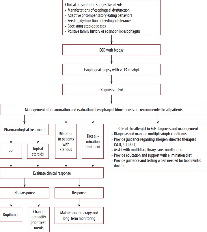
In 2015, Bousquet et al. reported on the phenomenon of multimorbidity in terms of allergic and non-allergic diseases: allergic rhinitis, asthma, and atopic dermatitis, noting the similar immunological and non-immunological pathomechanisms of these conditions [62, 63]. The authors emphasized that multimorbidity differs from the atopic march and noted that co-morbidities adversely affect one another, worsening their respective courses [62, 63]. Due to the frequent co-occurrence of EoE with allergic rhinitis, allergic conjunctivitis, or asthma, allergists diagnosing these patients can contribute to early detection of the disease. Allergists should be on the lookout for subtle signs of dysphagia and refer patients for gastroscopy with biopsy. In light of the growing incidence of EoE and its conceptualization within the atopic march and multimorbidity, clinicians in both academic and community practice should anticipate increased involvement in multidisciplinary care alongside gastroenterology specialists. Figure 1 illustrates the diagnostic and therapeutic algorithm for eosinophilic esophagitis, emphasizing the key role of the allergist in the diagnostic and management pathway.
FIGURE 1
Diagnostic and therapeutic algorithm for eosinophilic esophagitis for allergists. Adapted from Dellon ES, Muir AB, Katzka DA, et al. ACG Clinical Guideline: Diagnosis and Management of Eosinophilic Esophagitis. Am J Gastroenterol 2025; 120: 31-59 [34] and McGowan EC, Wright BL, Ruffner MA, et al. Eosinophilic esophagitis: an allergy and immunology perspective on the updated guidelines. J Allergy Clin Immunol 2025; 156: 252-8 [40]
EGD – esophagogastroduodenoscopy, EoE – eosinophilic esophagitis, eos/hpf – eosinophils per high-power field, PPI – proton pump inhibitors.