

INTRODUCTION
Affecting almost 50 million people worldwide, epilepsy is a chronic neurological condition marked by repeated, unprovoked seizures [1]. Despite advances in anti-seizure medications (ASMs), up to 30% of patients remain pharmacoresistant [2]. Importantly, drug resistance in epilepsy is often a consequence of the underlying etiology rather than a separate phenomenon, as seen in conditions such as tuberous sclerosis, Dravet syndrome, and structural brain lesions [3]. This resistance has a significant impact on patients’ quality of life, underscoring the urgent need for improved diagnostic and therapeutic strategies. The search for reliable biomarkers that can aid in diagnosis, predict treatment response, and monitor disease progression is a significant focus in epilepsy research [4, 5]. A biomarker is a measurable biological indicator used to assess the presence, severity, progression, or response to treatment of a disease. In the context of epilepsy, biomarkers can serve multiple purposes, including confirming diagnosis, predicting treatment response, assessing disease severity, monitoring disease progression, and guiding therapeutic decisions [6].
The pathophysiology of epilepsy involves a disrupted balance between excitatory and inhibitory neurotransmission, primarily mediated by glutamate and GABA. The regulation of neuronal excitability depends critically on neuropeptides, either as modulators of neurotransmitters or as receptors for them [4]. Recent studies have highlighted the potential of endogenous peptides, including ghrelin and nesfatin-1, not only in metabolic regulation but also in neuroprotection and seizure modulation [7, 8].
Ghrelin is an orexigenic peptide predominantly produced in the stomach, acting via the growth hormone secretagogue receptor 1a (GHSR1a). Ghrelin exists in two forms: acylated ghrelin (AG, often referred to as ghrelin) and desacylated ghrelin (DAG) [4]. While initially recognized for its role in appetite and energy balance, ghrelin is also expressed in the central nervous system (CNS), where it interacts with neuropeptide Y and agouti-related peptide pathways [9-11]. Since its discovery in 1999, interest in ghrelin’s potential role in epilepsy has grown significantly. Experimental studies have reported both its anticonvulsant and proconvulsant effects, and clinical data on its levels in epilepsy patients are still conflicting [7, 12].
Although ghrelin is primarily produced peripherally in the stomach, it crosses the blood–brain barrier and can also be synthesized locally in the hypothalamus and hippocampus. The peptide exerts central effects by binding to GHSR1a receptors, which are widely distributed throughout the brain, including regions critical for seizure gene-ration and propagation, such as the hippocampus, amygdala, and cortex. This peripheral-central communication axis allows ghrelin to influence neuronal excitability and susceptibility to seizure [13-15].
Nesfatin-1, derived from the precursor protein NEFA/nucleobindin2 (NUCB2), was first identified in the hypothalamus and is implicated in appetite suppression and stress response. Research on nesfatin-1 in epilepsy has been spurred by its capacity to regulate neuronal excitability and its altered levels in various neurological diseases [16]. Studies have shown that nesfatin-1 can induce hyperpolarization in most nigral dopaminergic neurons, suggesting a potential role in regulating neuronal excitability [17]. This has attracted more attention to nes-- fatin-1’s involvement in neurological conditions, like epi-lepsy [18].
Nesfatin-1, initially identified in hypothalamic neurons, is also expressed peripherally in the gastric mucosa, pancreatic β-cells, and adipose tissue. Like ghrelin, nesfatin-1 can cross the blood–brain barrier, establishing bidirectional communication between peripheral organs and the CNS. Its receptors are expressed in brain regions involved in epileptic activity, including the hippocampus and substantia nigra, enabling it to modulate neuronal excitability and potentially influence seizure threshold [17-19].
Given the complex and dynamic roles of ghrelin and nesfatin-1 in neurological functioning, these neuro-peptides have emerged as promising candidates for biomarker discovery in epilepsy. However, results from different studies vary, and the clinical value of these findings remains unknown.
This systematic review aims to consolidate present knowledge on the roles of ghrelin and nesfatin-1 in human epilepsy and evaluate their potential as biomarkers for: (1) differentiating epilepsy types (focal vs. generaliz-ed), (2) predicting treatment response to ASMs, (3) assessing disease severity and drug resistance, (4) monitoring treatment effects, particularly metabolic side effects, and (5) providing prognostic information. By identifying these specific applications, we aim to guide future research and clarify the clinical utility of these neuropeptides in the management of epilepsy.
METHODS
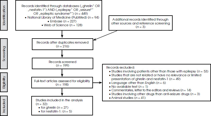
This systematic review was conducted in accordance with the Preferred Reporting Items for Systematic Reviews and Meta-Analysis (PRISMA) guidelines [20]. Figure I outlines the study’s flowchart, following these guidelines. Details of the protocol for this systematic review were registered in PROSPERO before initiation, under the ID CRD42024567659 (www.crd.york.ac.uk/PROSPERO/display_record.asp?ID=CRD42024567659).
Search strategy
A comprehensive systematic search was conducted across three primary databases: PubMed (National Library of Medicine), Embase, and Web of Science in June 2025. Two independent reviewers applied the search term (“ghrelin” OR “nesfatin-1”) AND (“epilepsy” OR “seizure*” OR “epileptic syndrome*”) consistently across all platforms, with sources indexed through June 2025. The only filter applied was a restriction to English-language publications.
Quality assessment
The methodological quality of the studies included was evaluated using the Newcastle-Ottawa Scale for three domains: (1) Selection, (2) Comparability and (3) Outcome. Each study was awarded a maximum of nine stars, with scores of ≥ 7 considered high quality, 5-6 moderate quality, and ≤ 4 low quality. Two independent reviewers performed the assessments, and study inclusion required a minimum score of 5 points.
Selection of studies and evaluation
Studies underwent systematic evaluation at three levels: title, abstract, and full-text review according to predefined criteria. Inclusion criteria encompassed original research papers and case reports involving human subjects (both adult and pediatric) with a confirmed epilepsy diagnosis and reported ghrelin or nesfatin-1 measurements. Given the limited literature explicitly examining these peptides in epilepsy contexts, broad inclusion parameters were maintained.
Exclusion criteria encompassed commentaries, letters to editors, and review articles, studies with unavailable full texts, research involving non-epilepsy patients, animal or cell culture studies, research with limited or no relevance to ghrelin or nesfatin-1, pharmacological interventions other than ASMs, and non-English publications (Figure I).
Two independent reviewers screened titles, abstracts, and full-text articles for adherence to the inclusion criteria. Subsequently, two reviewers (O.Y. and M.Z.) conducted a supplementary manual search, reference and citation tracking to finalize the selection of papers. Any discrepancies or disagreements were resolved through collaborative discussion and consensus.
RESULTS
General findings
The summaries of studies evaluating ghrelin and nesfatin-1 in epilepsy patients are presented in Tables 1 and 2, respectively. A total of 449 records were identified through database searches. Three additional articles were identified through reference screening during the manuscript preparation process. After removing duplicates, 216 studies remained for title and abstract screening. Of these, 199 articles underwent full-text review, and 32 studies met all inclusion criteria and were included in the systematic review. The study selection process is detailed in the PRISMA flowchart (Figure I).
Table 1
Summary of studies investigating ghrelin levels in human epilepsy
| Author, year, country [Ref.] | Group size | Drugs | Study design | Aim of the study | Methodology (ghrelin form, type of analysis) | Main findings | Other findings |
|---|---|---|---|---|---|---|---|
| Durgut et al. 2025, Turkey [37] | 57 pediatric pts (29 resistant, 28 mild epilepsy), 31 HC | VPA, LEV | Prospective, cross-sectional study | To investigate the relationship between ghrelin and inflammatory markers | Serum ghrelin after 8-12 hours fasting, ELISA | ↑ Ghrelin levels in the resistant group than in HC | ≠ Ghrelin levels between mild epilepsy and HC Ghrelin levels are influenced by the VPA but not by the LEV |
| Yasguclukal et al. 2023, Turkey [26] | 38 pediatric pts (14 EE-SWAS, 24 SeLFE), 21 HC | NI | Multi-center, prospective, cross-sectional study | To investigate the relationship between EE-SWAS and neuropeptides, such as ghrelin | Serum ghrelin after one-night fasting, ELISA | ↑ Ghrelin levels in the SeLFE group than in HC | |
| Costa et al., 2022, Italy [27] | 50 nd pediatric pts | CBZ, LEV, LCZ | Prospective, multicenter study | To evaluate the plasma levels of total ghrelin, ghrelin, and DAG before and after the ASM treatment in children with epilepsy | Serum total ghrelin, AG and DAG levels after one-night fasting, ELISA,1-year follow-up | ≠ Total ghrelin and DAG levels before and after the treatment ↓ Total ghrelin levels in children with GE compared to those with combined FE and GE | ↓ AG-to-DAG ratio in GE compared to all the other types of epilepsy ≠ Ghrelin levels with ASMs (CBZ, LEV, LCZ) |
| Erkec et al. 2021, Turkey [25] | 58 adult TLE (24 drug-resistant, 34 not drug- resistant), 28 HC | NI | Cross-sectional study | To evaluate the serum levels of ghrelin in TLE pts with and without drug resistance and to compare them to HC | Serum ghrelin after one-night fasting, ELISA | ↓ Serum ghrelin levels in patients with drug-resistant TLE than in HC | |
| Marchio et al. 2019, Italy [40] | 16 pediatric pts (refractory epilepsy) | Poly-therapy, KD | Cohort | To find out how the KD changed the plasma levels of ghrelin and DAG in children with refractory epilepsy who received treatment for up to ninety days | Plasma ghrelin and DAG, ELISA, 0-30-90 days follow-up | ↓ Ghrelin and DAG plasma levels in children affected by pharmacoresistant epilepsy and maintained on the KD | These changes became significant after 30 days and were maintained for 90 days |
| Marchio et al. 2019, Italy [41] | 6 pediatric pts with refractory epilepsy | CBZ, LEV, TPM, NZP, VPA | Prospective | To evaluate ghrelin and growth during 1 year of KD | Serum ghrelin after one-night fasting, ELISA, 1-year follow-up | ↓ Ghrelin plasma levels in children with refractory epilepsy and maintained on the KD | This change was associated with low growth indexes in the majority of pts |
| De Amicis et al. 2019, Italy [42] | 25 pediatric, 5 adult pts with refractory epilepsy | Poly-therapy, KD | Multicenter, cohort, longitudinal study | To assess how a 1-year KD altered ghrelin concentrations in younger children, teens, and adults with DRE or GLUT1 deficiency syndrome | Serum ghrelin after one-night fasting, ELISA, 1-year follow-up | ≠ Ghrelin levels | There are no correlations between ghrelin changes and nutritional status and body composition |
| Mohamed et al. 2019, Egypt [23] | 40 adult pts (16 FE, 24 GE), 40 HC | NI | Prospective, case-control | To evaluate the ghrelin effect in adult epileptic patients and the response to ASMs | Fasting serum AG, DAG, and urine AG levels by ELISA | ↑ Serum AG, DAG, and urine AG levels in pts than HC ↑ Levels in polytherapy and GE pts ↑ Level of serum AG among GE pts | ↑ Serum AG and DAG in epileptic pts, especially with prolonged epilepsy duration and good ASM response |
| Marchio et al., 2018, Italy [44] | 114 pediatric pts (77 ASM responsive, 37 ASM resistant) 59 HC | Observational, case-control | To investigate the potential correlation between plasma levels of ghrelin and DAG in children with epilepsy and the response to ASMs | Serum ghrelin and DAG after one-night fasting, ELISA | ↑ Ghrelin and DAG plasma levels are higher in ASM responders than in HC | ↑ AG and DAG plasma levels in pts who positively respond to AEDs | |
| Cicek et al., 2018, Turkey [35] | 48 pediatric pts with idiopathic epilepsy (25 VPA, 23 TPM), 25 HC | VPA, TPM | Prospective, case-control | To compare insulin, leptin, neuropeptide Y, and ghrelin levels in children with epilepsy on monotherapy of TPM and VPA | Serum ghrelin after one-night fasting, ELISA kits | ≠ Ghrelin levels in the patient groups compared to HC | |
| Hasaneen et al. 2016, Egypt [36] | 20 idiopathic nd pediatric FE pts, 22 HC | LEV | Prospective comparative case-control | To examine changes in body weight, BMI, and composition in children with idiopathic FE using bioelectrical impedance analysis, serum ghrelin level, and insulin resistance | Serum ghrelin after one-night fasting, ELISA, 6 months follow-up | ≠ Ghrelin serum levels of children (6-10 years) with epilepsy receiving LEV as monotherapy | |
| Varrasi et al. 2014, Italy [46] | 56 adult pts (19 IGE, 18 CFE, 19 symptomatic FE), 68 HC | VPA, CBZ | Cross-sectional | Separately evaluate AG and DAG levels and understand their correlations with clinical features | Serum AG and DAG after one night of fasting, ELISA | ≠ Interictal ghrelin levels in pts and HC ↑ AG-to-DAG ratio in pts | Levels of ghrelin were in direct proportion to disease duration (both AG and DAG) |
| Ozcelik et al. 2014, Turkey [38] | 20 pediatric prepubertal pts (14 FE, 6 IGE) | TPM | Prospective, case-control, cross-sectional | To assess how ghrelin levels are affected by TPM and how these factors relate to the pathophysiology of weight loss in prepubertal epileptic children | Serum ghrelin, RIA, 0-3-6 months follow-up | ≠ Ghrelin levels | Levels of ghrelin did not change significantly in the early stages of TPM treatment |
| Taskin et al. 2014, Turkey [45] | 30 pediatric prepubertal IGE pts, 30 prepubertal HC | VPA | Prospective, case-control, cross-sectional | To determine the serum, urine, and saliva levels of AG and DAG in the newly diagnosed pediatric IGE patients in the pretreatment period and the third month of VPA treatment | Serum, saliva, and urine AG and DAG, 3 months follow-up, saliva and urine with RIA, and blood with ELISA | ↑ AG and DAG in the blood, urine, and saliva, but not in the post-therapy body mass index | |
| Tokgoz et al. 2012, Turkey [58] | 20 prepubertal pediatric pts with idiopathic epilepsy | VPA | Prospective, case-control, cross-sectional | To assess plasma ghrelin levels in kids receiving VPA treatment for epilepsy, and to evaluate these markers for early atherosclerosis | Serum ghrelin after one-night fasting, ELISA and CAIMT before the treatment and after a follow-up period of 6 to 12 months | ≠ Ghrelin levels after 6 and 12 months | In children treated with VPA for epilepsy, there was no discernible increase in the risk for early atherosclerosis, according to CAIMT |
| Cansu et al. 2012, Turkey [34] | 76 pediatric pts (prepubertal 22 with OXC, 22 VPA, pubertal 14 OXC, 18 VPA) | OXC, VPA | Prospective, comparative | To assess how children with idiopathic epilepsy respond to VPA or OXC monotherapy in terms of their linear growth | Blood serum ghrelin after overnight fasting with RIA, 0, 6 and 18 months follow-up | ≠ Ghrelin levels | |
| Cansu et al. 2011, Turkey [32] | 18 pediatric pts (9 prepubertal, 9 pubertal) newly diagnosed with FE and IGE with VPA and 18 controls (9 prepubertal, 9 pubertal) | VPA | Prospective comparative case-control | To investigate the cause of VPA-associated weight gain in children | Blood serum ghrelin after overnight fasting with RIA, 18 months follow-up | ↓ Ghrelin in weight-gain patients | Ghrelin’s serum levels are not directly linked to weight gain |
| Aydin et al. 2011, Turkey [43] | 70 adult male pts (18 FE, 16 GE, 16 functional seizure), 20 HC | VPA, PHT, CBZ | Prospective, comparative study | To compare serum levels of nesfatin-1 and ghrelin with HC and examine the relationship between these markers measured at various periods following an epileptic seizure or non-epileptic incident | Blood serum AG, DAG, and ASM were stopped 48 h before study initiation, 5 min, 1, 24, and 48 hours after the seizure, RIA | ↓ Ghrelin levels after seizures 5 min and 1 h of a seizure compared to paroxysmal events and HC, but increased after 48 hours | These findings could help distinguish epileptic patients from functional seizure pts |
| Cansu et al. 2011, Turkey [39] | 22 pediatric pts (nd with IFE) | OXC | Prospective, comparative | To investigate whether OXC monotherapy causes weight gain in epileptic children | Serum ghrelin with RIA, in 0, 6 and 18 months follow-up | ≠ Ghrelin levels between initial values (month 0) and those in the 6th and 18th months after OXC therapy | |
| Prodam et al. 2010, Italy [50] | 34 prepubertal pediatric pts (12 CBZ (4 PGE, 8 FE), 22 VPA, PGE), 18 prepubertal HC | VPA, CBZ | Prospective comparative case-control | To assess whether VPA or CBZ medication affects ghrelin secretion in young prepubertal epileptic children | Blood plasma ghrelin with RIA | ↓ Ghrelin in epileptic pts than in HCs | ↓ Ghrelin in the CBZ group than in the VPA group |
| Dag et al. 2010, Turkey [22] | 91 adult (F+M) pts (10 nd PGE, 18 PGE, 37 PE) and 26 HC | VPA, PHT, CBZ | Prospective comparative case-control | The study aims to compare ghrelin levels in serum and saliva in epilepsy pts | Saliva and serum, total ghrelin, ELISA | ↓ Serum and saliva ghrelin levels in all pts than HC ↑ In FE groups than in the other pts | Saliva showed ghrelin concentrations similar to those in serum Saliva may be a non-invasive alternative |
| Güzel et al. 2009, Turkey [31] | 25 pediatric pts (14 M, 11F) (24 IGE, 1 IFE) | VPA | Prospective comparative study | To investigate the relationship between weight gain, serum ghrelin levels in children taking VPA | Serum ghrelin after one-night fasting, ELISA, 1-year follow-up | ↓ Serum ghrelin levels in children receiving VPA | 12 pts developed obesity |
| Tomoum et al. 2009, Egypt [33] | 24 pediatric pts (22 M, 12 F) (8 IGE, 7 cryptogenic, 19 symptomatic epilepsy), 40 HC | VPA | Prospective, cross-sectional, comparative | To investigate the relationship between growth metrics, VPA level and epileptic condition features, as well as the impact of VPA on ghrelin levels in children with epilepsy | Serum ghrelin after one-night fasting, ELISA | ↑ Ghrelin levels in pts than in HCs (significant) | None of the pts developed obesity |
| Aydin et al. 2009, Turkey [21] | 73 adult (F+M) pts (8 nd with GE, 21 IGE, 24 FE), 20 HC | VPA, PHT, CBZ | Prospective comparative case-control | To investigate differences in ghrelin concentrations in saliva and serum samples between epilepsy pts and HC and whether salivary glands produce nesfatin-1 | Saliva and blood serum, ELISA and RIA, DAG and AG forms of ghrelin | ↓ Serum and saliva ghrelin levels in epileptic pts before treatment than in HC | ↑ Ghrelin levels in newly diagnosed primary GE pts but ↓ with treatment |
| Gungor et al. 2007, Turkey [29] | 35 pediatric pts (3-15 years) | VPA | Prospective, cohort, case-control | To investigate how VPA affects ghrelin and its possible consequences on growth and weight gain in epileptic children | Serum ghrelin after one night fasting, ELISA 0-6 months, follow-up | ↑ Ghrelin in prepubertal children after VPA treatment | ↑ Ghrelin levels during the first few months of VPA may be linked to weight gain |
| Berilgen et al. 2006, Turkey [24] | 35 adult (F+M) pts with epilepsy (20 GE, 15 FE), 30 HC | VPA, PHT, CBZ | Prospective, case-control | To evaluate serum levels of ghrelin in adult epilepsy pts | Commercial RIA kit, blood serum total ghrelin levels (AG, DAG) | ↑ Serum ghrelin in all kinds of epilepsy pts | ↑ Serum ghrelin levels in the FE than the GE |
| Greco et al. 2005, Italy [30] | 40 adult F pts (31 GE, 5 absence, 9 FE), 40 HC | VPA | Prospective comparative case-control | To evaluate serum levels of ghrelin in epilepsy pts | Blood serum ghrelin, ELISA, 2-year follow-up | ↓ Ghrelin in weight-gain pts | 15 epileptic pts (37.5%) developed obesity No significant correlation between serum VPA concentration and its daily dosage and ghrelin concentrations |
[i] ↑ – increased, ↓ – decreased, ≠ – unchanged, AG – acylated ghrelin, ASM – antiseizure medications, CAIMT – common carotid artery intima media thickness, CBZ – carbamazepine, CFE – cryptogenic focal epilepsy, DAG – des-acyl ghrelin, DRE – drug-resistant epilepsy, EE-SWAS – epileptic encephalopathy with spike-and-wave activation in sleep, ELISA – enzyme-linked immune sorbent assay, F – female, FE – focal epilepsy, GE – generalized epilepsy, IFE – idiopathic focal epilepsy, IGE – idiopathic generalized epilepsy, HC – healthy controls, KD – ketogenic diet, OXC – oxcarbazepine, pts – patients, nd – newly diagnosed, M – male, NI – no Information, NZP – nitrazepam, pts – patients, RIA – radioimmunoassay, SeLFE – self-limited focal epilepsy of childhood, TLE – temporal lobe epilepsy, TPM – topiramate, UAG – unacylated ghrelin, VPA – valproic acid
Table 2
Summary of studies investigating nesfatin-1 levels in human epilepsy
| Author, year, country [Ref.] | Group size | Drugs | Study design | Aim of the study | Method | Main findings | Other findings |
|---|---|---|---|---|---|---|---|
| Erkec et al. 2021, Turkey [25] | 58 adult TLE (24 drug-resistant, 34 not drug-resistant), 28 HC | NI | Prospective, cross-sectional study | To evaluate the serum levels of nesfatin-1 in TLE pts with and without drug resistance, and to compare them to HC | Serum nesfatin-1 after one night fasting, ELISA | ↑ Serum nesfatin-1 in the TLE group, whereas they had ↓ in the DR-TLE group when compared to HC (both with no significance) | |
| Yu et al. 2021, China [47] | 54 adult pts (34 M, 20 F), 54 HC | NI | Prospective, comparative study | To investigate the clinical utility of EEG monitoring and the application of silver nanoparticles to measure blood nesfatin-1 levels in evaluating the degree and prognosis of epilepsy | 1-year follow-up, fasting serum nesfatin-1, ELISA | ↑ Nesfatin-1 in epilepsy pts than in HCs ↑ Nesfatin-1 in the seizure group than the non-seizure group | There is no significant correlation between EEG abnormalities and biomarker serum levels |
| Chen et al., 2019, China [48] | 43 adult pts with pr | NI | Prospective, comparative study | To investigate the dynamic variation of serum nesfatin-1 in pts with SE and its clinical values in evaluating the illness condition and short-term prognosis | 0, 1 week, 2 weeks, 1 month after therapy, and 1-year follow-up | ↓ Nesfatin-1 gradually with the prolongation of time | ↑ Serum nesfatin-1 was a risk factor for the one-year survival prognosis |
| Aydin et al., 2011, Turkey [43] | 70 adult male pts (18 FE, 16 GE, 16 PNES), 20 HC | VPA, PHT, CBZ | Prospective, comparative study | To compare nesfatin-1 serum levels with HC and examine the relationship between these markers measured at various periods following an epileptic seizure or non-epileptic incident | Blood serum nesfatin-1 and ASM were stopped 48 h before study initiation, 5 min, 1, 24, and 48 hours after the seizure, enzyme immunoassay, ELISA | ↑ Serum nesfatin-1 in FE and GE pts | ↓ Nesfatin-1 in epileptic pts after a seizure These findings could help distinguish epileptic pts from functional seizure pts |
| Aydin et al. 2009, Turkey (21) | 73 adult (F+M) pts (8 nd with GE, 21 IGE, 24 FE), 20 HC | VPA, PHT, CBZ | Prospective comparative case-control | To investigate differences in nesfatin-1 concentrations in saliva and serum samples between epilepsy pts and HC and whether salivary glands produce nesfatin-1 | Salivary gland tissue samples were analyzed for nesfatin-1 expression by immunochemistry and ELISA | ↑ Serum and saliva nesfatin-1 in untreated pts than in HC ↓ Saliva and serum nesfatin-1 with ASM (still higher than HC) | Salivary glands produce their nesfatin-1 but around 12-fold less than brain tissues in epilepsy pts |
[i] DRTLE – drug-resistant temporal lobe epilepsy, ELISA – Enzyme-Linked ImmunoSorbent Assay, FE – focal epilepsy, FMJ – first myoclonic jerk, GCS – generalized clonic seizures, GSH – glutathione, GPx –glutathione peroxidase, GR – glutathione reductase, NES-1 – nesfatin-1, NI – no Information, NO – nitric oxide, PHT – phenytoin, PGE – primary generalized epilepsy, pts – patients, PTZ – pentylenetetrazole, SE – status epilepticus, SOD – superoxide dismutase, TGE – tonic generalized extension, TLE – temporal lobe epilepsy
Exclusions were based on the following criteria:
studies involving non-epileptic patients (n = 53),
studies with limited or no relevance to ghrelin and nesfatin-1 (n = 49),
non-English language publications (n = 6),
unavailable full texts (n = 1),
commentaries, letters to the editors and reviews (n = 14),
studies involving drugs other than ASMs (n = 3),
animal studies (n = 41).
The studies included comprised 27 on ghrelin and five on nesfatin-1, published between 2005 and 2025. Study designs included prospective case-control, cross-sectional, and cohort studies, with sample sizes ranging from 6 to 114 participants. All studies were hospital-based. Most studies compared epilepsy patients to healthy controls (HC), while some included additional sub- groups (e.g., generalized epilepsy, focal epilepsy, resistant or mild epilepsy). The main characteristics of the studies included are summarized in Table 1 (ghrelin) and Table 2 (nesfatin-1).
Ghrelin in epilepsy
This systematic review identified 27 studies investigating ghrelin in epilepsy patients conducted between 2005 and 2025. The majority focused on pediatric po-pulations (70.4%, n = 19), with eight studies (29.6%) involving adults. ELISA was the predominant measurement method (66.6%, n = 18), with blood samples collected after overnight fasting as the standard protocol.
Ghrelin levels showed heterogeneous patterns in epilepsy patients: 37.1% (10/27) of the studies reported decreases, 29.6% (8/27) reported increases, and 33.3% (9/27) reported no change – Aydin et al. [21], noted that although ghrelin levels in serum and saliva increased after treatment in newly diagnosed patients, they remained lower than in HC [21]. Notably, the correlation between saliva and serum ghrelin concentrations highlights the potential of saliva and urine as non-invasive alternatives for hormone measurement [22].
Ghrelin levels varied by epilepsy type [22-25]. Patients with generalized epilepsy (GE) demonstrated higher AG and DAG levels compared to HCs [23]. On the other hand, focal epilepsy (FE) patients showed higher plasma ghrelin levels than both GE patients [22, 24] and HC [26]. Another study identified lower total ghrelin and reduced ghrelin-to-DAG ratios in children with GE compared to other epilepsy types [27].
ASM effects on ghrelin levels showed variation across studies. Research on valproic acid (VPA) yielded conflicting results regarding weight gain mechanisms [28, 29], with some studies linking elevated ghrelin to increased appetite. In contrast, others found reduced levels in VPA-treated patients who developed obesity [22, 23, 30-33]. Age- related differences emerged, with varying ghrelin responses between prepubertal and pubertal children [24, 34]. Other ASMs, including levetiracetam, oxcarbazepine (OXC)and topiramate, showed minimal effects on ghre-lin levels [35-39].
Three studies examined ghrelin during ketogenic diet (KD) treatment. Two reported reduced ghrelin and DAG levels in children with drug-resistant epilepsy [40, 41], while one found no significant changes after 12 months in mixed pediatric and adult populations receiving the diet [42]. Notably, lower serum ghrelin levels were observed in drug-resistant epilepsy patients, suggesting ghrelin’s potential involvement in the pathophysiology of drug-resistant epilepsy [25, 37].
Ghrelin levels demonstrated dynamic changes following seizures, with decreases observed immediately after the seizure, followed by increases at 48 hours [43]. Higher ghrelin levels were associated with positive ASM responses, suggesting potential as biomarkers for treatment efficacy prediction [23, 44-46]. Lower ghrelin levels in drug-resistant epilepsy patients indicated possible involvement in treatment-resistant pathophysiology [25, 37].
Finally, ghrelin’s relationship with comorbidities in epilepsy has been explored. For example, OXC treatment has been associated with increased linear growth in children, whereas VPA reduced ghrelin levels without significantly affecting growth parameters [34].
Nesfatin-1 in epilepsy
Five studies investigated nesfatin-1 in epilepsy patients between 2009 and 2021, primarily focusing on adult po-pulations using prospective designs. All studies measured serum nesfatin-1 via ELISA using fasting samples, with one study additionally examining saliva samples [21].
Four studies (80%) reported elevated serum nesfatin-1 levels in epilepsy patients compared to HCs, parti-cularly in untreated patients or those with active seizures [21, 25, 43, 47]. Aydin et al. [21] demonstrated that while nesfatin-1 levels decreased following ASM treatment, they remained high compared to HC in both serum and saliva samples.
Yu et al. [47], observed higher nesfatin-1 levels in patients with frequent seizures compared to those with fewer seizures. In drug-resistant temporal lobe epilepsy (TLE), patients showed lower nesfatin-1 levels than non-drug- resistant patients, though this difference was not statistically significant [25].
Chen et al. [48], identified serum nesfatin-1 as a potential prognostic marker, with higher levels associated with poorer one-year survival outcomes in status epilepticus patients.
Additionally, nesfatin-1 demonstrated potential as a differential biomarker, with epileptic patients showing consistently elevated levels compared to patients with functional seizures (previously termed psychogenic non-epileptic seizures), who had lower levels. However, this finding was limited to male participants only [43]. The same authors also demonstrated that salivary glands produce nesfatin-1 at levels significantly lower than those in brain tissue, providing insight into its peripheral sources [21].
DISCUSSION
The complex interplay between the endocrine system and epilepsy has been the subject of extensive research, motivated by frequent reports of hormonal abnormalities in epilepsy patients. While neuropeptides such as prolactin, vasopressin, and oxytocin have been shown to increase during seizures, recent attention has turned to ghrelin and nesfatin-1 as potential biomarkers and the-rapeutic targets in epilepsy. Their multifaceted roles in appetite regulation, metabolic homeostasis, and neuroprotection position them as particularly relevant candidates for advancing epilepsy care [49].
Currently, the therapeutic landscape for epilepsy includes only one neuropeptide, adrenocorticotropic hormone (ACTH), for infantile spasms, highlighting a significant gap in neuropeptide-based treatments. This limitation underscores the urgent need for expanded investigation into neuropeptides as both diagnostic tools and therapeutic interventions for epilepsy [4].
This systematic review synthesizes evidence from 32 studies investigating ghrelin and nesfatin-1 in epilepsy, revealing distinct biomarker profiles with clinical implications. Our analysis demonstrates that while ghrelin shows context-dependent variability, nesfatin-1 exhibits more consistent elevation patterns, positioning both neuropeptides as promising candidates for epilepsy management.
Ghrelin in epilepsy
The observed heterogeneity in ghrelin levels – 37.1% of studies reporting decreases, 29.6% increases, and 33.3% no change – reflects its complex interplay with epilepsy subtypes, ASM effects, and metabolic factors. Notably, increased ghrelin levels were particularly evident in patients with FE or SeLFE compared to those with GE or HC [24, 26]. This pattern aligns with emerging evidence of subtype-specific neuroendocrine dysregulation in epi-lepsy networks.
The relationship between ASMs and ghrelin levels remains one of the most clinically relevant yet inconsistent findings in our review. While some medications like levetiracetam and topiramate showed minimal effects on ghrelin levels, others, particularly VPA and carbamazepine, demonstrated more pronounced but variable impacts [50]. The observation that patients with better ASM responses tend to have higher ghrelin levels suggests a potential role for ghrelin as a biomarker for treatment efficacy. However, this requires validation in larger, controlled studies.
The relationship between VPA and ghrelin-mediated weight gain warrants particular attention due to its clinical significance. As an orexigenic hormone, ghrelin stimulates appetite via neuropeptide Y and agouti-related peptide pathways, theoretically contributing to VPA- associated weight gain [14]. However, our review reveals conflicting evidence: some studies link elevated ghrelin levels during early VPA therapy with increased appetite and weight gain [29], while others associate VPA-induced obesity with paradoxically reduced ghrelin levels [30, 31]. These discrepancies may stem from patient age, treatment duration, or metabolic factors.
The three studies examining ghrelin during KD treatment provide intriguing insights into metabolic adaptation in epilepsy. Standardized procedures are clearly needed in future KD research, as this discrepancy may reflect variations in study length, patient populations, or the degree of ketosis achieved.
Additionally, ghrelin levels were shown to decrease shortly after seizures but rebounded within 48 hours, indicating a possible role in postictal recovery [43, 51]. The strong correlation between saliva and serum ghrelin levels underscores the potential for non-invasive diagnostic tools [22].
Nesfatin-1 in epilepsy
With 80% of the studies reporting increased levels in epilepsy patients compared to HC, nesfatin-1 shows more consistent patterns than ghrelin, despite being represented by just five studies (Table 2). This consistency, combined with its correlation with seizure frequency and potential prognostic value, positions nesfatin-1 as an up-and-coming biomarker candidate.
The correlation between nesfatin-1 levels and seizure frequency suggests a direct relationship with epileptic activity, potentially reflecting the neuropeptide’s role in neuronal excitability or stress response pathways [52]. However, decreases were noted in drug-resistant TLE and following prolonged ASM use, indicating dynamic regulation in response to drug therapy and disease progression [25, 47].
The preliminary evidence linking elevated nesfatin-1 levels to poor survival outcomes in status epilepticus, while requiring validation, indicates that this neuropeptide may serve as a marker of disease severity and prognosis [48]. Furthermore, identifying salivary nesfatin-1 production at levels lower than those in brain tissue opens avenues for the development of non-invasive biomarkers [21].
Perhaps most clinically significant is the emerging evidence that nesfatin-1 levels differ between epileptic seizures and functional seizures. This finding, though currently limited to male patients, suggests potential utility in differential diagnosis – a persistent challenge in epilepsy care where misdiagnosis rates remain substantial [43].
Comparative analysis
There are significant distinctions between ghrelin and nesfatin-1 in terms of their possible clinical uses. While ghrelin exhibits inconsistent and frequently conflicting patterns, which may reflect its complex role in energy homeo-stasis and multiple regulatory pathways, nesfatin-1 demonstrates a more consistent elevation in epilepsy patients. This disparity may indicate their unique physiolo-gical functions: ghrelin’s principal role in hunger and energy equilibrium may be more influenced by confounding variables such as pharmacological effects, body composition, and feeding conditions, whereas nesfatin-1’s function in stress response and neuronal regulation may more accurately represent epileptic pathophysiology.
Both neuropeptides show potential for non-invasive assessment, with demonstrated correlations between serum and saliva concentrations. This capability may enhance point-of-care testing and continuous monitoring, addressing current limitations in epilepsy biomarker development.
Clinical implications and practical applications
The findings of this systematic review have several immediate and potential future clinical applications:
Immediate clinical applications can be: (1) Monitoring VPA-associated weight gain: ghrelin measurement could help identify patients at risk for significant weight gain early in VPA treatment, allowing for proactive dietary counseling and potentially dose adjustment or medi-cation switching [53-55]. (2) Non-invasive moni-toring: the demonstrated correlation between saliva/urine and serum levels of both peptides offers practical advantages for pediatric patients and repeated monitoring, avoiding frequent blood draws [21, 22]. (3) Differential diagnosis: nesfatin-1 shows promise in distinguishing epileptic seizures from functional seizures, which remains a significant clinical challenge affecting 20-30% of patients in epilepsy monitoring units [43].
Potential future applications can be: (1) Treatment response prediction: elevated ghrelin levels in responders to ASMs suggest potential use in personalizing medication selection and predicting treatment success before clinical response is evident [23, 44]. (2) Drug resistance identification: lower ghrelin and nesfatin-1 levels in drug- resistant epilepsy may help identify candidates for alternative therapies (surgery, neurostimulation, KD) earlier in the disease course [25, 37]. (3) Prognostic biomarker: nesfatin-1 levels correlating with seizure frequency and survival in status epilepticus suggest utility in risk stratification and monitoring [47, 48]. (4) Therapeutic target: understanding the mechanisms by which these peptides influence seizure threshold could lead to novel neuropeptide-based therapies, expanding beyond ACTH (currently the only neuropeptide treatment for epilepsy) [4, 14].
Although these applications are promising, several barriers to clinical implementation persist, including the lack of standardized measurement protocols, the absence of established reference ranges, cost considerations, and the need for validation in diverse populations. These challenges underscore the need for translational research that bridges laboratory findings to bedside applications [56, 57].
Limitations
Several limitations must be acknowledged when interpreting these findings. The methodological quality assessment revealed that some studies lack control groups, primarily due to ethical constraints in epilepsy research, limiting the strength of comparative conclusions [27, 40, 42, 58]. The predominance of cross-sectional studies limits causal inference; heterogeneity in study populations, epilepsy types, measurement methodologies, ASM regimens, and the geographical concentration of studies may affect the generalizability of findings. Additionally, many studies had relatively small sample sizes and short follow-up periods, which is particularly concerning for understanding long-term relationships between neuropeptide levels and epilepsy outcomes.
Currently, measurement of ghrelin and nesfatin-1 in clinical epilepsy care remains research-based rather than routine practice. While our review identifies potential applications, translation to clinical utility requires several unmet prerequisites.
Biomarker development progresses through three phases: discovery, validation, and clinical utility [55, 56], with current evidence indicating that these neuropeptides are currently in the early stages. The substantial gap between biomarker discovery and clinical implementation is well-recognized [59], with key requirements for clinical adoption including: (1) standardized, cost-effective assays; (2) clinically meaningful cutoff values validated across diverse populations [60, 61]; (3) prospective demonstration that biomarker-guided decisions improve patient outcomes [61]; and (4) cost-effectiveness evidence [62].
Epilepsy biomarker development faces unique challenges, including clinical heterogeneity and complex pathophysiology. Therefore, while the theoretical foundation is established through the studies reviewed here, practical clinical application awaits further translational research that bridges the critical gaps between laboratory findings and bedside implementation [6, 63].
We also acknowledge that meta-analysis and meta- regression methods, along with forest plot visualization, could have strengthened our findings. However, the significant heterogeneity in study designs, measurement methods (ELISA vs. RIA), patient populations (pediatric vs. adult, various epilepsy types), ASM regimens, timing of measurements, and outcome definitions precluded meaningful quantitative synthesis. The included studies used different cutoff values, units of measurement, and control group definitions, making statistical pooling unreliable and potentially misleading. Future studies with standardized protocols would enable robust meta-analytic approaches.
Future research
This review reveals some areas of research priority. First, large-scale, multicenter, prospective cohort studies with standardized measurement protocols are needed to validate the potential of biomarkers and establish reference ranges. Second, studies investigating which seizures influence the regulation of ghrelin and nesfatin-1 could provide insights into potential therapeutic targets. Third, intervention studies examining whether neuropeptide modulation affects seizure control or epilepsy-related comorbidities could establish therapeutic potential. Fourth, validation of non-invasive measurement techniques could facilitate clinical implementation.
Consensus rules for neuropeptide measurement in epilepsy research would benefit the field by establishing standardized procedures for sample collection, processing, and analysis, thereby enhancing comparability across subsequent studies and facilitating significant meta- analyses.
CONCLUSIONs
This systematic review establishes ghrelin and nesfatin-1 as promising biomarkers in epilepsy management. Ghrelin monitoring shows particular value for managing VPA- associated weight gain and predicting ASM treatment response, though its variable patterns require careful interpretation. Nesfatin-1 demonstrates more consistent elevations, correlates with seizure frequency and offers potential for differentiating epileptic from functional seizures. Additionally, the strong correlation between saliva and serum levels of both neuropeptides supports the development of non-invasive diagnostic tools.
Finally, the exploration of ghrelin and nesfatin-1 as therapeutic targets warrants attention. Given that ACTH remains the only neuropeptide-based therapy for epilepsy, expanding this field may lead to novel treatments. These neuropeptides show clinical potential for monitoring ASM side effects, differentiating seizure types, and predicting drug resistance. However, validation studies demonstrating clinical utility and cost-effectiveness are needed before routine implementation.
Future studies should investigate the longitudinal changes in ghrelin and nesfatin-1 levels in response to different ASMs and explore their potential as biomarkers for treatment efficacy across various epilepsy types.








