
Purpose
Adenoid cystic carcinoma (ACC), a rare epithelial malignancy, accounts for approximately 1% of all head and neck cancers and less than 5% of all orbital tumors [1, 2]. Characterized by its indolent yet relentless course, ACC is notorious for perineural invasion, local recurrence, and late distant metastases, making long-term disease control challenging [3-5]. Orbital ACC, particularly when arising from the lacrimal gland or periorbital structures, presents a unique therapeutic dilemma due to the anatomical constraints imposed by adjacent critical structures, including the optic nerve, globe, and lens.
Surgical resection, followed by adjuvant radiotherapy, remains the standard approach for localized malignant tumors of the paranasal sinus, skull base, and orbital regions [6-8]. However, in recurrent cases where surgery cannot be performed due to high-risk location of the tumor, especially in previously irradiated patients, this standard procedure poses a therapeutic challenge. The risk of toxicity to organs at risk (OARs) often precludes the delivery of curative doses in such scenarios using external beam radiotherapy (EBRT) alone.
Re-irradiation strategies using advanced modalities, such as stereotactic body radiotherapy (SBRT), interstitial brachytherapy (ISBT), and particle therapy, have shown promise in selected head and neck cancers, including ACC [9, 10].
The rationale for combining SBRT and ISBT, a hybrid radiotherapeutic strategy, lies in overcoming the limitations of each modality when used in isolation. ISBT alone may inadequately cover the entire residual tumor because of geometric constraints and potential geographical misses near lateral margins. Conversely, SBRT in a re-irradiation setting, carries both the increased integral dose and the risk of late-tissue toxicities, which often limit safe dose escalation near radiosensitive OARs. A hybrid approach leverages the steep dose gradients and conformality of ISBT to deliver a high-dose boost to the central tumor while utilizing SBRT for volumetric coverage. Compared with monotherapy, this combined strategy enables improved target coverage while maintaining cumulative OAR doses within safe thresholds.
In this technical note, we presented a case with recurrent orbital adenoid cystic carcinoma, treated with a hybrid technique combining 3D-printed template-based interstitial brachytherapy and stereotactic body radiotherapy. This case illustrated the feasibility and clinical rationale of this personalized approach, highlighting its dosimetric advantages and encouraging clinical outcomes with acceptable toxicity in a highly dose-constrained setting.
Material and methods
A 39-year-old male was initially diagnosed in 2017 with ACC of the right skull base. Following sub-total surgical resection, he received post-operative intensity-modulated radiotherapy (IMRT) to a residual tumor measuring 3.3 cm × 2.5 cm × 2.8 cm. The 2017 gross tumor volume (GTV) encompassed a right-sided skull base lesion, involving the cavernous sinus, Meckel’s cave, and pterygopalatine fossa, with extension through the base of skull foramina into the infratemporal fossa. Treatment plan delivered 66 Gy in 30 fractions to high-dose volume planning target volume (PTV) 66, defined by a 0.3-cm margin around GTV. Simultaneously, an elective volume PTV 54 received 54 Gy in 30 fractions, utilizing a 1.0-cm clinical target volume (CTV) expansion from GTV and a 0.5-cm PTV margin. During this course, the right optic nerve received a maximum dose (Dmax) of 53.74 Gy (D0.1cc = 50.50 Gy), and the right eye received 43.65 Gy (D0.1cc = 0.12 Gy). These baseline exposures necessitated highly constrained planning for the current recurrence. The patient remained disease-free for over 6 years.
In 2023, he presented with progressive right-sided nasal obstruction and epistaxis. Magnetic resonance imaging (MRI) revealed a mass measuring 6.5 cm × 3.4 cm × 5.7 cm, involving the right nasal cavity, medial orbital wall, and adjacent ethmoid sinus. Biopsy confirmed recurrent adenoid cystic carcinoma. The patient underwent endoscopic debulking surgery; however, R0 resection was not feasible due to the tumor involvement of the medial orbital wall, leaving a post-operative residual lesion of 1.8 cm × 1.6 cm × 1.2 cm. Further surgical resection was precluded by the proximity of critical structures. Considering prior irradiation, a hybrid re-irradiation approach was adopted, with radiation treatment delivered in two phases: Phase I – ISBT, and Phase II – SBRT.
Phase I: Interstitial brachytherapy
Pre-planning
To ensure accurate catheter placement, a dummy pre-plan was generated. The patient was simulated in the supine position on a flat couch, using a Philips CT scanner with 1-mm slice thickness (CT-1). Diagnostic MRI was co-registered with a simulation CT, and GTV along with the right eye, lens, and optic nerve were delineated. Two dummy catheters were placed virtually within GTV, spaced 0.5 cm apart, and a plan was generated for a prescription dose of 8 Gy in 2 fractions, with D90 covering 7 Gy. After obtaining an optimal plan, the catheter spacing, insertion depth, and angle of placement were recorded.
Template preparation
Using the DICOM dataset from CT-1, a customized 3D-printed template was designed with the aid of 3D printing technology. The template was fabricated using DSM SOMOS WaterClear Ultra 10122 material (Figure 1) to enable precise catheter placement while minimizing risk to orbital structures. The template was contoured to fit securely along the periorbital region, extending from the right eye to the medial canthus of the left eye. It incorporated two 3-mm diameter catheter channels, spaced 5 mm apart, to ensure accurate and reproducible insertion of flexible interstitial brachytherapy catheters.
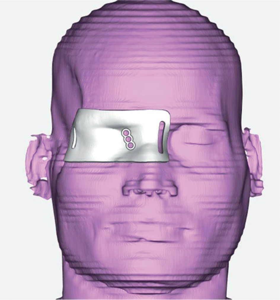
Interstitial brachytherapy
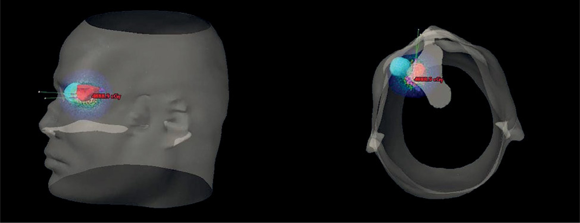
Under general anesthesia and aseptic precautions, two flexible catheters were inserted through 3D-printed template into the tumor (Figure 2). A post-insertion contrast-enhanced CT simulation was performed with 1-mm slice thickness, extending from the vertex to C5 vertebra. Catheters were identified and reconstructed (Figure 3). CT scan showed disease progression, involving the medial orbital bone and extending into the nasal canal. GTV for the ISBT phase was 2.9 cm3; however, the treatment specifically targeted high-risk medial 1.0 cm3 of the tumor bed. Organs at risk, including the right eye, lens, and optic nerve, were also contoured.
Fig. 2
Intra-operative photograph showing customized 3D-printed template positioned over periorbital region, with two flexible HDR brachytherapy catheters inserted into the tumor under general anesthesia. Catheter placement follows pre-planned angles and depths from CTbased dummy plan
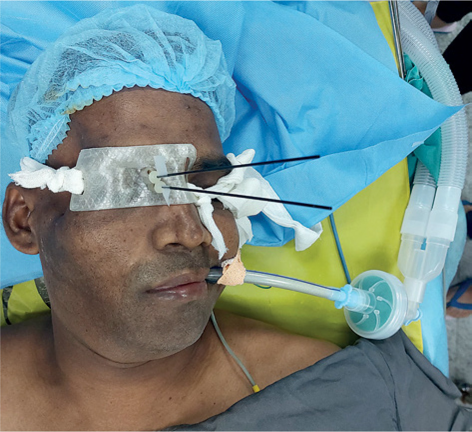
Fig. 3
3D reconstruction of brachytherapy plan showing: Catheter positions (green), gross tumor volume (GTV, red), and isodose distribution (Gy) covering intra-orbital component near medial orbital wall. Planning was performed on Monaco v. 5.11 TPS, with CT slice thickness of 1 mm
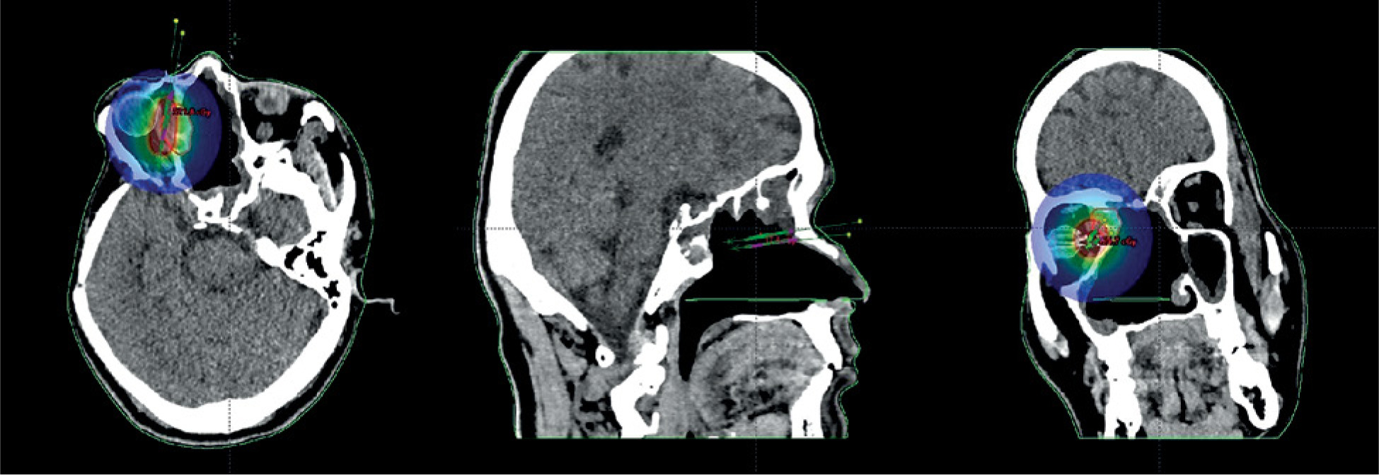
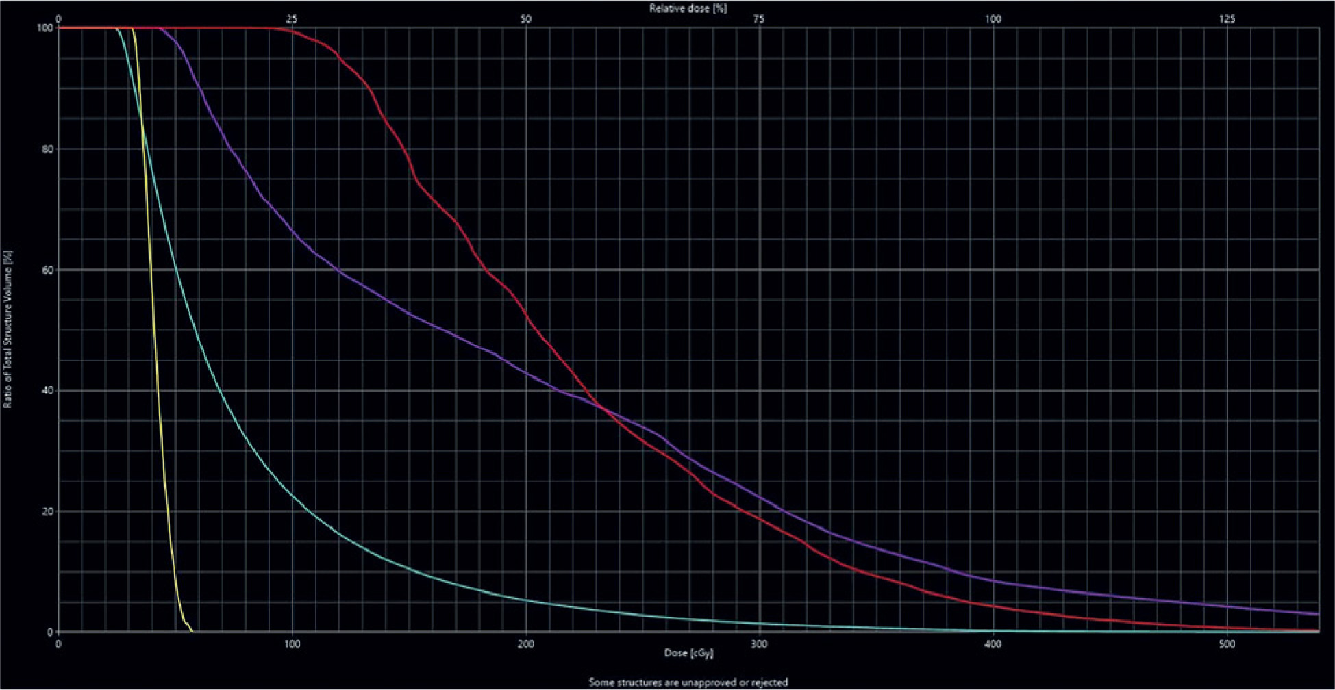
Treatment was planned to deliver 4 Gy per fraction in 2 fractions (total 8 Gy), with D90 covering 90% of the target volume, and an 8-hour interval between fractions. Dose optimization was performed to maintain OAR dose constraints (Figures 4, 5). The plan was executed using an HDR Iridium-192 (192Ir) source. Upon completion of treatment, the catheters were removed, and the patient was discharged. The procedure was well-tolerated without any acute complications.
Phase II: Stereotactic body radiotherapy
As part of the planned hybrid approach, SBRT was initiated one week after completion of brachytherapy. The intent was to cover residual disease regions, which might have been underdosed by ISBT due to the inverse square law. The patient was immobilized with an SRT head cast in the supine position, and a contrast-enhanced planning CT (1-mm slice thickness) was acquired from vertex to C5 vertebra.
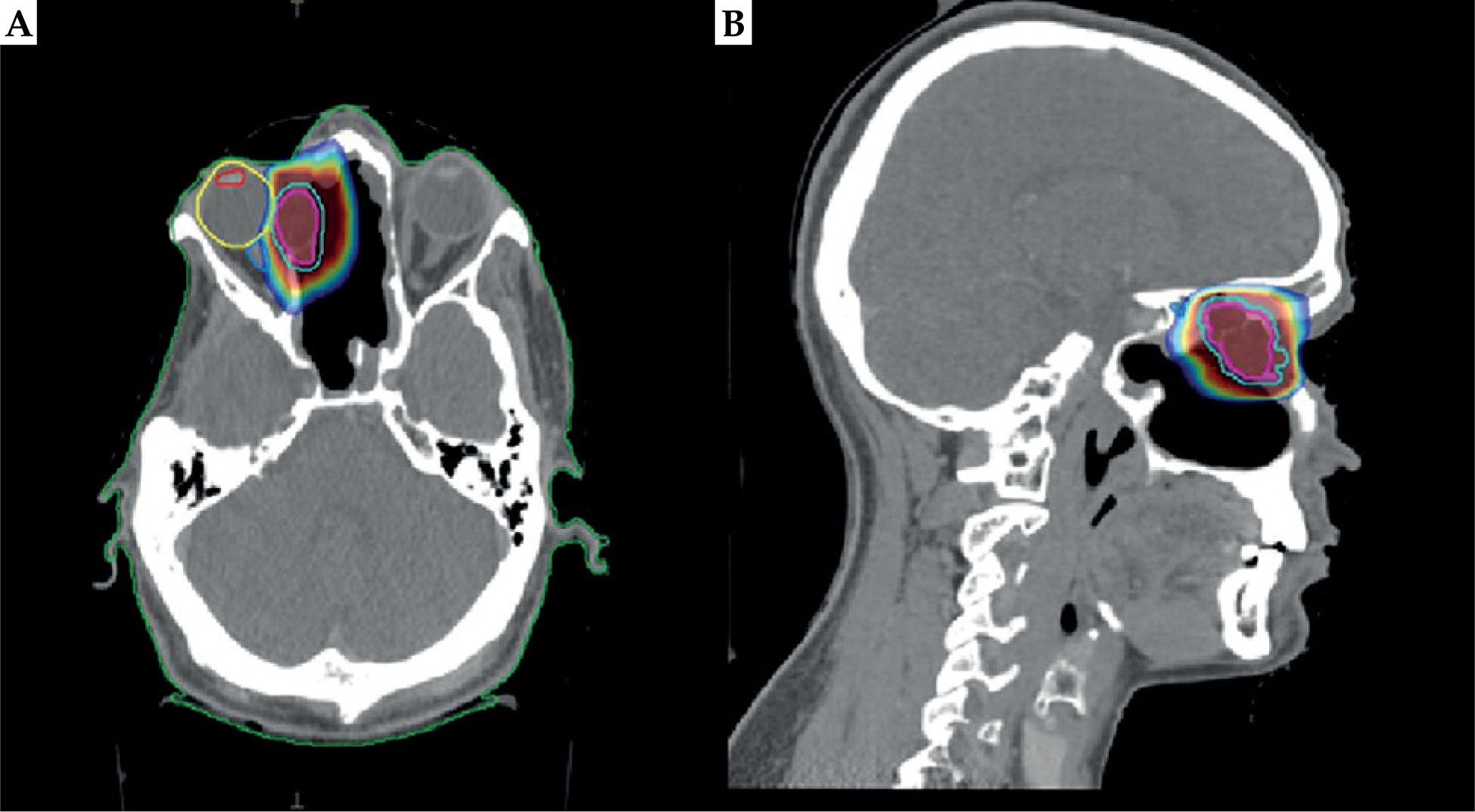
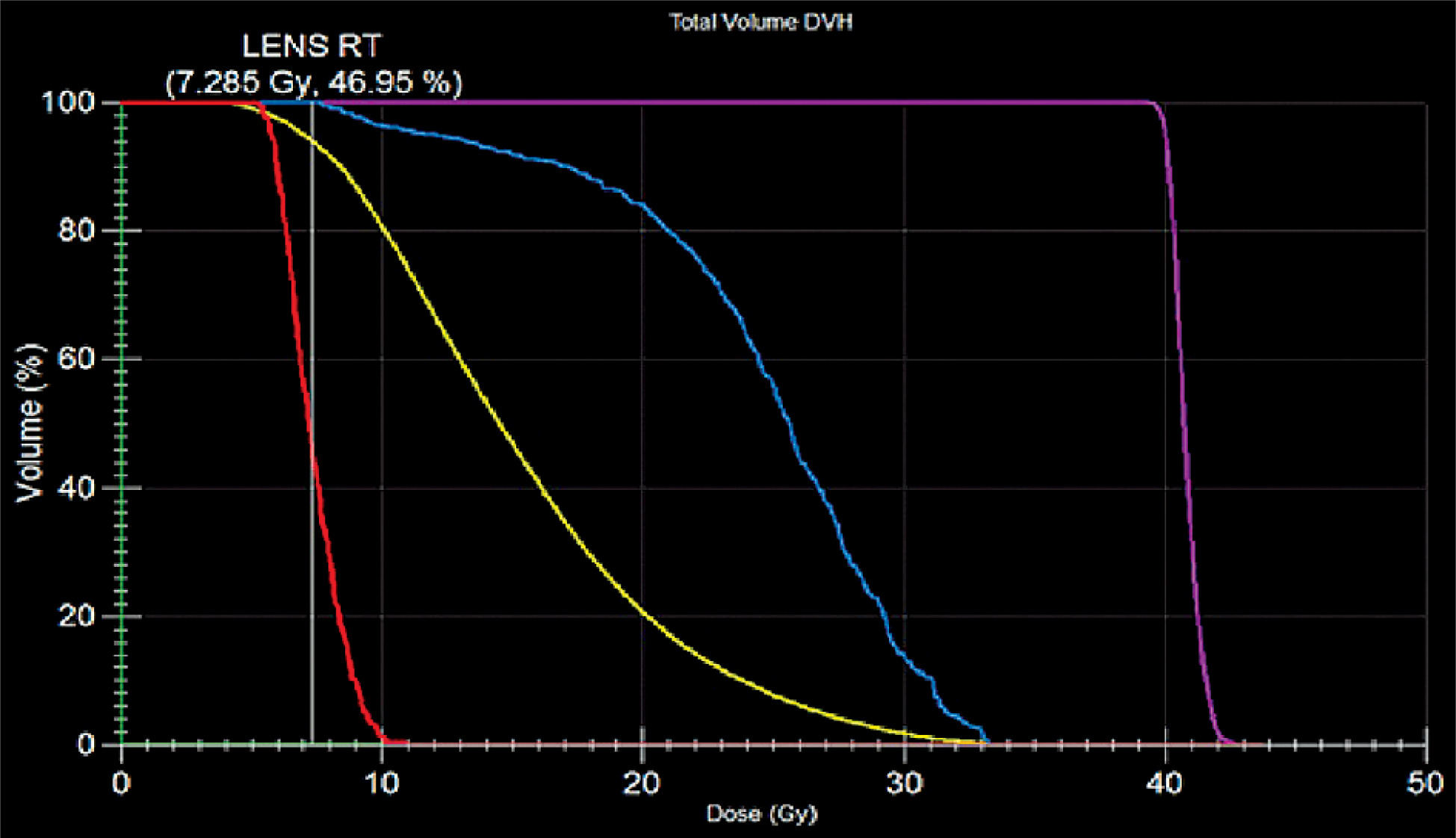
Gross tumor volume was defined as the contrast-enhancing residual lesion along the medial orbital wall, as visualized on pre-brachytherapy MRI. Planning target volume was generated with a uniform 2-mm margin around GTV. GTV for the SBRT phase was 4.5 cm3, covering both the medial and lateral components of the tumor. To prioritize the sparing of adjacent, previously irradiated OARs, a CTV expansion was omitted (Figure 6). This strategy was specifically chosen to deliver an ablative dose to the gross disease, while ensuring that the cumulative dose to the optic nerve remained within safe tolerance. The right eye, lens, and optic nerve were delineated as OARs. Treatment was planned to a dose of 40 Gy in 10 fractions using volumetric modulated arc therapy (VMAT) in Monaco v. 5.11 treatment planning system, achieving PTV D95 of 97% (Figure 7). SBRT was delivered once a day, five days per week. Daily image guidance using cone-beam computed tomography (CBCT) was performed before each fraction to verify target alignment and ensure precise dose delivery.
Results
Hybrid re-irradiation phase: ISBT and SBRT
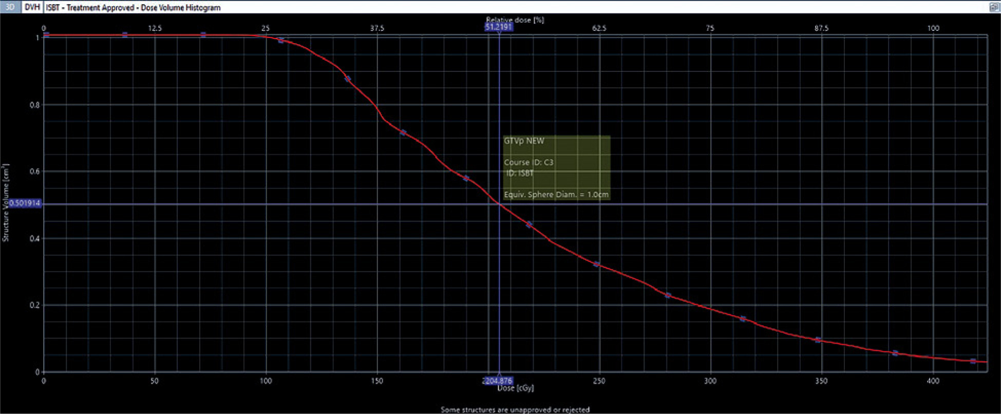
The patient completed the hybrid regimen without interruption. During the ISBT phase, the target volume achieved a D90 of 1.32 Gy per fraction. To ensure maximal sparing of the previously irradiated orbital apex, V100 was intentionally limited to 4.3%, representing a targeted boost of 2 Gy to the medial recurrence site, while the subsequent SBRT phase provided comprehensive coverage (Table 1 and Figure 8).
Table 1
Brachytherapy (ISBT) dosimetry
| ISBT parameter | Value |
|---|---|
| HR-CTV D90 | 1.32 Gy per fraction |
| HR-CTV V100 | 4.3%* |
| Dose homogeneity index (DHI) | 1% |
Fig. 8
DVH illustrates intentional medial-boost strategy for GTV. In this phase, V100 was restricted to 4.3% to deliver a localized 2 Gy boost to medial portion of tumor adjacent to orbital apex. This intentional underdosage in ISBT phase was designed to spare previously irradiated optic apparatus, remaining volumetric target coverage achieved during subsequent SBRT phase
In the SBRT phase (40 Gy in 10 fractions), the PTV achieved a D95 of 97.5%. The plan demonstrated high precision with a conformity index (CI) of 0.72, a heterogeneity index (HI) of 1.07, and a gradient index (GI) of 1.16, facilitating a sharp dose fall-off to protect adjacent critical structures (Table 2).
Cumulative dosimetry and OAR sparing
The hybrid re-irradiation course provided a cumulative physical dose of 41.82 Gy (ISBT D90 of 2.64 Gy, and SBRT D95 of 39.18 Gy). Due to the focal nature of the ISBT boost (V100 = 4.3%), a single cumulative tumor EQD2 was not derived. The cumulative lifetime EQD2 to the right optic nerve was 82.04 Gy (α/β = 3 Gy). High-precision gradients from the hybrid approach limited the current re-irradiation contribution to an EQD2 of 31.54 Gy (Table 3), maintaining the total exposure within acceptable limits for this high-risk salvage setting. No grade 3 acute toxicities were observed, while mild conjunctival irritation and transient dry eye symptoms were managed conservatively.
Table 3
Cumulative biological effective dose (EQD2) for OARs
Discussion
Recurrent ACC involving the orbit is clinically challenging due to its proximity to critical structures and prior high-dose irradiation [11]. In this case, a hybrid radiotherapy approach combining SBRT and template-guided ISBT achieved excellent local control with minimal toxicity. This strategy enabled delivery of an ablative dose to the residual tumor, while respecting cumulative dose constraints of nearby OARs, particularly the optic nerve and lens.
Re-irradiation of orbital ACC is primarily constrained by previous normal tissue exposure. In this case, the recurrence’s medial location near the optic nerve prevented safe dose escalation with SBRT alone, while ISBT monotherapy posed risks of lateral geographical misses. In order to resolve this, 3D-printed template-guided ISBT delivered a localized medial boost with a steep fall-off at the orbital apex, while SBRT ensured volumetric coverage of lateral margins and microscopic extensions. This spatial partition, medial escalation via ISBT, and lateral coverage via SBRT, offered a superior therapeutic index compared with monotherapy, achieving an ablative dose while maintaining cumulative OAR doses within safe thresholds.
The ISBT phase delivered a D90 of 1.32 Gy per fraction (EQD2 = 2.19 Gy), while the SBRT phase delivered D95 of 39.18 Gy (EQD2 = 57.96 Gy). Due to the heterogeneous target coverage of the ISBT boost (V100 = 4.3%), these biological doses were evaluated independently rather than as a single cumulative estimate. The cumulative EQD2 of the hybrid course to OARs remained within tolerance, i.e., optic nerve: 31.54 Gy, lens: 7.6 Gy, and right eye: 33.73 Gy. The one-year disease-free interval with no grade 3 toxicity supports the safety and feasibility of this hybrid strategy.
This approach aligns with emerging evidence, favoring highly conformal salvage techniques in recurrent ACC. The dosimetric advantage of brachytherapy in achieving steep dose gradients is well-documented. Chen et al. reported an 85.7% objective response rate and 100% disease control in 21 patients with recurrent head and neck adenoid cystic cancers, treated with iodine-125 (125I) stereotactic ablative brachytherapy using 3D-printed templates [12]. Similarly, Huang et al. presented 5-year local control and OS rates of 59% and 65%, respectively, in recurrent or unresectable ACCs [13]. In a larger series, Ji et al. [14] demonstrated that CT-guided 125I seed implantation achieved a 5-year local control rate of 26.6% in 101 recurrent cases. Our patient’s favorable outcome may be attributed to early salvage at a low residual tumor volume (1.8 cm × 1.6 cm × 1.2 cm).
While particle therapy has shown encouraging results, limited accessibility and high costs often preclude its use [15-17]. Hybrid SBRT-ISBT may offer a similar biological advantage by utilizing widely available, highly conformal technology. The integration of 3D printing in ISBT planning further enhances implant precision and reproducibility in anatomically complex regions.
In summary, this case reinforces the paradigm of individualized, conformal salvage re-irradiation in recurrent ACC. The combination of SBRT and template-guided HDR-ISBT, enables safe dose intensification within a high-risk orbital sub-site. This multidisciplinary approach represents a promising direction for extending curative potential in selected re-irradiation candidates.
Limitations, implications, and future directions
This case highlights the clinical utility of combining SBRT and template-guided ISBT for recurrent, previously irradiated orbital ACC, achieving dose escalation while respecting OAR constraints through image-guided planning and patient-specific 3D-printed applicators. A significant clinical challenge was the five-month interval between surgical excision and treatment completion, primarily due to multiple patient-related defaults. This delay resulted in interval of tumor progression, between the initial CT simulation and the eventual treatment delivery. Consequently, the custom 3D-printed template designed for a smaller, post-operative volume, provided incomplete coverage of the enlarged lesion. In this scenario, the hybrid addition of SBRT was essential to re-optimize dosimetric plan, providing necessary volumetric coverage for the expanded margins, while maintaining cumulative OAR doses within safe thresholds.
Despite the encouraging outcome, this report presents a single-patient experience with a relatively short follow-up. Long-term outcomes, including late recurrence and delayed radiation toxicities, such as optic neuropathy, remain to be evaluated. Furthermore, the broader applicability of this hybrid strategy depends on institutional resources, specifically expertise in interstitial brachytherapy, access to 3D printing for customized templates, and close multidisciplinary collaboration.
Future implementation should focus on adaptive image-guided planning and advanced radiobiological modeling to further personalize salvage therapy. Prospective studies and multi-institutional registries are warranted to define optimal dose-volume constraints, and evaluate long-term local control, late-effect profiles, and patient quality of life.
Conclusions
In the context of recurrent adenoid cystic carcinoma within a previously irradiated orbital region, this case illustrates the feasibility, safety, and efficacy of hybrid radiotherapy approach combining SBRT and template-guided ISBT. Through integration of MRI-based planning, 3D-printed patient-specific templates, and precision of dosimetric optimization, safe dose escalation was achieved while maintaining OAR constraints.
This tailored strategy resulted in complete radiologic response and absence of ≥ grade 3 toxicity, demonstrating that carefully planned hybrid re-irradiation can offer a potentially curative option in challenging, inoperable recurrences. Our experience reinforces the importance of multidisciplinary collaboration, advanced imaging, and individualized treatment design in extending the therapeutic window for patients with recurrent head and neck ACCs.











