
Purpose
The main therapeutic options for localized prostate cancer are surgery, radiotherapy, and hormonal therapy, alone or combined, according to risk group [1]. Despite definitive treatment, 20-50% of patients develop biochemical recurrence (BCR) within 10 years [2], defined as prostate-specific antigen (PSA) > 2 ng/ml above nadir post-radiotherapy (Phoenix criterion) or a PSA increase of 0.1-0.4 ng/ml above nadir after radical prostatectomy [3].
In patients with BCR after prostatectomy, early salvage radiotherapy achieves 5-year biochemical control rate above 85% [4-6], with benefits in overall survival and metastasis-free survival (MFS) at 15 years [7]. Despite this, a significant proportion of these patients develop a second recurrence, the risk of which depends on multiple prognostic factors, initial treatment, and use or omission of hormonal therapy [8].
Advanced imaging, with positron emission tomography (PET) and multiparametric magnetic resonance imaging (mpMRI), has improved early detection of local recurrence (LR). Choline PET shows sensitivity of 62-73% depending on PSA level [9], while PSMA PET demonstrates higher sensitivity at lower PSA values [10-13]. After radiotherapy, mpMRI accurately detects LR [14-16], supporting diagnostic and therapeutic planning through MRI fusion techniques [17-20].
Multiple salvage options exist for LR after external beam radiotherapy (EBRT), including radical prostatectomy, high-intensity focused ultrasound (HIFU), cryotherapy, stereotactic body radiotherapy (SBRT), and brachytherapy (BT), using either permanent ultra-low-dose-rate (uLDR) implants or high-dose-rate BT (HDR-BT), with comparable oncological outcomes.
Prognostic factors after salvage BT include PSA nadir after initial radiotherapy, disease-free interval, and PSA doubling time [21, 22], as highlighted in contemporary expert-endorsed reviews, such as the uroGEC consensus by Gomez-Iturriaga et al. [23].
Re-irradiation approaches can achieve acceptable genitourinary (GU) and gastrointestinal (GI) toxicity profiles in appropriately selected patients; however, reported outcomes vary substantially depending on technique, dose, fractionation, and patient selection [24-26].
Focal salvage BT targeting nodules in the prostate bed has been explored in small retrospective series with limited follow-up, reporting favorable oncological outcomes and acceptable toxicity [27-31].
Salvage BT has also been applied after prostatectomy and adjuvant or salvage EBRT. Small uLDR BT studies reported good safety [32-34], while two HDR-BT series (10 and 15 patients) with follow-up under 4 years described 3-year biochemical recurrence-free survival (BRFS) rates above 60% and grade ≥ 2 GU toxicity in approximately one third of patients [35, 36].
The aim of this study was to evaluate clinical and biochemical outcomes of HDR-BT for LR after surgery and adjuvant or salvage radiotherapy, and to explore potential prognostic factors in this complex clinical setting.
Material and methods
A retrospective observational study was conducted at the Radiation Oncology Department of Hospital Meixoeiro (Vigo, Spain), and included men older than 18 years treated between 2015 and 2024 for LR of prostate adenocarcinoma after radical prostatectomy and EBRT. Baseline staging to exclude distant metastatic disease was performed using choline PET. Patients had no evidence of distant disease, whereas LR was confirmed by pelvic MRI imaging and/or choline PET. Eligibility criteria included international prostate symptom score (IPSS) < 15, Eastern Cooperative Oncology Group (ECOG) performance status 0-2, anesthetic suitability, and informed consent.
Clinical target volume (CTV) was defined as a MRI-visible recurrent lesion within the prostatic bed, with a margin adapted to anatomical boundaries, based on pre-implant multiparametric MRI fused with real-time transrectal ultrasound. Urethra and rectum were considered organs at risk.
Follow-up was performed every 3-6 months during the first two years and annually thereafter, assessing PSA, imaging, and urinary and GI toxicity according to CTCAE v. 5.0. Imaging follow-up with MRI was performed based on clinical suspicion, PSA kinetics, or biochemical progression. Primary endpoint was local recurrence-free survival (LRFS), while secondary endpoints were BRFS, regional recurrence-free survival (RRFS), MFS, overall survival (OS), and toxicity.
Local recurrence-free survival was defined as the time from completion of salvage HDR-BT to radiological evidence of LR within the treated region. LR was assessed primarily by multiparametric MRI and/or PET imaging, based on clinical suspicion or after biochemical progression. Histological confirmation was not mandatory and was not systematically performed.
Biochemical recurrence-free survival was defined as the time from completion of salvage HDR-BT to biochemical recurrence, according to institutional criteria applied for the re-irradiation setting. RRFS was specified as the time from completion of salvage HDR-BT to detection of radiological evidence of LR outside the treated region and/or regional nodal disease on imaging. MFS was defined as the time from completion of salvage HDR-BT to detection of distant metastatic disease on imaging. OS was specified as the time from completion of salvage HDR-BT to death from any cause.
High-dose-rate BT was planned by fusing prior pelvic MRI with real-time transrectal ultrasound (Hitachi EUB-7000HV, Amertek BT stepper, and Elekta rotational system) for target and organs at risk delineation. Transperineal implantation was performed under ultrasound guidance using Elekta ProGuide® needles. A virtual plan was generated and adjusted in real-time in Oncentra Prostate®. Planning objectives were V100 (percentage of the target volume receiving at least 100% of the prescribed dose) > 95% and D90 (minimum dose received by 90% of the target volume) ≥ 100%, minimizing urethral dose, with intra-vesical hyaluronic acid administered as a protective measure. Treatment was delivered using an HDR MicroSelectron® system (Elekta, iridium-192 source) in two weekly fractions of 2 × 8.5 Gy or 2 × 10 Gy.
Statistical analysis included descriptive evaluation of clinical and dosimetric variables, Kaplan-Meier survival estimates, and comparisons using log-rank test, with p < 0.05 considered statistically significant. A stratified analysis was done to identify factors associated with local and/or BCR after salvage HDR-BT, evaluating pathological stage ≥ pT3, nodal involvement, positive surgical margins, and high-grade Gleason score. Patients were grouped as low-risk (one factor) or high-risk (≥ 2 factors), and oncologic outcomes were compared accordingly. All statistical analyses were performed using Python® (version 2.7). Log-rank tests were used to compare survival outcomes, BRFS, RRFS, MFS, and OS, between pre-defined sub-groups, such as Gleason score categories and risk groups. Given the limited sample size, all stratified assessments were considered exploratory in nature. The study protocol was approved by the Institutional Ethics Committee.
Results
Demographic characteristics and primary treatment
Seventeen patients treated with salvage HDR-BT between 2015 and 2024 were included in the study. The median age at prostate cancer diagnosis was 59 years (range, 45-69 years). The baseline characteristics are summarized in Table 1.
Table 1
Baseline characteristics of the study’s patients
In terms of primary treatment, 12 patients (70.6%) received EBRT with early salvage intent after prostatectomy, while 5 patients (29.4%) received adjuvant EBRT. The median dose delivered to the surgical bed was 66.36 Gy (range, 63.84-72 Gy) in a median of 28 fractions (range, 28-35 fractions). One patient received a boost up to 72 Gy due to residual disease.
Seven patients (41.2%) had pelvic nodal irradiation, performed in selected patients with high-risk disease features, according to institutional practice at the time of treatment, with a median dose of 46.39 Gy (range, 43.4-52.5 Gy). Three patients received concomitant ADT with EBRT, with a mean duration of 72 months and a median duration of 84 months (6, 84, and 126 months). The PSA nadir after EBRT was 0.055 ng/ml (range, 0.02-0.39 ng/ml) at a median of 14 months after completion of radiotherapy.
The median age at salvage HDR-BT was 69 years (range, 50-78 years). The median interval between initial EBRT and HDR-BT was 4 years (range, 1-14 years). Baseline staging to exclude distant metastatic disease was performed using choline PET in all but one patient, who was staged with conventional imaging. Local recurrence was identified on multiparametric MRI in all patients.
Salvage treatment and dosimetry
In the salvage HDR-BT setting, 9 patients (52.9%) received 2 fractions of 8.5 Gy, and in 8 patients (47.1%), 2 fractions of 10 Gy were delivered. The median CTV was 1.35 cm3 (range, 0.32-12.12 cm3). Dosimetric analysis showed a median V100 of 99.4% and a median D90 of 99.98%. The median maximum urethral dose was 110.45% of the prescribed dose.
The median time from HDR-BT to initiation of subsequent ADT was 15 months (range, 6-48 months). At the last follow-up, 13 patients were receiving ADT and 3 patients concomitant ADT. In all cases, ADT was initiated after salvage HDR-BT due to biochemical and/or radiological progression, and was not administered systematically at the time of salvage treatment. The median follow-up after HDR-BT was 74 months (range, 6-100 months). The mean PSA level before HDR-BT was 1.0 ng/ml. At 6 years, 11 patients (64.7%) remained recurrence-free within the HDR-BT-treated region, and 3 patients (17.6%) were disease-free. Eight patients (47.0%) developed loco-regional recurrence (6 local [35.2%] and 2 in other pelvic sites), and 7 patients (41.1%) developed metastatic disease. The management after recurrence included androgen receptor pathway inhibitors (ARPIs) in 3 patients, an LHRH agonist in 1 patient, SBRTs in 2 patients, and conventional EBRT in 1 patient.
Survival
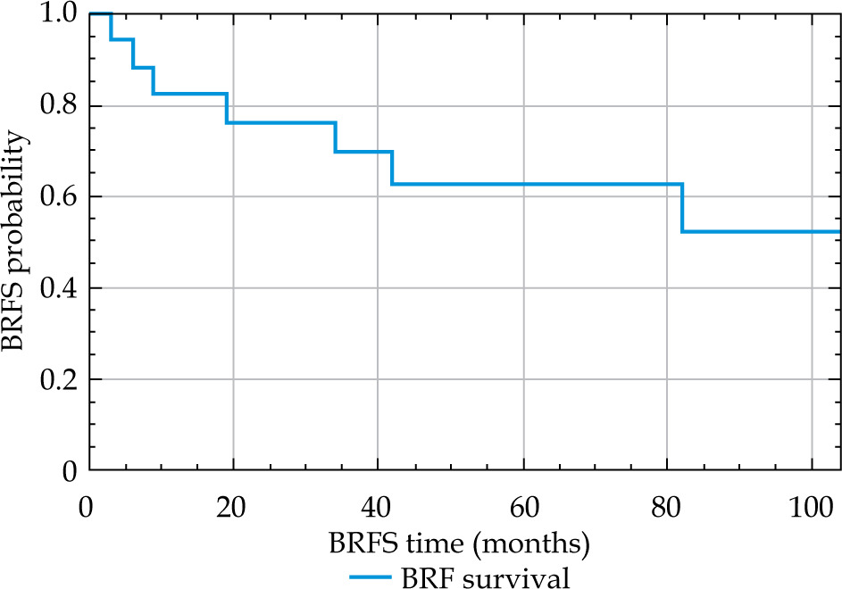
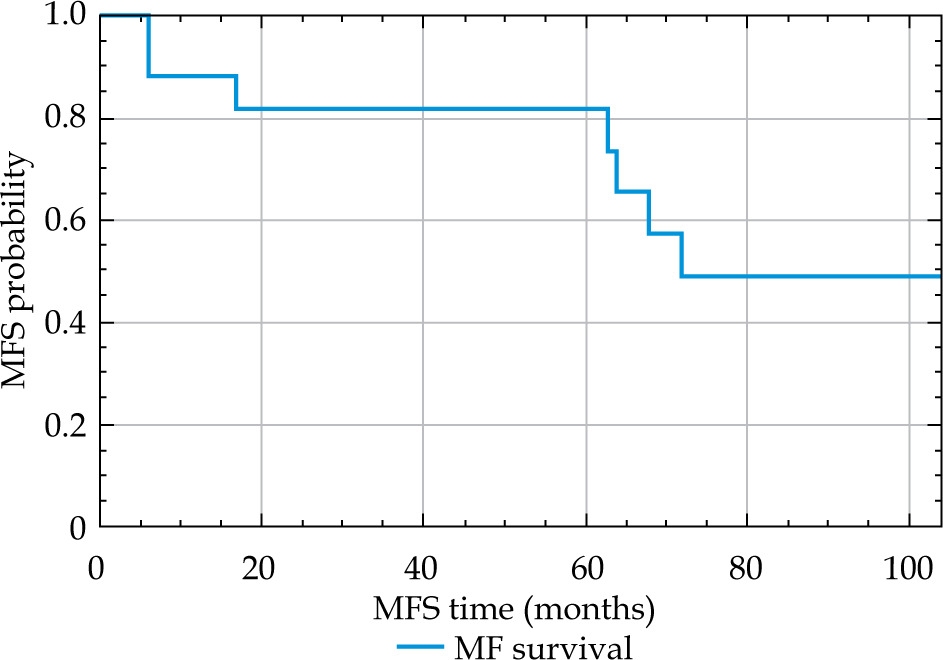
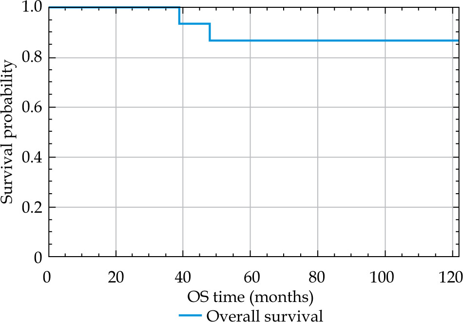
After a median follow-up of 74 months, 15 patients remained alive. BRFS was 69.7% at 36 months (95% CI: 42-86%), 62.7% at 60 months (95% CI: 35-81%), and 52.3% at 120 months (95% CI: 23-75%) (Figure 1). The median time to metastasis was 6.0 years. MFS was 81.9% at both 3 and 5 years (95% CI: 54-94%) and 49.2% at 10 years (95% CI: 21-72%) (Figure 2). No deaths were recorded at 3 years, yielding an OS of 100%. OS at both 5 and 10 years was 86.7% (95% CI: 56-96%) (Figure 3).
Analysis by Gleason score
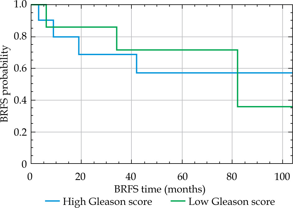
Comparison between high-grade (HG) and low-grade (LG) Gleason groups showed no statistically significant differences in oncological outcomes. BRFS in the HG group was 68.6% at 3, 5, and 10 years (95% CI: 21-91%). In the LG group, the median was 6.8 years, with probabilities of 70.0% at 3 years (95% CI: 33-89%), 58.3% at 5 years (95% CI: 23-82%), and 29.2% at 10 years (95% CI: 2-69%). Differences between groups were not statistically significant (p = 0.448) (Figure 4). Local recurrence-free survival did not reach the median in either group. The probability at both 3 and 5 years was 60.0% in the HG group (95% CI: 25-83%) and 71.4% in the LG group (95% CI: 26-92%), with similar values at 10 years. MFS did not reach the median in the HG group, while in the LG group, it was 72 months. The probability at both 3 and 5 years was 85.7% in the HG group (95% CI: 33-98%) and 80.0% in the LG group (95% CI: 41-95%). At 10 years, the probabilities were 51.4% and 48.0%, respectively. No significant differences were identified (p = 0.899). The log-rank test did not show significant differences (p = 0.483), and OS also did not reach the median in either group. At both 5 and 10 years, OS was 77.8% in the HG group (95% CI: 36-94%) and 100% in the LG group (p = 0.234).
Analysis by risk group
In this cohort, patients with one or two risk factors demonstrated comparable biochemical and LRFS, OS, and MFS during follow-up, with no statistically significant differences between risk groups across any of the analyzed endpoints.
Toxicity
Five patients (29.4%) developed grade 1 acute GU toxicity. The median time to peak acute GU toxicity was 3 months (range, 1-5 months), while the median time to peak chronic GU toxicity was 17 months (range, 8-27 months). One patient experienced grade 4 chronic GU toxicity. In this patient, for a prescribed dose of 8.5 Gy per fraction delivered in two fractions, the maximum point dose to the urethra was 103.58% in the first fraction and 112.43% in the second fraction. The recurrent lesion was located anterior to the urethra. Macroscopic hematuria developed one year after BT and persisted despite transurethral resection two years later, ultimately requiring radical cystectomy. All the remaining chronic GU toxicities were grade 2 or lower. No acute or late GI toxicities due to HDR-BT were observed.
Discussion
Salvage BT after radical prostatectomy and EBRT remains a relatively underexplored therapeutic modality, with limited evidence primarily derived from small retrospective series [26, 32-37]. Nevertheless, these studies demonstrate that the procedure is feasible and provides effective local control. Our study, based on patients treated with salvage HDR-BT after radical prostatectomy and external beam radiotherapy, provides a median follow-up of 6 years.
This represents a distinctive strength, as the available literature rarely exceeds a median follow-up of 48 months [35, 36]. In localized disease, most BCRs occur within the first 5-10 years [2]; thus, this extended follow-up offers a more reliable view of disease evolution and long-term treatment safety.
The dose schedules used in our patients (2 fractions of 8.5 Gy and 2 fractions of 10 Gy) were lower than those described in contemporary series [27, 35, 36, 38]. Despite this, our outcomes in terms of local control and BRFS are comparable, suggesting that less intensive hypofractionated approaches may be adequate while offering potential advantages in toxicity reduction. When this technique was first implemented at our center, dose standards ranged from 10 to 13 Gy per fraction. Therefore, our experience reflects an early phase in the shift towards more moderately hypofractionated regimens.
In the setting of permanent seed implants (uLDR BT), published studies show encouraging results, with favorable biochemical responses observed in patients in the short-term. High rates of local control and cancer-specific survival with minimal toxicity have also been described, although follow-up was limited in some studies [29, 31-34].
High-dose-rate BT has also been explored for LR after prostatectomy. Buchser et al. [27] reported a series of 13 patients treated with HDR-BT, with or without EBRT, guided by MRI-ultrasound fusion, achieving favorable biochemical responses in all cases and no significant late toxicity. Similarly, Ni et al. [30] analyzed 8 patients with palpable post-prostatectomy recurrence treated with HDR (delivered either as two fractions of 9.5 Gy or a single-fraction of 15 Gy, with or without EBRT), reporting 100% local control and 3-year BRFS of 43%, with only one patient experiencing grade ≥ 3 toxicity.
Soror et al. [35] demonstrated outcomes of 10 patients treated by salvage HDR-BT, with a median follow-up of 34 months, achieving biochemical relapse-free survival rates of 80%, 60%, and 60% at 1, 3, and 4 years, respectively. In comparison, our cohort showed probabilities of remaining free from BCR of 69.7% at 3 years, 62.7% at 5 years, and 52.3% at 10 years, indicating sustained long-term disease control and slightly higher BRFS rates than those reported in most previous series. The extended follow-up of up to 120 months enables evaluation of long-term biochemical control, something which earlier smaller studies with 20-49 months of follow-up could not provide.
On the basis of these single-institution series, our findings can also be contextualized within larger, multicenter experiences. Our results are consistent with those reported in the multicenter HDR-REPOPRA study [38], which analyzed 90 patients treated with HDR-BT after radical prostatectomy and adjuvant or salvage EBRT. In that series, the 5-year LRFS was 81%, BRFS was 63%, and grade ≥ 3 GU toxicity occurred in fewer than 9% of patients. OS at 72 months was 95.87% and MFS at 5 years was 77.46%, in line with our findings. The median time from HDR-BT to LR in the HDR-REPOPRA study was 37 months, while the median time to biochemical relapse was 31 months.
Our outcomes are similar in terms of safety and efficacy. After a median follow-up of 72 months, we observed a 6-year LRFS of 65%, loco-regional control of 64.7%, MFS of 82% at 5 years, and OS of 86.7% at 10 years.
Overall, our series reported only one case of grade 3 GU toxicity and no significant GI toxicity, which is consistent with previous studies and even lower than in series using higher per-fraction doses. The low incidence of severe adverse events supports the feasibility and safety of the regimens employed.
An important consideration in interpreting these results is the absence of consensus on defining BCR after multimodal approaches. The Phoenix criterion (an increase of 2 ng/ml above the PSA nadir), validated mainly for radiotherapy, may not accurately reflect PSA dynamics post-prostatectomy, highlighting the need for specific, standardized criteria in the prostatic bed re-irradiation.
At the time these treatments were performed, concomitant ADT was not systematically used. Current evidence, however, indicates that ADT improves MFS in the salvage setting [3]. In line with contemporary guidelines, patients considered for re-irradiation should receive ADT, particularly those with adverse features.
Although most patients eventually required androgen deprivation therapy, salvage HDR-BT provided a median ADT-free interval of 15 months. It should be noted that, under current evidence and treatment standards, all of the patients would likely have received ADT at the time of salvage therapy.
Advanced imaging is essential for patient selection and treatment planning. In our study, PET-MRI accurately identified recurrent nodules in the prostatic bed, optimizing CTV delineation while sparing healthy tissues. Previous research, such as Buchser et al. [27], have similarly shown the value of MRI-guided HDR-BT in salvage settings, with favorable biochemical outcomes. PSMA-PET demonstrates high sensitivity for detecting BCR, with detection rates increasing from ~45% at PSA < 0.5 ng/ml to > 90% at PSA ≥ 2 ng/ml, varying according to primary treatment [39, 40]. Local detection in the prostatic bed may be limited by urinary tracer activity; however, integration with multiparametric MRI overcomes this limitation by combining metabolic sensitivity with high tissue resolution, improving staging accuracy and CTV definition. Several studies, including the FORECAST trial [41, 42], confirmed that MRI-targeted biopsies reliably detect recurrence but may miss microscopic disease, supporting PET-MRI fusion for comprehensive mapping. This approach enables targeted salvage treatment while minimizing morbidity. Furthermore, multicenter data from the IAEA-PSMA study showed that PSMA-PET detected recurrence in most patients and influenced therapeutic decisions in over half of cases [43].
Overall, the combined use of PSMA-PET and mpMRI provides a robust strategy for accurate localization, patient selection, and personalized planning of salvage BT, reinforcing its role as a safe and effective option in well-selected patients.
This study has several limitations that should be considered when interpreting the results, including its retrospective design, limited sample size, absence of a control group, and heterogeneity in prior external beam radiotherapy (adjuvant versus salvage) as well as variability in the use and duration of ADT, which may represent a potential confounding factor for survival outcomes. In addition, imaging follow-up with MRI was performed based on clinical suspicion, PSA kinetics, or biochemical progression rather than at fixed pre-defined intervals. Therefore, time to LR should be interpreted with caution, as it may reflect detection timing rather than true biological onset. Nonetheless, to the best of our knowledge, this study provides one of the longest available follow-up series of salvage HDR-BT in patients previously treated with both radical prostatectomy and external beam radiotherapy, addressing a particularly underexplored clinical setting.
Conclusions
Salvage HDR-BT may represent a feasible option for selected patients with LR after radical prostatectomy and external beam radiotherapy. In this cohort, with the longest follow-up reported in the literature, the oncological outcomes observed are comparable to those reported in series using higher doses, suggesting that lower-intensity hypofractionated regimens may be sufficient to maintain therapeutic efficacy while minimizing toxicity. However, given the limited sample size and the frequent use of androgen deprivation therapy, definitive conclusions regarding the independent oncological efficacy of salvage HDR-BT cannot be drawn.
The absence of standardized criteria for defining BCR continues to hinder comparison across studies, emphasizing the need for a specific definition applicable to re-irradiation scenarios.
Also, the incorporation of advanced molecular imaging and its fusion with multiparametric MRI is expected to play an important role in future clinical practice and research protocols, as this approach may refine patient selection, improve target delineation, and optimize treatment personalization in this challenging clinical setting.











