Introduction
Mast cells play a crucial role in the regulation of immune responses and the maintenance of tissue homeostasis. As tissue-resident cells primarily localised in barrier sites, such as the skin and the mucosal surfaces of the respiratory and gastrointestinal tracts, they constitute an important component of the first line of defence against environmental stimuli. Their ability to rapidly respond to external signals and release a wide array of biologically active mediators makes them key players in both innate and adaptive immunity [1–4]. In recent years, increasing attention has been focused on the complex role of mast cells in pathological conditions [5]. While their involvement in allergic diseases is well established and primarily associated with IgE-mediated type I hypersensitivity reactions, their role in cancer is considerably more complex and highly context-dependent [2–4, 6, 7]. Mast cells can influence tumour development through interactions with the tumour microenvironment, modulation of immune responses, and participation in angiogenesis and tissue remodeling processes [3, 4, 8, 9].
Understanding the mechanisms underlying mast cell function in different disease contexts is significant not only for elucidating disease pathogenesis but also for identifying novel therapeutic targets. The analysis of shared molecular and cellular pathways may contribute to the development of more effective mast cell-targeted therapeutic strategies in both allergology and oncology [1, 10–13].
The aim of this review is to present the shared mechanisms of mast cell activity in allergic diseases and cancer, as well as their clinical significance.
Biology of mast cells
Mast cells originate from haematopoietic progenitor cells in the bone marrow, which migrate through the bloodstream to peripheral tissues, where they undergo final differentiation under the influence of local microenvironmental factors. A key role in this process is played by stem cell factor (SCF) and its receptor KIT (CD117), which regulate mast cell survival, proliferation, and maturation [10, 11, 14].
Mature mast cells exhibit significant phenotypic and functional heterogeneity depending on their tissue localisation. Among the main subtypes are MCT mast cells (tryptase-positive mast cells), which predominantly contain tryptase, and MCTC mast cells (tryptase- and chymase-positive mast cells), which contain both tryptase and chymase. These subtypes differ in their mediator profiles and biological functions [10, 15, 16].
Mast cells express a wide range of surface receptors that enable their activation. The most important of these is the high-affinity receptor for immunoglobulin E, FcεRI, whose cross-linking by antigen leads to rapid cellular activation. In addition, mast cells express receptors for cytokines and chemokines, as well as pattern recognition receptors (PRRs), including Toll-like receptors (TLRs), allowing them to participate in both innate and adaptive immune responses [10, 11, 17]. Upon activation, mast cells release a broad spectrum of mediators that can be classified into three main groups: preformed granule-associated mediators (e.g. histamine, tryptase, heparin), newly synthesised lipid mediators (e.g. prostaglandins, leukotrienes), and cytokines and chemokines (e.g. TNF-α, IL-4, IL-6). The ability to rapidly release these substances makes mast cells key effectors of the early immune response [6, 10, 15].
Mast cells in allergic diseases
Mast cells play a central role in the pathogenesis of allergic diseases, acting as primary effector cells in IgE-mediated type I hypersensitivity reactions [2, 6, 7]. This process begins with the binding of allergen-specific IgE antibodies to FcεRI receptors on the surface of mast cells. Subsequent exposure to the allergen leads to receptor cross-linking, which initiates intracellular signalling cascades resulting in mast cell degranulation [6, 18]. Mast cell degranulation represents a key event in allergic reactions and leads to the immediate release of mediators such as histamine, tryptase, and other pro-inflammatory factors. Histamine plays a major role in increasing vascular permeability, inducing smooth muscle contraction, and stimulating sensory nerve endings, thereby contributing to the typical symptoms of allergic responses [6, 18].
Mast cells are critically involved in the pathogenesis of various allergic diseases, including asthma, allergic rhinitis, and anaphylaxis. In asthma, they contribute to chronic airway inflammation and bronchial hyperresponsiveness, whereas in anaphylaxis, they are responsible for a rapid and systemic reaction that may be life-threatening [2, 6, 19].
A characteristic feature of mast cell-mediated responses in allergy is their rapid onset and high intensity, resulting from the presence of preformed mediators. In addition, mast cells participate in the late-phase allergic response through the production of cytokines and chemokines that recruit other inflammatory cells, such as eosinophils and T helper type 2 (Th2) lymphocytes [2, 7, 16]. Given that the mechanisms of mast cell activation and function in allergic diseases are well characterised and extensively discussed in the literature, they are not analysed here in detail. Instead, this section provides a concise overview to support comparison with their role in cancer. Comprehensive discussions of this topic are available in previous review articles [19–22].
Mast cells in cancer
In the context of cancer, mast cells represent an important component of the tumour microenvironment (TME), where they interact with both tumour cells and other stromal elements [3, 4, 15]. Their role is complex and depends on tumour type, disease stage, and local microenvironmental conditions. Mast cells may promote tumour progression through the secretion of pro-angiogenic factors, such as vascular endothelial growth factor (VEGF), which stimulate the formation of new blood vessels required for tumour growth. In addition, they contribute to extracellular matrix remodeling through the release of proteases, thereby facilitating tumour invasion and metastasis [3, 9, 23–27].
An important aspect of mast cell function is their influence on the immune system. By secreting immunosuppressive cytokines, such as IL-10 and TGF-β, mast cells can inhibit anti-tumour immune responses and promote tumour immune evasion [4, 8, 28–31]. These cytokines suppress the activity of cytotoxic T lymphocytes and natural killer (NK) cells, while also promoting the expansion of regulatory T cells (Tregs), thereby creating an immunosuppressive microenvironment that supports tumour growth. Additionally, mast cells can interact with myeloid-derived suppressor cells (MDSCs) and tumour-associated macrophages, further enhancing immune suppression. Conversely, mast cells may also exert anti-tumour effects, for example through the production of cytotoxic mediators and the enhancement of immune responses directed against tumour cells. They can release pro-inflammatory cytokines such as TNF-α and chemokines that recruit dendritic cells and cytotoxic lymphocytes to the tumour site, thereby promoting anti- tumour immunity. In certain contexts, mast cells may also enhance antigen presentation and support the activation of adaptive immune responses, highlighting their functional plasticity within the tumour microenvironment [4, 8, 26, 29, 30, 32].
Molecular mechanisms underlying mast cell function in allergic diseases and cancer
Mast cells contribute to the pathogenesis of both allergic diseases and cancer through a range of shared, evolutionarily conserved molecular and cellular mechanisms. A key feature of their function is the ability to rapidly respond to environmental stimuli, integrate signals from the surrounding microenvironment, and release a broad spectrum of biologically active mediators that modulate immune responses and disease progression [4, 10–12]. A central mechanism of mast cell activation is mediated by the high-affinity IgE receptor (FcεRI), whose cross-linking by antigen triggers intracellular signalling cascades involving tyrosine kinases such as Lyn and Syk. This leads to the phosphorylation of adaptor proteins and activation of downstream pathways, including mitogen-activated protein kinases (MAPK; ERK, JNK, p38) and phosphoinositide 3-kinase (PI3K)/Akt [11, 14, 17]. A key convergence point of these signalling pathways is the transcription factor NF-κB, which regulates the expression of genes involved in inflammation, cell survival, and cytokine production. NF-κB activation occurs in response to both IgE-dependent stimuli in allergic diseases and to signals present in the tumour microenvironment, such as pro-inflammatory cytokines and stress-related factors [4, 11, 33]. In cancer, additional receptors, including KIT (CD117), activated by stem cell factor (SCF), and Toll-like receptors (TLRs), play an important role. These receptors enable mast cells to respond to tissue damage signals and tumour-derived factors, thereby maintaining an activated phenotype within the tumour microenvironment [3, 4, 8].
Effector mechanisms of mast cells in allergic diseases and cancer
Degranulation and cytokine-mediated immunomodulation represent key, interconnected mechanisms of mast cell function in both allergic diseases and cancer. Degranulation is one of the hallmark processes of mast cell activation, leading to the rapid release of preformed mediators such as histamine, tryptase, and heparin, which exert potent effects on surrounding cells [6, 10, 15]. In allergic diseases, this process is responsible for the rapid onset of clinical symptoms, including increased vascular permeability, smooth muscle contraction, and activation of sensory nerve endings. In the context of cancer, these mediators may influence vascular permeability and extracellular matrix remodeling, thereby facilitating tumour cell migration and invasion [3, 4, 9]. In addition to preformed mediators, mast cells synthesise lipid mediators de novo (e.g. prostaglandins and leukotrienes), as well as cytokines and chemokines that sustain and amplify inflammatory responses [10, 15, 33].
Mast cells are also an important source of pleiotropic cytokines, including TNF-α, IL-4, IL-6, IL-10, and TGF-β [10, 15, 33]. In allergic diseases, these cytokines promote a Th2-type immune response, supporting IgE production and the recruitment of eosinophils [2, 7, 22]. In cancer, the same mediators may exert immunosuppressive effects; for example, IL-10 and TGF-β inhibit the activity of cytotoxic T lymphocytes and natural killer (NK) cells, thereby facilitating tumour immune evasion. TNF-α, depending on the context, may act either as a pro-inflammatory and anti-tumour factor or promote tumour progression through the induction of chronic inflammation [4, 8, 28]. The ability of mast cells to modulate immune responses is largely determined by their interactions with various immune cell populations, including T lymphocytes, dendritic cells, and macrophages, highlighting their role as key immunoregulatory cells in both disease contexts [10, 15, 23, 24].
Cellular interactions, angiogenesis, and tissue remodeling
Cellular interactions and microenvironmental regulation represent key aspects of mast cell function in both allergic diseases and cancer. Mast cells act as integrators of signals derived from the microenvironment and propagate these signals through complex networks of cellular interactions, including interactions with T lymphocytes [10, 11, 15]. In allergic diseases, they interact with Th2 lymphocytes, eosinophils, and epithelial cells, thereby amplifying inflammatory responses [2, 17]. Within the tumour microenvironment, mast cells interact with tumour cells, cancer-associated fibroblasts (CAFs), endothelial cells, and immune cells, forming a dynamic signalling network that supports tumour cell survival, angiogenesis, and immunosuppression [3, 4, 8, 28].
A closely related mechanism is the ability of mast cells to promote angiogenesis and tissue remodeling. Mast cells secrete pro-angiogenic factors, such as vascular endothelial growth factor (VEGF), fibroblast growth factor 2 (FGF-2), and tryptase, which stimulate endothelial cell proliferation and the formation of new blood vessels [3, 4, 9, 23, 26, 27]. Although angiogenesis is more prominent in tumour progression, similar processes may also occur in chronic inflammatory conditions, including allergic diseases, where tissue remodeling and increased vascular permeability are observed [2, 13, 15]. In addition, mast cells contribute to extracellular matrix remodeling through the release of proteases, such as tryptase and chymase, thereby influencing tissue structure and cell migration [3, 4, 19].
Integration of mechanisms – a unified model
Analysis of the mechanisms described above indicates that mast cells function as central effector and regulatory cells, integrating immunological, inflammatory, and environmental signals [10, 11, 15]. In both disease contexts, the following key mechanisms can be identified:
Despite distinct clinical outcomes – namely allergic reactions or tumour progression – the fundamental mechanisms of mast cell function exhibit substantial similarities. These differences are primarily determined by the microenvironmental context and the nature of dominant regulatory signals [3, 4, 10]. A schematic overview of these mechanisms is presented in Figure 1.
FIGURE 1
Shared and distinct mechanisms of mast cell function in allergic diseases and cancer. The schematic illustrates mast cells as central effector cells integrating immunological and environmental signals. In allergic diseases (left panel), mast cell activation occurs via IgE-dependent cross-linking of the high-affinity FcεRI receptor, leading to activation of Lyn and Syk kinases and downstream MAPK signalling pathways (ERK, JNK, p38). This results in rapid degranulation and the release of mediators such as histamine, tryptase, and leukotrienes, contributing to bronchoconstriction, increased vascular permeability, and Th2-type inflammation. In the tumour microenvironment (right panel), mast cells are activated in a context-dependent manner via KIT (CD117), Toll-like receptors (TLRs), and cytokines. This leads to activation of NF-κB, PI3K/ Akt, and MAPK signalling pathways and the release of mediators such as VEGF, TGF-β, and IL-10, promoting angiogenesis, immunosuppression, and extracellular matrix remodeling. The central panel highlights shared mechanisms of mast cell function, including receptor-mediated activation (FcεRI, KIT, TLRs), activation of key signalling pathways (NF-κB, MAPK, PI3K/Akt), cytokine secretion (e.g. TNF-α, IL-6), de novo synthesis of lipid mediators, and extracellular matrix remodeling via proteases (tryptase, chymase). Created based on [1–4, 10, 11, 15]
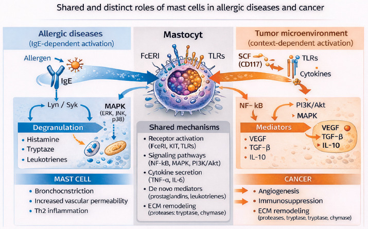
Table 1 illustrates a unified mechanistic model of mast cell function, highlighting both shared pathways and context-dependent differences between allergic diseases and cancer.
TABLE 1
Comparison of mast cell function in allergic diseases and cancer
Clinical significance and future perspectives
Given their significant role in the pathogenesis of both allergic diseases and cancer, mast cells represent a promising therapeutic target [1, 10, 19]. In allergic conditions, treatment strategies include mast cell stabilisers that inhibit degranulation, as well as agents that block the effects of released mediators, such as histamine receptor antagonists [1, 6].
In recent years, increasing attention has been directed toward the potential application of mast cell-targeted therapies in oncology. These approaches include the inhibition of surface receptors, such as KIT, as well as the modulation of mediator release affecting the tumour microenvironment [3, 4, 19, 27].
Future research should focus on the development of targeted therapeutic strategies that enable selective modulation of mast cell function depending on the disease context, which may improve treatment efficacy while minimising off-target effects. Moreover, the integration of molecular and clinical data may enhance the understanding of mast cell heterogeneity and their context-dependent roles in different pathological conditions, facilitating the identification of reliable biomarkers and supporting the advancement of personalised medicine. A deeper insight into the complex biology of mast cells and their interactions within the tissue microenvironment will be essential for the development of novel therapeutic strategies in both allergic diseases and cancer.
Discussion
The role of mast cells in allergic diseases is relatively well established and supported by a substantial body of experimental and clinical evidence. Their function as key effector cells in IgE-mediated type I hypersensitivity reactions is well defined, and the underlying mechanisms, including FcεRI activation, degranulation, and mediator release, have been extensively characterised [2, 7, 10].
In contrast, the role of mast cells in cancer remains considerably more complex and less clearly defined. The available literature presents conflicting findings regarding their impact on tumour progression. Some studies suggest a pro-tumourigenic role associated with angiogenesis, immunosuppression, and extracellular matrix remodeling [3, 9, 19, 26, 32, 34, 35], whereas others indicate potential anti-tumour activity, mediated by the enhancement of immune responses and the production of cytotoxic mediators [4, 15, 36].
These discrepancies likely reflect differences in tumour type, microenvironment, and mast cell phenotype. First, the type of tumour appears to be a critical determinant because the presence of mast cells has been associated with poor prognosis in certain cancers, such as pancreatic cancer and melanoma, while in others it may correlate with improved immune responses [23, 28]. Second, the localisation of mast cells within the tumour is of importance because stromal mast cells may exert different effects compared to those infiltrating tumour parenchyma [3, 8]. Third, phenotypic heterogeneity and the activation state of mast cells may significantly influence their biological function [11, 16, 37].
Increasing attention has been directed toward the tumour microenvironment as a key factor shaping mast cell activity. Interactions with tumour cells, fibroblasts, and immune cells create a dynamic signalling network that may drive mast cells toward either pro-tumourigenic or anti-tumour phenotypes [3, 4, 16, 38, 39]. In this context, mast cells should be considered not as a uniform population of effector cells, but rather as versatile regulators within a complex biological system [10, 11]. Another important issue is the methodological variability among studies investigating mast cells. Differences in experimental models, detection techniques, and markers used to identify mast cells may contribute to inconsistent findings and limit direct comparisons across studies [11, 33]. The lack of standardisation remains a significant challenge in this field.
Despite these limitations, accumulating evidence indicates that mast cells play an important role in both inflammatory and neoplastic processes. Their ability to integrate multiple signals and modulate the tissue microenvironment highlights their potential as therapeutic targets. However, further research is required to better understand their context-dependent functions and to translate this knowledge into clinical applications [10, 40].
Conclusions
Mast cells play a crucial role in the pathogenesis of both allergic diseases and cancer, functioning as both effector and regulatory cells of the immune response [5, 10, 18]. In both disease contexts, their activity is based on shared mechanisms, including degranulation, the release of inflammatory mediators, and the modulation of other immune cell functions [10, 15, 16]. In allergic diseases, the role of mast cells is well defined and closely associated with IgE-mediated type I hypersensitivity reactions [2, 10, 13]. In contrast, in cancer, their function is highly context-dependent and may involve both pro- and anti- tumour activities, influenced by interactions within the tumour microenvironment and the phenotypic heterogeneity of these cells [3, 4, 8].
From a clinical perspective, mast cells represent a promising therapeutic target [1, 19]. Strategies aimed at modulating their activity has potential applications in both allergic and oncological settings [1, 3, 19]. However, their successful translation into clinical practice requires further research, particularly in the identification of reliable biomarkers and a better understanding of the context-dependent functions of mast cells [10, 40].