Purpose
Non-melanoma skin cancer (NMSC), encompassing primarily basal cell carcinoma (BCC) and cutaneous squamous cell carcinoma (SCC), stands as the most frequently diagnosed malignancy in Caucasian populations worldwide [1]. While exact figures are difficult to ascertain due to underreporting to central cancer registries, its incidence is known to be rising annually, representing a significant and growing global healthcare burden [2]. Despite a generally favorable prognosis with low rates of metastasis and mortality, the sheer volume of cases imposes substantial demands on healthcare systems [1, 2]. A critical feature of NMSC is its predilection for sun-exposed areas, as over 95% of cases occur in the head and neck region, including the nose, ears, eyelids, and lips. This anatomical distribution fundamentally elevates the importance of treatment outcomes beyond mere oncologic control, placing a profound emphasis on the preservation of function and cosmetic appearance [1-3].
The cornerstone of curative-intent treatment for NMSC is surgical excision. Mohs micrographic surgery, in particular, is considered the gold standard for many high-risk or cosmetically sensitive lesions, boasting exceptional local control rates of 95% to 99% [4]. However, surgery alone may be insufficient for a subset of tumors harboring high-risk features that predict a greater likelihood of local or regional recurrence [2]. In these instances, post-operative adjuvant radiotherapy serves as a critical component of a multidisciplinary management strategy to eradicate microscopic residual disease and improve loco-regional control [5].
The generally accepted indications for adjuvant radiotherapy in the management of NMSC are well-established and form the clinical context for this review. One primary indication is positive or close surgical margins, which applies when a tumor is present at the inked margin of resection, or when the margin is narrow (typically < 1-2 mm) and further surgical re-excision is not feasible or would result in unacceptable morbidity [6]. Additionally, high-risk pathologic features serve as a strong indication. The presence of specific histologic characteristics is associated with a more aggressive tumor biology and an increased risk of recurrence; these features prominently include perineural invasion (PNI), particularly of large-caliber nerves or with clinical symptoms, lymphovascular space invasion (LVSI), poorly differentiated histology, and deep invasion into underlying structures, such as muscle, cartilage, or bone [4, 7]. Finally, recurrent disease warrants intervention, as tumors that have recurred after a previous treatment modality are often considered for adjuvant radiotherapy after re-excision to address the higher risk of subsequent relapse [2].
For adjuvant treatment, two principal radiotherapy techniques are commonly employed: external beam radiotherapy (EBRT) with electrons and high-dose-rate (HDR) brachytherapy, which encompasses a rapidly evolving spectrum of techniques [8, 9]. Electron beam therapy is a long-established and widely accessible form of EBRT utilized for treating superficial targets. Electrons are subatomic particles, which deposit their energy over a finite and predictable range in tissue, followed by a rapid dose fall-off. This characteristic makes them suitable for treating the skin and subcutaneous tissues while relatively sparing deeper structures [8, 10-12]. However, the physical properties of electrons also present several clinical challenges. Low-energy electron beams exhibit a “build-up” effect, where the maximum dose (Dmax) is deposited several millimeters below the skin surface, resulting in relative skin sparing. Furthermore, treating small or irregularly shaped fields with electrons can be dosimetrically complex. The construction of custom lead cutouts to shape the beam can be cumbersome, and electron beams are subject to dose limitation and output variations in small fields, which can introduce dosimetric uncertainty [8, 11, 12].
Alternatively, HDR brachytherapy is a highly conformal radiation technique. Historically relying on handcrafted surface moulds (SMB), the field has expanded significantly to address complex anatomies and varying tumor depths [13, 14]. The defining physical characteristic of brachytherapy is its adherence to the inverse square law, resulting in an extremely sharp dose gradient [14, 15]. This allows the delivery of a high, therapeutic dose to the target volume while minimizing exposure to adjacent healthy tissues and underlying organs at risk (OARs) [4, 13, 16].
An evaluation of contemporary NMSC management requires consideration of both established and emerging brachytherapy modalities [17]. Although surface mould brachytherapy (SMB) remains clinically relevant, three-dimensional (3D)-printed brachytherapy is being increasingly utilized [18]. Derived from patient-specific computed tomography (CT) or surface scanning data, 3D-printed applicators achieve conformal apposition with irregular anatomical contours; this minimizes air gaps and improves dosimetric homogeneity relative to manual moulds [19]. Moreover, interstitial brachytherapy remains indicated for thicker or deeply invasive lesions (> 5 mm depth), where surface approaches may not deliver an adequate dose to the deeper target volume [20]. For complex post-operative facial defects necessitating surgical reconstruction, CT-based flap brachytherapy employs specialized applicators positioned directly over or within the reconstructed tissue, facilitating targeted adjuvant radiation delivery [21].
The primary objective of this systematic review was to critically appraise and synthesize the available evidence addressing whether this broader spectrum of modern HDR brachytherapy (including surface, 3D-printed, flap, and interstitial techniques) has clinically significant improvements in local control and cosmetic outcomes than EBRT with electrons in the adjuvant setting for surgically excised NMSC.
Material and methods
This systematic review was conducted in accordance with the preferred reporting items for systematic reviews and meta-analyses (PRISMA) guidelines. A literature search was performed across five electronic databases and publisher platforms (i.e., PubMed, Google Scholar, Springer, Semantic Scholar, and Wiley Online Library), identifying relevant literature published up to May 7, 2026; the search was updated on that date during revision of the manuscript. To ensure comprehensive coverage of established and contemporary treatment techniques, database-specific search strings combined terms related to non-melanoma skin cancer with intervention terms for surface mould or surface brachytherapy, HDR brachytherapy, 3D-printed or customized applicator brachytherapy, flap brachytherapy, interstitial brachytherapy, electronic skin surface brachytherapy, external beam radiotherapy, electron beam therapy, and adjuvant radiotherapy. Outcome-related terms included local control, cosmetic outcome, toxicity, organ preservation, and dosimetry. Boolean operators and MeSH/free-text terms were adapted to each database’s syntax. The complete search strategy and number of records retrieved from each source are summarized in Table 1.
Table 1
Article search strategy
Two reviewers (W.K.A. and H.) independently selected studies by initially screening titles and abstracts, followed by full-texts assessment. Disagreements were resolved by consensus or consultation with a third reviewer (R.). Eligible research included original clinical or dosimetric studies evaluating modern brachytherapy modalities and/or EBRT in patients with NMSC. Both definitive and post-operative/adjuvant treatment settings were considered, and comparative evidence was extracted where available. Excluded were reviews, editorials, non-human or phantom-only studies without clinically relevant outcomes, single-case reports without extractable cohort data, non-NMSC studies, non-brachytherapy studies, duplicate or overlapping publications, or conference abstracts without sufficient extractable data. Methodological quality and risk of bias of the included studies were evaluated using the Joanna Briggs Institute (JBI) critical appraisal checklist, while level of evidence was classified according to the Oxford Centre for Evidence-Based Medicine (CEBM) guidelines.
Results
Study selection and modality characteristics
Following implementation of the expanded inclusion criteria, the updated literature search yielded 23 studies for the final synthesis (Figure 1). The revised cohort encompassed a broader spectrum of advanced brachytherapy modalities utilized in the management of non-melanoma skin cancer (NMSC). The included studies evaluated SMB (n = 6), interstitial brachytherapy (n = 5), 3D-printed applicator brachytherapy (n = 5), flap brachytherapy (n = 2), electronic brachytherapy (n = 1), and comparative or dosimetric analyses of HDR brachytherapy vs. EBRT (n = 4). Modality selection across the literature was primarily determined by tumor depth, complex anatomical topography, and institutional infrastructure (Table 2) [8, 13, 16, 19, 21-39].
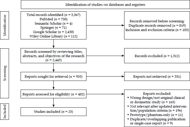
Table 2
Study characteristics, oncological efficacy, cosmetic outcomes, and toxicity profiles (n = 23)
| Author [Ref.] | Study design and modality | Sample size | Local control (LC) | Cosmesis and quality of life (QoL) | Toxicity profile |
|---|---|---|---|---|---|
| Bandyopadhyay et al. [22] | Retrospective; CT-based surface mould | 28 | 100% (long-term follow-up) | Rated excellent/good in all cases | Mild acute toxicity; negligible late complications |
| Brovchuk et al. [23] | Retrospective; interstitial and surface | 751 (534 BCC, 217 SCC) | > 96% complete response | Good to satisfactory | Severe complications (e.g., skin necrosis) < 2% |
| Budrukkar et al. [16] | Retrospective; surface mould | 35 | 92% for skin lesions (5-year) | 88% good cosmesis | Grade 3 toxicities rarely observed |
| Budrukkar et al. [24] | Retrospective; periocular interstitial | N/A (periocular cohort) | > 90% (high organ preservation) | Excellent structural preservation | Optimal lens sparing; radiation cataracts avoided |
| Casey et al. [25] | Retrospective; surface mould | 59 (65 lesions) | 98% (median follow-up: 1-2 years) | Good in > 90% of patients | Predominantly acute grade 1-2 erythema |
| Chatzikonstantinou et al. [26] | Prospective; 3D-printed mould | N/A (early clinical phase) | Maintained with high conformality | Precise facial contour adaptation | Minimized due to strict OARs avoidance |
| Cisek et al. [27] | Retrospective; periocular interstitial | N/A | > 90% long-term control | Excellent | Minimal corneal/lens toxicity |
| Diefenhardt et al. [28] | Retrospective; individual 3D moulds | N/A | High oncological efficacy | Excellent anatomical fit | Tolerable grade 1-2 acute skin reactions |
| Esmati et al. [29] | Retrospective; surface mould | N/A (head and neck tumors) | Comparable with standard modalities | Acceptable cosmesis | Transient localized cutaneous side effect |
| Gogineni et al. [21] | Retrospective; flap brachytherapy | 50 (53 lesions) | > 95% (15-month follow-up) | Satisfactory on curved surfaces | Severe toxicity rare in sensitive facial areas |
| Haseltine et al. [30] | Comparative; HDR vs. EBRT | 61 | Equivalent between HDR and EBRT | Marginal cosmetic trend favoring HDR | Similar safety profile, shorter HDR treatment time |
| Hommadi et al. [31] | Case series; flap applicator | N/A | No early local recurrence | Good | Acute skin reactions resolved promptly |
| Krengli et al. [32] | Retrospective; eyelid interstitial | 60 | 95% (5-year follow-up) | 92% good/excellent cosmesis | Epiphora and eyelid retraction uncommon |
| Kuo et al. [33] | Prospective; electronic brachytherapy | 34 | 100% (short-term follow-up) | High QoL and cosmetic satisfaction | Mostly grade 1, with rapid resolution within 12 weeks |
| Licher et al. [19] | Dosimetric; 3D-printed moulds | 42 | Dosimetric study; optimal PTV | Theoretically excellent based on fit | Significant dose reduction to healthy tissues |
| Monge-Cadet et al. [34] | Retrospective; facial HDR | 67 | > 90% intermediate-term LC | QoL maintained | Low incidence of late toxicity |
| Placidi et al. [35] | Dosimetric; HDR vs. EBRT/VMAT | N/A | N/A (dosimetry focused) | N/A | HDR demonstrated superior dose conformality |
| Poltorak et al. [36] | Implementation; 3D printing | 5 | Precise target volume optimization | High adaptation for irregular lesions | Enhanced OARs protection via 3D precision |
| Prasiko et al. [37] | Case series; surface brachytherapy | 5 | Controlled at 40.5 Gy | Good | Expected erythema resolved in the first month |
| Rio et al. [38] | Retrospective; facial interstitial | 97 | > 90% (periorificial areas) | 85-90% satisfactory | Low risk of cartilage or bone necrosis |
| Sen et al. [8] | Dosimetric; EBRT vs. mould | 10 | N/A (dosimetry focused) | N/A | Mould therapy provided steeper dose fall-off |
| Vyas et al. [39] | Comparative; EBRT vs. HDR | 99 | EBRT: 98.7%, HDR: 100% | Equivalent cosmetic outcomes | HDR enabled greater hypofractionation (6 vs. 17) |
| Yadavalli et al. [13] | Retrospective; 3D surface mould | 15 | 90% complete response | Maintained QoL (DLQI scores) | Cutaneous toxicity effectively managed |
Synthesis of findings
Oncological efficacy and local control
Analysis of the extracted data indicated that brachy-therapy, across its modern technical iterations, provided sustained local control for NMSC. The reported local control rates consistently ranged between 90% and 100%, although with heterogeneous follow-up durations across the cohorts. Interstitial brachytherapy was specifically indicated for lesions with an invasion depth exceeding 5 mm or those situated in complex periorificial zones (Table 3).
Table 3
Analytical summary of oncological efficacy and local control by modality
| Brachytherapy modality | Representative studies [Ref.] | Local control (LC)/efficacy rates | Primary clinical indications and efficacy notes |
|---|---|---|---|
| Interstitial brachytherapy | Brovchuk et al. [23] Rio et al. [38] Krengli et al. [32] | > 96% complete response (Brovchuk); > 90% LC in periorificial zones (Rio); 95% at 5-year follow-up (Krengli) | Highly efficacious for nodular or infiltrative lesions with an invasion depth > 5 mm; demonstrates sustained efficacy in complex topographies (e.g., the nasal ala, periocular) |
| Surface mould and 3D-printed brachytherapy | Bandyopadhyay et al. [22] Budrukkar et al. [16] Casey et al. [25] | 100% LC in long-term follow-up (Bandyopadhyay); 98% LC at median 1-2 years (Casey); 92% at 5 years (Budrukkar) | Optimal for superficial lesions (≤ 5 mm depth); 3D printing ensures precise target volume coverage, mitigating the risk of marginal recurrences |
| Flap brachytherapy | Gogineni et al. [21] Hommadi et al. [31] | > 95% LC at 15-month follow-up (Gogineni); zero early local recurrences reported (Hommadi) | Effective for extensive, curved superficial lesions; provides uniform dose distribution across broad anatomical planes |
| Comparative: HDR vs. EBRT | Vyas et al. [39] Haseltine et al. [30] | HDR: 100% vs. EBRT: 98.7% (Vyas); equivalent LC rates between modalities (Haseltine) | HDR brachytherapy demonstrates non-inferior oncological efficacy compared with EBRT, with the added operational advantage of profound hypofractionation |
In these challenging presentations, interstitial techniques yielded complete response rates exceeding 90%, as evidenced in large-cohort analyses [23, 38]. For superficial lesions, flap brachytherapy and customized 3D-printed moulds demonstrated recurrence-free intervals comparable with conventional surgical excision and EBRT. Furthermore, comparative studies matching HDR brachytherapy against EBRT [30, 39], identified no statistically significant differences in local control, establishing the non-inferiority of brachytherapy while simultaneously offering the clinical advantage of accelerated, hypofractionated treatment schedules.
Cosmesis, quality of life, and anatomical preservation
Cosmetic outcomes and preservation of anatomical function remain critical metrics in the management of NMSC, given the predominant facial location of these malignancies. The integration of individualized 3D printing technology in applicator fabrication has mitigated historical challenges associated with anatomical irregularities (e.g., the nasal ala and auricle). This advancement minimizes air gaps, ensures uniform source-to-skin distance, and optimizes dose conformality, thereby translating into high rates of clinician- and patient-reported cosmetic satisfaction [26, 28, 36].
In the context of periocular lesions, interstitial brachytherapy facilitated exceptional rates of organ preservation. Studies specific to the eyelid and canthal regions documented minimal radiation exposure to the underlying cornea and lens. This precision effectively circumvents the risk of radiation-induced cataracts while maintaining structural integrity and preventing epiphora [24, 27, 32]. Validated quality of life assessments, such as the Skindex-16 and DLQI, corroborated the clinical findings, reflecting stable patient-reported outcomes following brachytherapy [13, 33, 34] (Table 4).
Table 4
Analytical summary of cosmesis, quality of life, and anatomical preservation
| Brachytherapy modality | Representative studies [Ref.] | Cosmetic outcomes | Functional and anatomical preservation | Patient-reported QoL metrics |
|---|---|---|---|---|
| 3D-printed and customized moulds | Chatzikonstantinou et al. [26] Diefenhardt et al. [28] Poltorak et al. [36] | Rated excellent due to precise contour adaptation; prevents irregular dose distribution (hot/cold spots) | Avoids structural damage to underlying cartilaginous frameworks (ear, nose) by minimizing air gaps and optimizing source-to-skin distance | High patient satisfaction reported; treatment adaptability mitigates psycho-social distress associated with facial disfigurement |
| Periocular interstitial | Budrukkar et al. [24] Cisek et al. [27] Krengli et al. [32] | 92% good/excellent cosmesis; minimal tissue fibrosis or contracture | Exceptional organ sparing; effectively prevents radiation-induced cataracts, epiphora, and cicatricial ectropion/eyelid retraction | Functional preservation of vision heavily correlates with sustained long-term quality of life |
| Electronic and surface brachytherapy | Kuo et al. [33] Yadavalli et al. [13] Monge-Cadet et al. [34] | High degree of cosmetic satisfaction; minimal long-term depigmentation or telangiectasia | Maintains the integrity of superficial dermal layers through highly conformal low-energy photon delivery | Validated scales (Skindex-16, DLQI) demonstrate transient early decline with rapid restoration to baseline QoL within 12 weeks |
Dosimetry and toxicity profiles
The safety profile across the evaluated brachytherapy modalities was well-tolerated. Morbidity was primarily limited to acute, transient dermatological reactions, specifically grade 1 and 2 erythema and desquamation, which predictably resolved within several weeks post-treatment. The incidence of severe or delayed complications, including radio-necrosis of the cartilage or bone, was consistently reported at less than 2% (Table 5).
Table 5
Analytical summary of dosimetry and toxicity profiles
| Metric/ parameter | Representative studies [Ref.] | Dosimetric advantages and physical characteristics | Acute toxicity profile | Late toxicity and complications |
|---|---|---|---|---|
| Dosimetric conformality | Placidi et al. [35] Sen et al. [8] Licher et al. [19] | Demonstrates a significantly steeper dose fall-off compared with electron-beam EBRT or VMAT; custom 3D printing further decreases unnecessary dose to attached healthy tissues | N/A (focus on physics and planning metrics) | N/A (focus on physics and planning metrics) |
| Skin/mucosal reactions | Kuo et al. [33] Prasiko et al. [37] Casey et al. [25] | Precise target delineation minimizes the irradiated volume of adjacent non-neoplastic epidermis | Predominantly grade 1-2 RTOG/CTCAE (erythema, dry/moist desquamation); typically peaks at 2-4 weeks and resolves spontaneously within 1-3 months | Persistent-localized alopecia or mild hypopigmentation (infrequent) |
| Severe and deep tissue toxicity | Brovchuk et al. [23] Rio et al. [38] Budrukkar et al. [16] | Constrained deep-tissue dose delivery inherent to the inverse square law of brachytherapy sources | Grade 3 acute toxicity is rarely observed, primarily limited to extensive fields or concurrent comorbidities | Incidence of severe late complications (e.g., osteoradionecrosis, chondronecrosis, or severe skin necrosis) is consistently reported at < 2% |
From the medical physics perspective, comparative dosimetric analyses highlighted the intrinsic physical advantages of HDR brachytherapy over electron-based EBRT. Brachytherapy demonstrated a substantially steeper dose gradient, allowing the delivery of ablative doses to the target volume with rapid dose fall-off in adjacent tissues [8, 35]. This dosimetric superiority was particularly pronounced when utilizing personalized 3D-printed moulds and interstitial vectors, which secured optimal planning target volume (PTV) coverage while strictly constraining exposure to contiguous organs at risk.
Discussion
This systematic review evaluates the oncological efficacy, toxicity profiles, and cosmetic outcomes of modern brachytherapy modalities compared with conventional EBRT in the management of NMSC. Based on the analysis of 23 eligible studies, contemporary brachytherapy variants (including interstitial, flap, and 3D-printed mould techniques) demonstrate local control rates between 90% and 100%, comparable with those of EBRT [23, 30, 39]. Furthermore, technological advancements in brachytherapy provide dosimetric advantages, which contribute to anatomical preservation and improved cosmetic outcomes while maintaining tumor control.
Improved cosmetic and organ preservation outcomes are associated with the transition from conventional moulds to personalized technologies, such as 3D-printed applicators and flap brachytherapy [19, 26]. Anatomically, NMSC lesions frequently present in facial regions with complex and irregular contours, including the ala nasi, auricle, and periorificial areas. The implementation of 3D printing minimizes air gaps between the applicator and the skin surface, ensuring a uniform source-to-skin distance [28, 36]. Optimizing this contact mitigates heterogeneous dose distributions, specifically the formation of hot and cold spots, which are the primary contributors to tissue necrosis and marginal recurrence, respectively. This dosimetric uniformity likely accounts for the consistently high patient satisfaction regarding cosmesis reported following treatment with 3D-printed applicators and flaps [21, 31].
These findings align with and expand upon existing literature comparing EBRT with conventional brachytherapy. Historically, electron- or photon-based EBRT has served as the standard adjuvant modality. However, the dosimetric analyses reviewed herein emphasize a fundamental physical property of brachytherapy: a substantially steeper dose fall-off gradient compared with electron beams or advanced photon techniques, such as volumetric modulated arc therapy (VMAT) [8, 35]. This rapid dose fall-off is particularly advantageous in periocular and eyelid cases, where interstitial brachytherapy reduces dose exposure to organs at risk (OARs), including the lens, cornea, and lacrimal glands [24, 27]. Consequently, late toxicities that frequently limit the application of EBRT, such as radiation-induced cataracts and dry eye syndrome, can be mitigated [32, 38]. Additionally, brachytherapy offers operational utility through hypofractionation, allowing a shortened overall treatment duration without compromising oncological efficacy [39].
The clinical implications of this review highlight the necessity of an individualized approach in dermatological radiation oncology. Adjuvant modality selection should extend beyond institutional equipment availability to incorporate considerations of tumor invasion depth, anatomical topography, and the patient’s functional priorities. For instance, interstitial brachytherapy may be considered the preferred approach for deeper tumor invasion (> 5 mm) or periorificial facial locations [13, 23]. Conversely, customized surface moulds (whether wax, thermoplastic, or 3D-printed) are well-suited for superficial lesions situated on curved anatomical planes [16, 29]. At the institutional level, these findings support investments in advanced treatment planning software and 3D printing technologies within radiotherapy departments to facilitate precise, patient-centered oncological care.
The findings of this review should be interpreted in the context of several limitations. The majority of the 23 ana- lyzed studies are retrospective, single-institution case series, which are inherently susceptible to selection bias [25, 37]. Heterogeneity in follow-up duration, variations in initial lesion size and depth, and inconsistencies in dose-fractionation prescriptions across the studies, precluded a quantitative meta-analysis. Furthermore, the use of patient-reported outcome measures (PROMs) to assess quality of life was inconsistent across the evaluated literature [34].
Future research should transition from retrospective analyses to large-scale, multicenter randomized controlled trials (RCTs) directly comparing EBRT with modern brachytherapy modalities. Subsequent studies would benefit from integrating standardized PROMs, such as the Skindex-16 or dermatology life quality index (DLQI), to provide reproducible assessments of functional and cosmetic outcomes from the patient’s perspective [13, 33]. Finally, investigations into the integration of artificial intelligence for an automated optimization of 3D applicator designs, alongside long-term health economic analyses comparing the cost-effectiveness of hypofractionated brachytherapy against standard EBRT, will be essential for informing future clinical guidelines [22].
Conclusions
In post-operative non-melanoma skin cancer management, both adjuvant modern HDR brachytherapy and electron EBRT achieve high local control rates. Non-randomized evidence indicates that modern HDR brachytherapy, including surface mould, 3D-printed, flap and interstitial techniques, offers superior dosimetric conformity, which improves critical structure sparing and clinician-reported cosmetic outcomes. Additionally, the abbreviated treatment course improves patient quality of life. These benefits must be weighed against selection bias in the current literature and technical constraints, such as elevated superficial skin dose. Consequently, optimal treatment selection requires an individualized approach balancing tumor characteristics, patient preferences, and institutional expertise. High-quality, prospective comparative studies using standardized patient-reported outcomes remain necessary to define the relative merits of each modality.