Introduction
Munchausen syndrome is a rare psychiatric disorder classified as a factitious disorder. Patients typically produce symptoms deliberately to maintain medical attention or adopt the role of a patient, manipulate diagnostic processes, or harm themselves [1]. Skin lesions are a common physical manifestation of factitious disorders [2]. Adolescence is a critical period, during which such behaviours may first emerge [1, 2].
This case report presents an adolescent with Munchausen syndrome, who presented with atypical rashes and unexplained symptoms despite the exclusion of numerous systemic diseases.
Case report
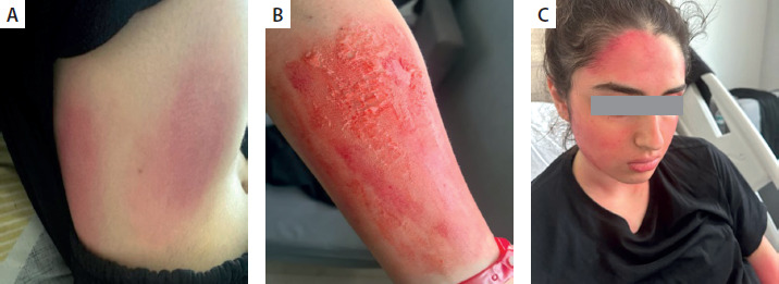
A 16-year-old female patient presented to the paediatric allergy clinic with erythematous rash that had been present for 1 week and was reported to be spreading. The lesions were located on the forehead, cheeks, neck, abdomen, and flexor surfaces of the upper extremities and hands. They were ill defined, non-blanching, macular in appearance, with areas of erosion and crusting, and varied in size and shape across the affected surfaces (Figure 1). The patient’s general condition was good, and vital signs were stable. No other systemic examination findings were noted. The patient’s medical history included body itching without urticaria or angioedema for the past 3 years. In addition, the patient experienced sneezing, facial flushing, and shortness of breath approximately 1 h after drinking cold tea for the first time in the past month, followed by facial redness, itching, and difficulty swallowing within 1 h after consuming cold baklava in the subsequent weeks, and finally, the patient presented to the emergency department with sneezing and skin rash complaints within hours of consuming cream-filled packaged biscuits at school, and adrenaline was administered during this third episode due to suspect of anaphylaxis.
FIGURE 1
Lesion/rash-like appearance on the patient’s face, left forearm and anterior and posterior costo-vertebral angle after lipstick staining
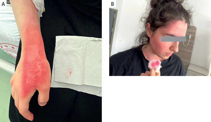
The patient admitted to our clinic was found to have leukocytosis in the admission tests: white blood cells (WBC): 18.25 k/µl (n: 4–10), neutrophils:16.68 k/µl (n: 2–7), and anaemia: Hb:10.4 g/dl (n: 11–16). Routine biochemical tests were within normal limits. Rheumatological evaluation, conducted with a suspected diagnosis of systemic lupus erythematosus, revealed negative results for ANA and anti-ds DNA. Thyroid function tests (TSH, T4, and T3) were normal; anti-thyroglobulin (18 IU/ml, n: 0–115) and anti-TPO (9.2 IU /ml, n: 0–34) were negative. In the infectious disease evaluation, C-reactive protein (CRP): 3 mg/l (n: 0–5) was within normal limits, while sedimentation rate (37 mm/h, n: 0–20) was elevated. Anti-Parvovirus B19 IgM, IgG, and PCR were negative; anti-CMV IgM was negative; EBV- VCA IgM was negative; and EBV- VCA IgG and EBV- EBNA IgG were positive (indicating a previous infection). Anti-Rubella IgG was negative. A viral respiratory panel was obtained from the patient consulted for infection, but no specific findings were identified. Allergic evaluation: skin prick test (inhalant panel) was negative: spIgE levels (latex, grass, dust mite, pollen, strawberry, orange, soy) were < 0.10 IU/ml. Total IgE level was 70 IU/ml (n: 0–100). Serum immunoglobulin levels were IgG: 1310 mg/dl (n: 830–1820), IgM: 303 mg/dl (n: 75–198.5), and IgA: 120 mg/dl (n: 46.5–221), indicating elevated IgM levels. Complement levels were C3c: 1.15 g/l (n: 0.9–1.8), C4: 0.22 g/l (n: 0.1–0.4), C1 esterase inhibitor function: 120 (n: 70–130%), and C1 esterase inhibitor antigen: 0.32 g/l (n: 0.23–0.41), which were within the normal range. The tryptase level measured to assess mast cell activity was 0.87 µg/l (normal range: 0–11.4), which was within normal limits. During follow-up after admission, the skin lesions were observed to fluctuate in severity on a daily basis. Intermittent bleeding was reported in the lesion in the following days, but no bleeding was observed during follow-up examinations. The patient was started on famotidine 1 mg/kg/day, prednisolone 2 × 0.5 mg/kg/day, and pheniramine 2 × 1 mg/kg/dose for the management of so-called erythematous rash. The patient reported improvement within the first day of treatment but noted that the lesions increased again afterward. During the treatment period, the patient’s caregiver intermittently requested referral. As the patient’s symptoms did not improve during clinical follow-up, local steroids were added to the current treatment. Because the food-related episodes and symptoms could not be associated with a systemic disease, adrenal and abdominal ultrasound was performed to rule out pheochromocytoma, and no pathology was observed. Concurrently, catecholamine metabolites were measured — metanephrine: 73.6 pg/ml (n: 0–90), adrenaline: 125.4 pg/ml (n: 20–190), dopamine: 32.4 pg/ml (n: 0–52), and normetanephrine: 101 pg/ml (n: 0–180) were within the reference range. During the physical examination, the patient reported redness on the skin surfaces in contact with the ultrasound gel, and the lesions appeared atypical in colour and shape. During the change of the intravenous line, it was observed that the lesion pigment transferred to the cotton due to the skin area being wiped with alcohol (Figure 2). Considering the dramatic course of symptoms, inconsistencies in the history, multiple consultations with various physicians, and diagnostic tests that failed to establish a diagnosis, an artificial disorder was suspected, and a diagnosis of Munchausen syndrome was made. Despite being offered, child psychiatry was refused. Verbal consent from the mother and written consent from the adolescent was obtained for the photographs used in this presentation.
Discussion
Munchausen syndrome is characterised by the deliberate feigning of illness, reporting of false symptoms, or self-harm. In these patients, frequent hospital visits, dramatic and convincing stories, symptoms resistant to diagnosis, and a need for attention are prominent [3]. Dermatological findings observed in Munchausen syndrome may manifest as skin lesions intentionally created by the individual themselves or by their caregiver. These lesions can present in various clinical forms, such as superficial scratches, ulcerative areas, burns, and deep tissue damage, and often complicate differential diagnosis due to their unusual appearance and atypical location. These patients often fail to respond to treatment. However, inconsistencies in the history and the lack of correlation between symptoms and the clinical picture raise suspicion of artificial disorders [1, 3].
Sirka et al. reported numerous dermatological findings in a 15-month-old girl, including bullous lesions characterised by recurrent ulcerations on the scalp and cheek area, irregularly shaped skin areas, alopecia associated with localised scarring on the scalp, and ectropion of the left upper eyelid. Initially, the patient was diagnosed with epidermolysis bullosa; however, further investigations revealed that these skin lesions more closely resembled those caused by intentional trauma. A detailed medical history also revealed that these lesions only appeared when the father was not at home, and each time, the mother was the first to notice the condition [4]. In this case, there were numerous lesions of various types and distributions, similar to our case.
In another recent case, Guez et al. investigated a 17-month-old girl with recurrent, healing, and unexplained skin lesions that had started at 2 months of age, but they were unable to obtain meaningful results from their investigations. Toxicological analyses revealed high levels of aluminium in skin swabs, and the lesions were determined to be cold burns resulting from prolonged exposure to deodorant spray. During the legal proceedings, hidden camera footage documented that the mother had intentionally injured the child with deodorant spray. This case highlights the difficulty in diagnosing Munchausen syndrome and the importance of considering spray-related burns as a possible cause [5].
In a case reported by Sung et al., a 14-year-old adolescent female patient was initially evaluated with a preliminary diagnosis of autoimmune bullous skin disease and presented with unexplained erosions in the left tibial region. Despite the application of immunosuppressive therapy, the lesions continued to change in shape and location, new biopsy findings indicated trauma-related epidermal damage, and moderate to severe depression was diagnosed in the mother. As a result, Munchausen syndrome was diagnosed. Separate interviews with the patient and her stepmother revealed that the patient had harmed herself and that the stepmother was aware of this but had not reported it [6]. Similarly, in our case, the lesions were intermittently changing location and did not respond fully to treatment. Additionally, the anamneses were inconsistent and contradictory.
The child may feel anxious because they have a distorted view of their health and may become distressed and preoccupied with their health status and vulnerability in this regard. They may experience conflict about their health status because they are repeatedly subjected to physical examinations, laboratory tests, and treatments even though they do not feel ill. They may start mimicking symptoms of illness or fall into the trap of silently mimicking illness, or they may eventually develop a somatoform disorder [7].
The perpetrators who cause factitious or induced disorders such as in Munchausen syndrome are usually mothers who report experiencing high levels of deprivation, neglect, and abuse during early childhood. More than half of perpetrators have somatoform or factitious disorders, and more than three-quarters have a comorbid personality disorder, particularly those in Cluster B (sociopathic, borderline, or histrionic) [7].
In Munchausen (fabricated or induced illness) syndrome, the caregiver may be overly eager for the patient to undergo various tests or surgical interventions. When healthcare professionals question the caregiver’s account, the caregiver may exhibit resistance or hostility or may request to transfer the patient to another facility [8].
In our case, numerous systemic diseases were ruled out, and no pathology was identified in specific tests. The fact that the lesions appeared inappropriate for possible diseases in terms of appearance and distribution, and that the patient experienced intermittent benefit from treatments but did not show improvement during follow-up, complicated the diagnostic process. In such cases, a multidisciplinary approach and patient clinical follow-up play a fundamental role.
Conclusions
In adolescent patients presenting with skin lesions resistant to treatment and significant inconsistencies between clinical and laboratory findings, artificial disorders such as Munchausen syndrome must be considered in the differential diagnosis. Delayed diagnosis may lead to unnecessary tests and treatments, as well as the risk of iatrogenic harm, so these cases should be managed with caution.