Purpose
Choroidal melanoma is the most common primary intraocular malignancy in adults [1]. Local tumor control is commonly achieved with ruthenium-106 (106Ru) brachytherapy, involving surgical implantation of a radioactive plaque [2]. Although this technique is generally safe and effective, several potential complications may occur [3]. In this report, we described the development of an orbital hematoma immediately following 106Ru plaque implantation for choroidal melanoma, and discussed clinical and dosimetric implications.
Case report
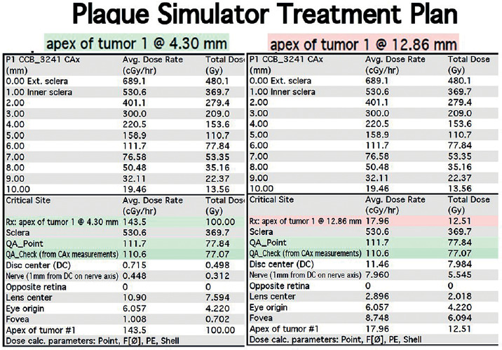
A 65-year-old female underwent 106Ru brachytherapy for a choroidal melanoma, located in the superior temporal periphery of the right eye (oculus dexter – OD), with apical tumor height of 4.36 mm and basal diameter of 11.6 mm, both measured ultrasonographically (Figure 1). Previous ophthalmic history was significant for recurrent ipsilateral subconjunctival hemorrhages, whereas arterial hypertension was found in prior systemic history. The intended prescribed dose was 100 Gy to be delivered to the tumor apex at 4.36 mm. The procedure was performed under general anesthesia using a CCB 106Ru plaque (Eckert & Ziegler BEBIG GmbH, Berlin, Germany). Tumor location was identified with transpupillary transillumination under mydriasis using a fiberoptic light source. A transparent dummy plaque was employed to confirm plaque apposition and mark anchoring points to insert pre-placed sutures for radioactive plaque attachment. The plaque with a diameter of 20.2 mm, resulting in lateral margins of 4.3 mm, was placed at the superior temporal quadrant, approximately 6 mm posterior to the corneo-scleral limbus, without any muscle extirpation or any noticeable intraoperative complications. However, upon tracheal extubation, the patient responded with intense coughing. Subsequently, progressive periorbital ipsilateral ecchymosis and eyelid tension compatible with an orbital hematoma was noticed. In response to this situation, topical ice packs were applied, the patient was placed in reverse Trendelenburg posture, and intravenous acetazolamide and methylprednisolone were administered. A few hours later, visual acuity of 20/32 and intraocular pressure of 17 mmHg (OD) with normal pupillary responses but deficient right supraduction, were observed (Figure 2A). B-mode ultrasonography confirmed the presence of an orbital hematoma between the plaque and underlying sclera, with an approximate thickness of 8.5 mm (Figure 2B). Since vision of OD was uncompromised and there were no clinical signs of orbital compartment syndrome (no afferent pupillary defect, stable visual acuity, color perception, and acceptable intraocular pressure), the patient was followed closely during the planned treatment period, with a view to interrupt treatment and prematurely remove the plaque if the orbital hematoma deteriorated. Oral steroids to suppress orbital inflammation as well as oral acetazolamide to keep intraocular pressure under control were administered. The plaque was removed under general anesthesia on pre-determined post-operative interval of 63 hours to deliver 100 Gy at 4.36 mm with a plaque simulator (Eye Physics LLC., Los Alamitos, CA, USA). The procedure of plaque removal was particularly challenging due to developed orbital hematoma and the need to forcefully retract the eyelids using sutures and Desmarres lid retractors to reach plaque location. Upon plaque removal, a dense black hematoma prolapsed and was drained (Figure 2C, D). Post-operative follow-up revealed full functional recovery without lingering ocular sequelae (visual acuity of 20/20 OD, 15 days later). However, given the possibility of suboptimal radiation dose resulting from increased distance of radioactive source from the sclera, the patient remains under close follow-up with the intention of further treatment if necessary. Considering the 8.5 mm displacement of the plaque from the sclera inflicted by the formation of hematoma, a new dosimetric plan was calculated for an apex of 12.86 mm. According to this revised dosimetric simulation performed using plaque simulator software for supplementary dosimetric assessment, the apex dose for the given time of 63 hours was 12.51 Gy (Figure 3).
Fig. 1
Choroidal melanoma of the right eye. A) Wide field color photo demonstrating location of the lesion at the superior-temporal periphery, and B) respective B-scan and A-scan ultrasonograms, showing apical elevation of 4.36 mm
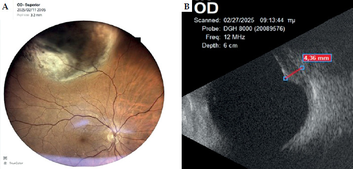
Fig. 2
A) Patient appearance on the first post-operative day, showing periocular ecchymoses and restricted eyelid opening, requiring digital traction. B) B-mode ultrasonography on the same day, indicating an orbital hematoma as a hypo-echoic mass (*) with a thickness of 8.5 mm. C) Intraoperative appearance immediately before plaque removal, showing the anterior edge of the silver-coated CCB plaque (blue arrow) and the anterior border of the underlying hematoma (*). D) Intraoperative appearance immediately after plaque removal, indicating draining of hematoma as dark black liquid (*). During the procedure, inability to insert a speculum necessitated utilization of traction sutures to forcefully retract the tight eyelids (white arrows, C and D)
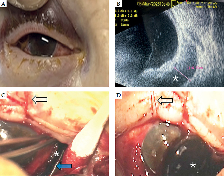
Fig. 3
Initial (left 3 columns) and revised (right 3 columns) dosimetry planning based on plaque simulator. The initially calculated dose for an apex of 4.30 mm was 100 Gy. For a presumed additional 8.5 mm height (due to the presence of hematoma between the plaque and sclera), resulting in an apex of 12.8 mm, the delivered dose would be reduced to only 12.51 Gy
Discussion
Ruthenium-106 plaque brachytherapy is a well-established modality for the treatment of choroidal melanoma, providing high rates of local tumor control while preserving the globe [2, 4]. However, optimal therapeutic efficacy depends critically on precise plaque apposition to the scleral surface, ensuring accurate dose delivery to the tumor apex. Any factor that alters spatial relationship between the radioactive source and the sclera may significantly affect the intended radiation dose distribution [5]. In the present case, the development of an acute orbital hematoma immediately following plaque implantation represents a previously unreported complication of 106Ru brachytherapy. While hemorrhagic complications, such as vitreous hemorrhage, have been previously described, the formation of a substantial extra-scleral hematoma in the immediate post-operative period appears to be uncommon, and to the best of our knowledge, has not been widely reported in the literature. The temporal association with forceful coughing during extubation suggests a Valsalva-induced increase in venous pressure as a plausible precipitating factor, particularly in a patient with a history of hypertension and recurrent subconjunctival hemorrhages.
From a brachytherapy perspective, the most critical consequence of this complication is the potential alteration of dosimetry. 106Ru is a β-emitting source characterized by a steep dose gradient, with rapid attenuation of radiation as a function of distance from the plaque surface [4, 5]. Consequently, even small increases in the separation between the plaque and sclera can lead to a substantial reduction in the dose delivered to the tumor apex [6]. In this case, ultrasonography demonstrated an approximate hematoma thickness of up to 8.5 mm, interposed between the plaque and sclera. Given the known physical properties of β radiation, such separation could result in a marked decrease of effective dose reaching the tumor. Published data indicate that dose fall-off for 106Ru plaques is highly sensitive to distance (as measured ultrasonographically), with significant reductions occurring even within the first few millimeters, given the steep and non-linear depth-dose gradient of 106Ru β radiation [6, 7]. As a practical estimate, the absorbed dose decreases by approximately one order of magnitude over 6-7 mm of tissue depth [8]. Therefore, an 8.5 mm displacement may theoretically reduce the delivered dose to a fraction of intended prescription, raising concerns regarding potential undertreatment and long-term tumor control. Despite this, short-term local tumor stability was observed at 6 months, suggesting that either partial dose delivery was sufficient in this case, or that the effective displacement may have varied during the treatment course. Nevertheless, this scenario highlights the importance of recognizing factors that may compromise plaque positioning and, by extension, dosimetric accuracy. Monte Carlo dosimetry studies of 106Ru ophthalmic plaques demonstrate that β radiation exhibits a very steep depth-dose fall-off within intraocular distances, with most of the energy deposited in the first few millimeters of tissue and a rapid exponential-like decline beyond ~5-7 mm. This sharp attenuation driven by the short range of β particles and accurately captured by Monte Carlo transport codes, results in high doses at the sclera and tumor base while substantially sparing deeper ocular structures, such as the retina and optic nerve, but also compromises the effectiveness of 106Ru brachytherapy for higher apices [8-10]. On the other hand, the absence of documented tumor progression during short-term follow-up may suggest that the effective dose delivered was higher than predicted by the revised static dosimetric calculation, possibly due to a dynamic behavior of the orbital hematoma, which may have resulted in varied distance of the plaque from the sclera during the follow-up period. Accordingly, dosimetric calculations in such clinical situations may only provide an approximation of the true dose delivered to the tumor, while the revised dosimetry represents a theoretical worst-case scenario assuming sustained maximal plaque displacement throughout treatment. Nevertheless, the short follow-up duration of 6 months for this single case report is insufficient to draw conclusions on eventual treatment outcome, which is a limitation of the present report.
Clinically, the absence of signs of orbital compartment syndrome allowed conservative management and continuation of the planned treatment schedule. However, this case underscores the need for heightened vigilance in the immediate post-operative period following plaque implantation. Early identification of orbital complications by clinical examination and ultrasonography is essential, as significant alterations in plaque position may warrant adjustment in treatment duration or early intervention. From a procedural viewpoint, strategies to minimize the risk of such complications should be considered. These include careful anesthetic management to avoid Valsalva maneuvers during extubation, optimization of systemic blood pressure, and awareness of patient-specific risk factors for hemorrhage. In cases where post-operative orbital hematoma is suspected, prompt imaging is recommended not only for diagnosis but also for assessing potential implications of plaque positioning and dose delivery. In our patient presented in this report, alternative decisions could have been made, including early plaque removal, repositioning, or even adaptive treatment based on the presumed change in plaque position. However, in an orbit with recent development of hematoma still under clinical evolution, the risk of hematoma expansion in an early intervention possibly resulting in compromised vision, was considered.
In conclusion, this case highlights a rare but clinically significant complication of 106Ru ophthalmic brachytherapy with potential direct implications for radiation dosimetry. Awareness of this entity is important for both ophthalmic surgeons and radiation oncologists, as early recognition and appropriate management may mitigate the risk of compromised treatment efficacy. From a practical perspective, a structured post-operative approach should be considered, including careful clinical assessment in the immediate post-implantation period and a low threshold for orbital imaging, particularly ultrasonography, when complications are suspected. In cases where plaque displacement is a concern, timely evaluation of plaque position and consideration of dosimetric impact may help guide decisions in terms of treatment continuation, adjustment, or intervention.