
Purpose
Cervical cancer remains a leading cause of cancer-related morbidity and mortality, particularly in low- and middle-income countries (LMICs). In India, the 5-year prevalence is 339,589 cases, with 79,906 deaths reported annually [1]. Majority of patients present with locally advanced disease, and the standard of care for these patients is definitive chemoradiation, with external beam radiation therapy (EBRT) followed by intracavitary and/or interstitial brachytherapy (BT) [2]. BT is a cornerstone in the treatment of cervical cancer, and attempts to replace this technique by newer EBRT advancements, such as stereotactic body radiation therapy (SBRT), have been met with significantly inferior outcomes [3-5]. The cervix, upper vagina, and uterus exhibit relatively high radiation tolerance, enabling dose escalation crucial for tumor control, an approach not feasible in most other anatomical sites. This capacity for safe, high-dose delivery makes BT necessary for achieving excellent outcomes.
EBRT is typically delivered using conventional fractionation of 1.8-2.0 Gy per fraction over 5-6 weeks to a total dose of 45 to 50 Gy to the gross disease and elective volumes. This is generally followed by BT administered to the primary tumor, planned using either point-based or volumetric approaches [6, 7]. Recent advancements in imaging and applicator designs have enabled the use of MRI-based planning, which has become ideal practice for image-guided adaptive brachytherapy (IGABT). Failure to adhere to strict timelines, leads to accelerated re-population, and loss of local control and overall survival by 1% per day of treatment extension [8].
In the low-dose-rate (LDR) era, entire BT dose was delivered in a single application. However, with the advent of HDR-BT, total dose was fractionated with radiobiological issues considered. Currently, multiple applications using single-application, single-fraction (SASF) approach is a standard [6, 9]. However, variations in applicator positioning with each application results in inconsistencies in geometry and dosimetry across applications [10, 11]. Additionally, prolonged overall treatment time (OTT) increasing hospital resource utilization, logistic challenges due to repeated patient admissions, and inter-fraction dosimetry variations, all contribute to the problems with SASF applications [12-14].
Several strategies have been employed to limit OTT, such as inter-digitation of SASF-BT applications, alongside EBRT single-application, two-fraction (SATF) BT after teletherapy using two applications, and single-application, multi-fraction (SAMF) BT using single application [15]. In this study, we analyzed outcomes of patients treated with SATF brachytherapy after EBRT and concurrent chemotherapy.
Material and methods
A retrospective review of 238 histopathologically proven squamous or adenocarcinoma, stage IB to IVA (FIGO 2018) uterine cervix patients was conducted between April 1, 2020 and December 31, 2022, following approval from the Institutional Review Board (IRB). Electronic medical records (EMR) and radiation oncology information system (ROIS) were utilized to screen and obtain data regarding treatment outcomes and adverse effects. All patients had received definitive chemoradiotherapy, followed by SATF brachytherapy at our institute. Patients with distant metastasis at diagnosis, a prior history of abdominal or pelvic radiotherapy, partial/total hysterectomy, immunocompromised status, psychological issues, those unsuitable for BT post-EBRT due to unsuitable anatomy/doubtful fitness for anesthesia due to medical comorbidities, or pregnant/lactating women, were excluded from this study.
Primary endpoint was 3-year disease-free survival (DFS), defined as the time (in months) from the date of diagnosis to date of relapse, second malignancy, or death due to malignancy. Secondary endpoints included 3-year local control (LC) rate, defined as the time (in months) from the date of diagnosis to date of local relapse, and 3-year overall survival (OS), defined as the time (in months) from the date of diagnosis to date of last follow-up or death due to any cause. Acute and late toxicity assessment was performed using the Radiation Therapy Oncology Group (RTOG) grading system. Late toxicities were analyzed with an actuarial method to determine the cumulative incidence of adverse events over time.
Following detailed history assessment and thorough clinical examination, patients were staged as per FIGO 2018 criteria. Clinical examination details were documented using GEC-ESTRO clinical diagrams [6]. Local staging in all patients included radiological evaluation with contrast-enhanced magnetic resonance imaging (MRI) of the pelvis. Metastatic workup was performed using contrast-enhanced computed tomography (CECT), or positron emission tomography (PET-CT).
Target volume was defined according to the ICRU 50/62 and ICRU 83 guidelines. EBRT contouring standards, planning aims for target and organs at risk (OARs) as well as bladder protocol principles, were followed as per the EMBRACE II protocol [15]. Eclipse treatment planning system (TPS) v. 16.0 was employed for contouring and planning. Megavoltage EBRT was delivered by a Varian TrueBeam linear accelerator using intensity-modulated radiotherapy (IMRT) or volumetric modulated arc therapy (VMAT) techniques. EBRT was delivered to a dose of 45 Gy in 25 fractions at 1.8 Gy per fraction over 4.5 to 5.5 weeks, with concurrent chemotherapy of weekly cisplatin at 40 mg/m2. In the presence of gross nodes, simultaneous integrated boost (SIB) was delivered with a total nodal dose of 55 Gy. Assessment and documentation of acute toxicities were done on a weekly basis, and were managed accordingly.
Following EBRT, patients were evaluated clinically for treatment response and BT suitability according to standard guidelines [3]. All patients underwent 3D-based planning using either CT- or MR-based imaging. A planning CT scan with 3 mm slice thickness was acquired in 3 dimensions for catheter reconstruction and planning. Accurate reconstruction of applicators on MRI was facilitated by dummy catheters containing water, placed in channels of the tandem and ovoids during image acquisition. To facilitate accurate disease mapping, T2-weighted sequences with 3 mm slice thickness were acquired in para-axial, para-sagittal, and para-coronal planes.
High-dose-rate (HDR) BT was performed using cobalt-60 (60Co) HDR afterloading therapy equipment (Elekta Flexitron HDR V2). Target volumes and OARs at BT were delineated according to the GYN GEC-ESTRO recommendations [6]. Planning aim was to deliver 85-95 Gy to D90 high-risk clinical target volume (HR-CTV; dose received by 90% of high-risk clinical target volume), > 60 Gy to D98 intermediate risk clinical target volume (IR-CTV; dose received by 98% of intermediate-risk clinical target volume), and > 65 Gy to point A. Dose constraints to OARs, namely bladder 2 cc (B2cc), rectum 2 cc (R2cc), and sigmoid 2 cc (S2cc) doses were kept at ≤ 90 Gy, ≤ 75 Gy, and ≤ 75 Gy, equivalent dose at 2 Gy fraction (EQD2), respectively, evaluated as per standard guidelines [15, 16]. Patients were treated with two BT applications using SATF approach that delivered 7 Gy in each fraction, with a minimum of 12 hours interval between fractions, i.e., 28 Gy delivered in 4 fractions during 2 applications.
Acute toxicity assessment using the RTOG grading system was accomplished during the first follow-up at 6 weeks. Subsequently, patients were followed up clinically every 3 months during the first 2 years and then every 6 months, with response assessment MRI performed 3-6 months after RT completion, followed by annual cross-sectional imaging of the abdomen and pelvis as per standard practice. A 3-year DFS, 3-year OS, and 3-year local control (LC) were estimated using the Kaplan-Meier method (Kaplan 1958). Time was measured from the date of diagnosis to the first date of failure (i.e., local, regional, or distant failure, or death due to any cause) or last follow-up. In addition to treatment, Cox regression model was utilized to analyze factors, associated with outcomes (Cox 1972). For the Cox proportional hazards model, FIGO 2018 stage and histological sub-type were selected as covariates to evaluate their impact on DFS and OS. Late toxicities were analyzed using the actuarial method to account for different follow-up durations among patients.
Results
The patient and tumor characteristics of the 238 patients are shown in Table 1. The median age of the cohort was 55 years (range, 27-81 years), with majority (77.7%) having a Karnofsky performance score ≥ 90 with no comorbidities. According to the American Society of Anesthesiologists (ASA), physical status of half of the patients was scored as ASA-I [17]. Squamous cell carcinoma was the predominant histology (96.6%), and at presentation, the majority of patients had locally advanced disease. FIGO stage IIB (47.5%) was the most common disease stage, while IIIC1r stage was seen in 67 (28.2%) patients.
Table 1
Patient and tumor characteristics
Intracavitary BT (ICBT) alone was the most common BT application technique in 213 (89.5%) patients. The median OTT was 55 days (range, 40-67 days), with 150 (63.0%) patients completing treatment within 56 days. CT scan was employed for BT planning in 84.5% of the cases. The mean dose to point A, D98 HR-CTV, D90 HR-CTV, and D98 IR-CTV were 77.7 ±5.7 Gy, 76.2 ±15.1 Gy, 88.0 ±5.2 Gy, and 69.4 ±2.4 Gy, respectively. The mean dose to B2cc, R2cc, and S2cc were 85.1 ±10.1 Gy, 66.0 ±7.9 Gy, and 69.9 ±7.5 Gy, respectively. The treatment details are shown in Table 2.
Table 2
Treatment details
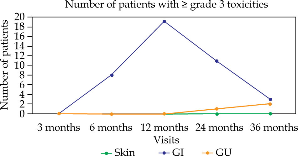
The majority of patients experienced grade 1 toxicities, including skin (n = 235, 98.7%), GI (n = 237, 99.6%), and GU (n = 235, 98.7%). Hematological toxicity was minimal, with 237 (99.6%) of patients reporting no toxicity; no acute grade 3 toxicity was observed. Late skin, GI, and GU toxicities were not seen in majority of patients. Cumulative ≥ grade 2 GI and GU toxicities after 24 months were observed in 29 (12.2%) and 4 (1.7%) cases, respectively. At 12 months, 19 (8.2%) patients had ≥ grade 3 GI toxicities; follow-up assessments at 18 and 24 months revealed a descending trend after conservative medical therapy (Figure 1).
At a median follow-up of 45 months (IQR: 38-51 months), 73 DFS events and 69 deaths were documented. The mean DFS for the entire cohort was 44.8 months (95% CI: 42.5-47.2), with estimated DFS rates of 91.1% ±1.9% at 1 year and 70.2% ±3% at 3 years. The mean OS was 46.4 months (95% CI: 44.2-48.6), with OS rates of 93.6% ±1.6% at 1 year and 72.2% ±3% at 3 years. Notably, based on FIGO staging, DFS and OS varied significantly (p = 0.007 and p = 0.003, respectively). In the univariate Cox regression analysis, stage IIA was associated with an 89% reduction in the hazard of recurrence (HR: 0.11, 95% CI: 0.01-0.92, p = 0.04). Stage IIB demonstrated significantly superior outcomes compared with stage IVA for both DFS (HR: 0.27, 95% CI: 0.11-0.65, p = 0.003) and OS (HR: 0.23, 95% CI: 0.10-0.57, p = 0.001). Additionally, stage IIIC1 showed significantly improved OS (HR: 0.38, 95% CI: 0.15-0.94, p = 0.04). Histological sub-type did not significantly influence hazards for DFS (p = 0.22) or OS (p = 0.21). Figure 2A and B shows the Kaplan-Meier curves of DFS and OS based on FIGO 2018 staging. Moreover, on multivariate analysis, local failure was higher, with OTT > 56 days vs. ≤ 56 days, accounting for 12 (85.7%) and 2 (14.3%) patients (p-value < 0.001), and with CT vs. MRI in 10 (71.4%) and 4 (28.6%) cases (p-value = 0.24), respectively. However, there was no significant difference in DFS and OS between the two groups. A recurrence was observed in sixty-nine of 230 patients with squamous cell carcinoma, and in 4 of 8 patients with adenocarcinoma. While the differences were not statistically significant, the 3-year DFS for squamous and adenocarcinoma histologies were 70.9 ±3.0% and 50.0 ±17.7%, respectively, and the 3-year OS were 73.3 ±3.0% and 50.0 ±17.7%, respectively. Figure 3A and B illustrates the Kaplan-Meier curves of DFS and OS based on histology. Table 3 demonstrates the univariate Cox regression analysis of FIGO 2018 and histology of patients using DFS and OS as endpoints.
Fig. 2
Kaplan-Meier estimation of patients based on FIGO 2018 staging, with A) disease-free survival (DFS) and B) overall survival (OS)
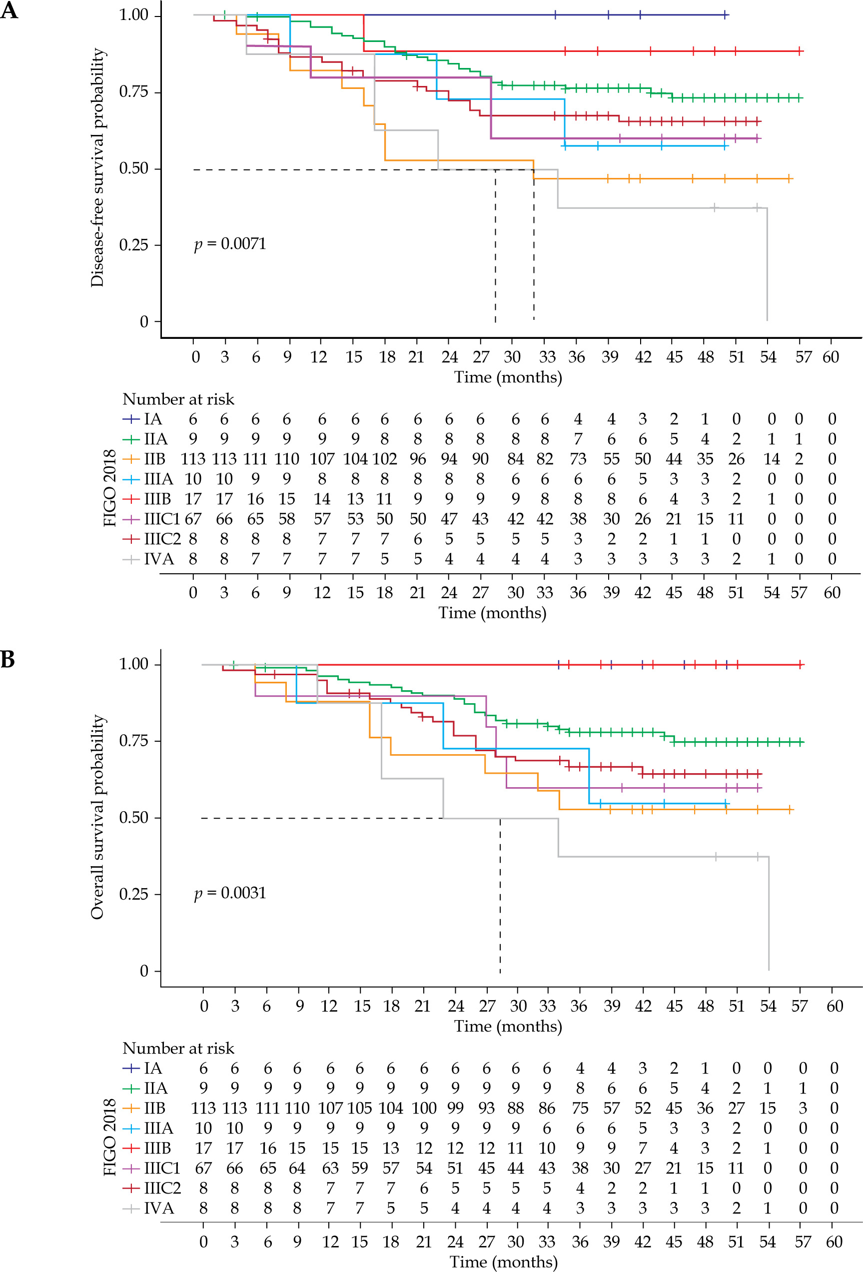
Fig. 3
Kaplan-Meier estimation of patients based on histology, with A) disease-free survival (DFS) and B) overall survival (OS)
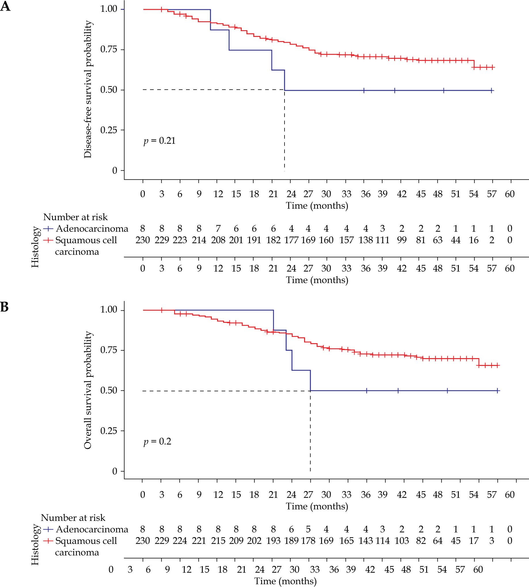
Table 3
Univariate Cox regression analysis of FIGO 2018 staging and histology of patients with disease-free survival (DFS) and B) overall survival (OS) as endpoints
Discussion
Low-dose-rate BT prescriptions were the outcomes of various dosimetric systems developed between 1920s and 1950s. The most commonly followed approach in BT was the Manchester system, where point A-based prescription provided predictable long-term local control and acceptable bladder and rectal toxicity. The earlier LDR systems involved implementation of BT treatment in fewer applications and fractions (fx.). With the introduction of HDR-BT, an important consideration was dose prescription that resulted in acceptable late tissue toxicities without compromising on local control. Patel et al. reported significantly better 3-year LC rates (81.3% vs. 65.1%, p = 0.04), culminating in an improved 3-year DFS rate (64.9% vs. 49.4%, p = 0.03) in patients receiving 9 Gy × 2 fx. vs. 6.8 Gy × 3 fx. [16]. Sood et al. obtained a 3-year LC and OS rates of 77% and 78%, respectively, without significantly increasing late toxicities in stage I-III cervical cancer patients receiving 9 Gy × 2 fx. delivered with HDR-BT [17]. Currently, the American Brachytherapy Society (ABS) recommends several HDR-BT schedules, namely 7 Gy × 4 fx. (EqD210 of 83.9 Gy), 6 Gy × 5 fx. (EqD210 of 84.3 Gy), 5 Gy × 6 fx. (EqD210 of 81.8 Gy), and 5.5 Gy × 5 fx. (EqD210 of 79.8 Gy) to point A. However, no recommendation has been proposed with regard to the number of applications and/or the most optimum dose regimen. ABS consensus recommends utilization of larger doses per fraction, such as 8 Gy × 3 fx., if necessary, for LMICs [18, 19].
Prolongation of OTT has a detrimental effect on cause-specific survival (CSS). In a study by Perez et al. among 1,330 patients, prolongation of OTT resulted in an increased failure rate of 0.59% per day in stage IB and IIA cases, and 0.86% per day in stage IIB disease [20]. As a result of clinical and radiobiological considerations, HDR-BT in cervical cancer treatment involves a larger number of fractions and increased number of applications. Considering the need for OTT reduction, one of the strategies is the delivery of more than one fraction in one BT application. The benefit of a reduced OTT, thereby minimizing re-population, is the most important radiobiological advantage of this approach, providing improved pelvic control. Chopra et al. reported a 5% detriment in LC if treatment was prolonged to > 8 weeks [21]. A study from India examining the impact of COVID-19 and delay in treatment for cervical cancer patients, projected a 2.5-3.8% life-time increase in death rate caused by cervical cancer, with treatment delays ranging from 9 weeks to 6 months compared with no delay in treatment [22]. The hypofractionated BT approach with either SATF or SAMF demonstrates additional logistical advantages for both the patients and hospital, with decreased number of hospital admissions and reduced sessions of general anesthesia, which may potentially lead to better resource utilization and capacity enhancement, especially in high-volume centers. This is of relevance in terms of large patient loads, fewer number of advanced cancer treatment facilities offering BT, and out-of-pocket expenses of patients coming from distant places for treatment, especially in LMICs. This method also reduces patient discomfort and travelling to the hospital on additional occasions, contributing to better compliance. However, changes in geometry between fractions and increased risk of deep vein thrombosis (DVT) and pulmonary vein thromboembolism (PTE) as a result of prolonged immobilization, can be considered a limitation of this approach [23]. Adequate and proper packing and placement of a restraining T bandage will help in the prevention of applicator displacement. Additional measures to be considered are marking on the skin of the thigh of patient, the position of lower end of applicator, repeat imaging before subsequent fraction for tracking applicator displacement, and using pressure stockings for preventing the development of PTE.
Studies have evaluated the role of using extreme hypofractionation with SAMF approach to reduce OTT. A phase II study (SIMBRACE) evaluated the feasibility and safety of SAMF under MRI guidance, with 9 Gy × 1 fraction on day 1 and 7 Gy × 2 fx. on day 2, with a minimum of 6 hours gap between treatment sessions. The authors reported a 2-year LC, DFS, OS of 90.1%, 85.0%, and 94.5%, respectively, with a feasibility of this treatment approach in 95% of patients [24]. However, in all patients of this study, hybrid intracavitary (IC) + interstitial (IS) BT was employed to meet OAR constraints effectively, hence it cannot be incorporated into routine practices and it is not possible to carry out this protocol in centers without access to hybrid applicators. While our study using the SATF approach reported the need for combined hybrid (IC + IS) applications in only 15.5% of cases, in terms of SAMF approach, the use of IC + IS application was seen in the range of 42-100% [14, 21, 24]. During the first application, the need to encompass slightly larger tumor volumes typically results in a little higher dose to OARs. However, tumor shrinkage before the second application enables improved optimization of both target coverage and OAR sparing, facilitating the accomplishment of planning objectives. This feature makes the SATF approach particularly feasible for centers with large patient load and logistic issues for both the patient and institution. In contrast, the SAMF approach allows only re-optimization during subsequent fractions for positional adjustments, while adequate tumor reduction is not anticipated during the repair window. In the SIMBRACE study, re-optimization was required in 6 out of 38 patients (15.7%) before the second and third applications to reduce bladder and rectal doses. Despite the adjustments, three patients developed grade ≥ II radiation proctitis during a 9-month follow-up. However, evaluation and comparison with more mature long-term data is needed.
In this study, the 3-year OS (72.2%) and DFS (70.2%) are consistent with the survival outcomes reported in landmark studies [25], such as retro-EMBRACE. Direct comparison with the EMBRACE I [15] study is not possible with the present research, as 5-year data is not yet mature. The outcomes suggest that the SATF protocol maintains oncological safety comparable with the international benchmarks. This is particularly noteworthy given that 75.7% of our cohort presented with locally advanced disease (stage IIB-IIIC1). Also, our 24-month late grade ≥ 2 GI (12.2%) and GU (1.7%) actuarial toxicity rates align with the retro-EMBRACE data [25], which demonstrated that transitioning to IGABT significantly reduces late morbidity compared with older point-based planning. By achieving these results with a single application, the SATF approach offers a balanced solution that maintains the safety standards of landmark protocols, while addressing the logistical constraints of high-volume centers.
The EMBRACE II study group recommends HR-CTV EQD2 of 80-85 Gy and 85-90 Gy for early and locally advanced cervical cancers, respectively, with a moderate hypofractionated schedule employing 7 Gy × 4 fx. delivered in 2 applications [15] – an approach similar to the one used in the present research. In our study, the median D90 HR-CTV was 88 Gy, and only one patient received dose below 85 Gy (73.2 Gy) due to larger tumor volume. A retrospective study by Houdou et al. reported the results of SAMF approach with 7 Gy × 4 fx. in single application. The 2-year LC, OS, and DFS rates were 79.4%, 77.7%, and 61.7%, respectively, with 25 (18%) patients experiencing grade 3 late complications [26]. Chopra et al. reported the outcomes of an abbreviated multi-fractionated BT schedule, with dose fractionation varying from 5.0-8.5 Gy per fraction, showing a 2-year DFS and OS of 65.5% and 81.3%, respectively. At a median follow-up of 16 months, with late genitourinary toxicity of 1.5%, late gastrointestinal grade ≥ 2 and ≥ 3 toxicities were 14% and 11%, respectively [21]. The present study reported better survival outcomes as compared with the SAMF technique, with 2-year DFS and OS of 78% and 85%, respectively [21].
Our cohort showed OAR doses similar to the SAMF approach, with a mean D2cc bladder of 87.1 Gy vs. 89 Gy, D2cc rectum of 66 Gy vs. 71 Gy, and sigmoid dose of 69.9 Gy vs. 67 Gy [21]. In the present study, the D2cc dose constraints for the bladder were < 90 Gy in 76% of patients, while those for the rectum and sigmoid < 75 Gy were 90.7% and 75.6% of patients, respectively. The observed 24-month late grade ≥ 2 GI and GU actuarial toxicity rates in this study were 12.2% and 1.7%, respectively. The majority of these toxicities showed a declining trend over time with conservative medical management, and did not warrant any surgical intervention or resulted in long-term morbidity. The observed toxicities are lower than those reported for many single-application multi-fraction (SAMF) schedules, with late GI grade ≥ 2 toxicities reaching 14%. This suggests that by allowing tumor shrinkage and re-optimization between applications, the SATF approach provides a safer therapeutic window for OAR sparing than extreme hypofractionation techniques, such as SAMF. This also indicates a relatively lower rectum and sigmoid tolerance using SAMF approach, but warrants modification in planning objectives with lower threshold while optimizing these plans. The late GU toxicities of grade ≥ II and ≥ III were both reported at 1.5%, which are similar to that in our study [21].
Conclusions
The present retrospective review of 238 patients of carcinoma cervix treated with chemoradiation and single-application two-fraction dose schedule HDR-BT, demonstrates favorable early outcomes with acceptable late toxicities. The study highlights that SATF is a more feasible and balanced approach than SASF as well as a safer approach compared with SAMF. Chemoradiation and SATF dose schedule HDR-BT is an effective treatment for carcinoma cervix, with acceptable toxicity and promising treatment outcomes, especially in a resource constrained settings of LMICs, within the boundaries of optimal OTT.











