Purpose
Up to one-third of patients with locally advanced or metastatic differentiated thyroid carcinoma (DTC) eventually develop radioactive iodine-refractory differentiated thyroid cancer (RAIR-DTC), which is associated with rapid disease progression and poor prognosis [1, 2]. According to the 2025 Chinese management guidelines for RAIR-DTC [3], the diagnosis is established when iodine-131 (131I) refractoriness is confirmed after excluding factors that may reduce iodine uptake shown on post-therapy whole-body scan, including residual thyroid tissue, inadequate thyroid-stimulating hormone (TSH) activity, or iodine contamination. RAIR-DTC is diagnosed when one of the two criteria are met: absence of iodine uptake in all DTC lesions, or presence of iodine uptake but disease progression within 12 months following 131I therapy. Worsening is indicated by at least one of the following factors: higher thyroglobulin (TG) or thyroglobulin antibody (TgAb) levels, larger existing lesions, new lesions, worsening of symptoms, new symptoms, or death.
For locally progressive metastatic lesions, iodine-125 seed implantation (125I-SI) is an effective treatment option with minimal systemic toxicity, offering rapid tumor control, pain relief, and improved quality of life. Most studies [4-6] reported a 6-month local effective rate (LER) of 60-80% and a local control rate (LCR) of 80-90%. However, local progression still occurs in about 10-20% of patients, highlighting the need for identification of response predictors and optimization of patient selection.
This study aimed to identify optimal positron emission tomography/computed tomography (PET/CT) metabolic parameters for predicting the effectiveness of 125I-SI in patients with RAIR-DTC. We compared parameters normalized to lean body mass (SULmax, SULpeak) with those normalized to body weight (SUVmax, SUVpeak) to minimize the confounding effect of adipose tissue. These were also compared with indices of tumor burden, such as metabolic tumor volume (MTV), total lesion glycolysis (TLG), and median tumor-to-background SUVmax ratio (TLR). This comparison will inform patient treatment and clinical decision-making. Although PET/CT metabolic parameters can project tumor prognosis in several cancers [7-10], their potential to predict the effectiveness of 125I-SI in RAIR-DTC remains unclear.
Material and methods
Patient selection
Clinical records of 48 patients treated with 125I-SI from November 2019 to June 2025 (Figure 1) were collected. This study was approved by the Ethics Committee of Jiangxi Cancer Hospital (approval number: 2025ky281), and all steps and procedures were followed according to relevant guidelines and regulations.
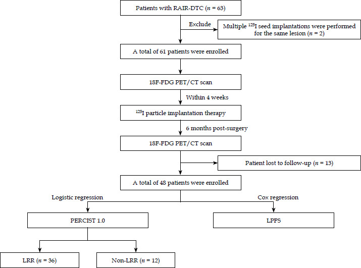
Inclusion criteria: 1. Diagnosis of RAIR-DTC as per the Chinese guidelines for diagnosis and management of radioactive iodine-refractory differentiated thyroid cancer (2025 edition); 2. PET/CT performed within 4 weeks prior to 125I-SI; 3. Local metastases unresponsive to radiotherapy [progressive disease according to the response evaluation criteria in solid tumors 1.1 (RECIST 1.1) based on contrast-enhanced CT assessment performed 12 weeks following radiotherapy completion], surgically inoperable local metastases, or patient’s refusal of surgery; 4. At least one measurable metastatic lesion suitable for 125I-SI; 5. At least one post-operative imaging follow-up; 6. Availability of complete clinical data.
Exclusion criteria: 1. Expected survival of less than 3 months or the Eastern Cooperative Oncology Group (ECOG) performance status greater than 2; 2. Systemic or local treatments interfering with efficacy evaluation within 3 months before seed implantation; 3. Concurrent treatments during the evaluation period, which could affect the implanted lesion.
18F-FDG PET/CT
18F-FDG PET/CT (Biograph mCT, Siemens) was performed according to standard operation protocol. Patients with blood glucose < 11.1 mmol/l received intravenous 18F-FDG (5.55 MBq/kg). After 60 min of rest, scans were acquired from orbits to upper thighs (3-5 min per bed position, reconstructed slice thickness of 3 mm). Low-dose CT (120 kVp, 140 mAs, slice thickness of 5 mm) was used for attenuation correction and anatomical localization. Two nuclear medicine specialists independently analyzed images blinded to clinical data. SUVmax, SUVpeak, SULmax, SULpeak, MTV, and TLG (threshold: 40% SUVmax) were assessed.
Iodine-125 implantation
Pre-implantation planning was performed using a treatment planning system (TPS) to ensure uniform seed distribution and adequate dose to lesions. Planning included delineation of gross tumor volume (GTV) and organs at risk (OARs), prescribed dose and seed activity specification, puncture needle trajectory simulation, seed number calculation, spatial distribution design, and dose calculation to GTV and OARs (large vessels, esophagus, and spinal cord). According to the Chinese expert consensus on radioactive 125I seed implantation for locally advanced differentiated thyroid carcinoma (2023) [11], for tumors ≤ 3 cm, radical treatment is recommended with a prescribed dose of 120-140 Gy; For tumors > 3 cm undergoing initial implantation, 100-120 Gy is recommended; For recurrent lesions after prior external beam radiotherapy, 80-100 Gy is recommended, with implantation performed 2-3 months after radiotherapy.
Follow-up
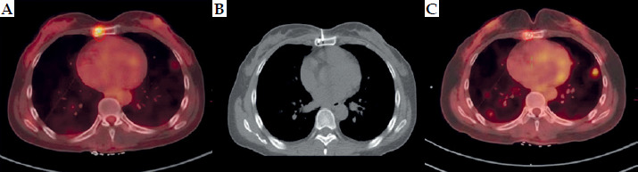
Patients underwent regular follow-up every 3 to 12 months, with clinical examination, neck ultrasound, CT, magnetic resonance imaging (MRI), and/or 18F-FDG PET/CT performed. Efficacy was assessed at 6 months using positron emission tomography response criteria in solid tumors 1.0 criteria (PERCIST 1.0) criteria. Image assessments were done independently by two attending physicians, but if the inter-rater correlation was < 0.9, a third physician was consulted. Local response rate (LRR) was defined as the proportion of patients with complete metabolic response (CMR) or partial metabolic response (PMR). The primary endpoint was local progression-free survival (LPFS) defined as the time from seed implantation completion to the target lesion progression or death. The treatment flowchart is presented in Figure 2.
Statistical analyses
Statistical analyses were performed with R software (version 4.3.1) and IBM SPSS Statistics version 26. (IBM Corp.). Categorical variables were compared using chi-square or Fisher’s exact test. Continuous variables with normal distribution were analyzed with independent sample t-tests, while non-normal variables were compared using Mann-Whitney U test. Univariable and multivariable logistic regressions were employed to assess predictors of LRR, while Cox regression identified predictors of LPFS. Model discrimination and calibration were evaluated using receiver operating characteristic (ROC) curves, area under the curve (AUC), decision curve analysis (DCA), and calibration curves. A two-sided p < 0.05 was set as the criterion for statistical significance.
Results
The baseline characteristics (Table 1) of the 48 RAIR-DTC patients showed a predominantly female cohort (70.8%), with a mean age of 50.23 years. All patients had received prior radioiodine therapy. Overall, the cohort represented a heavily pre-treated population with persistent disease despite conventional and systemic treatments other than TSH suppression. Table 2 characterizes the 48 treated lesions and corresponding PET/CT parameters. Both bone and lymph nodes were the predominant sites of implantation. At 6 months, 75% of patients achieved metabolic response (CMR or PMR), and no progressive disease was observed.
Table 1
Baseline characteristics of 48 patients with RAIR-DTC treated with 125I-SI
[i] RAIR-DTC – radioactive iodine-refractory differentiated thyroid cancer, 125I-SI – iodine-125 seed implantation, SD – standard deviation, PTC – papillary thyroid carcinoma, FTC – follicular thyroid carcinoma, Q1 – first quartile (25th percentile), Q3 – third quartile (75th percentile), ECOG – Eastern Cooperative Oncology Group performance status (0 = fully active, 1 = restricted in physically strenuous activity but ambulatory), TSH – thyroid stimulating hormone, TKI – tyrosine kinase inhibitor, 131I – iodine-131; Radiotherapy refers to external beam or other local radiotherapy
Table 2
Characteristics of lesions and PET/CT parameters
125 I-SI – iodine-125 seed implantation, PET/CT – positron emission tomography/computed tomography, SUVmax – standardized uptake value maximum, SUVpeak – peak standard uptake value, SULmax – maximum standardized uptake value normalized by lean body mass, SULpeak – peak standardized uptake value normalized by lean body mass, MTV – metabolic tumor volume, TLG – total lesion glycolysis, TLR – tumor-to-liver ratio, D90 – 90% of the dose received by gross tumor volume, SD – standard deviation, Gy – Gray (unit of absorbed radiation dose), MBq – megabecquerel (unit of radioactivity), Q1 – first quartile (25th percentile), Q3 – third quartile (75th percentile), CMR – complete metabolic response, PMR – partial metabolic response, SMD – stable metabolic disease, PMD – progressive metabolic disease, LPFS – local progression-free survival, Mann-Whitney U test for continuous variables, chi-square test for categorical variables
D90, MTV, and TLG differed significantly between LRR and non-LRR groups (p < 0.001, p = 0.006, p = 0.013, respectively), indicating that these factors may influence 6-month efficacy (Table 3).
Table 3
Comparison of characteristics of 48 patients with different 6-month efficacy after 125I-SI
[i] LRR – local response rate, defined as the percentage of patients achieving complete metabolic response (CMR) or partial metabolic response (PMR) at 6 months, non-LRR includes patients with stable metabolic disease (SMD) or progressive metabolic disease (PMD), 125I-SI – iodine-125 seed implantation, Q1 – first quartile (25th percentile), Q3 – third quartile (75th percentile), ECOG – Eastern Cooperative Oncology Group, TSH – thyroid-stimulating hormone, TKI – tyrosine kinase inhibitor, PET/CT – positron emission tomography/computed tomography, SUVmax – maximum standardized uptake value, SUVpeak – peak standardized uptake value, SULmax – maximum SUV normalized by lean body mass, SULpeak – peak SUV normalized by lean body mass, MTV – metabolic tumor volume, TLG – total lesion glycolysis, TLR – tumor-to-liver ratio, D90 – dose covering 90% of gross tumor volume, Gy – Gray, MBq – megabecquerel, SD – standard deviation, 125I – iodine-125
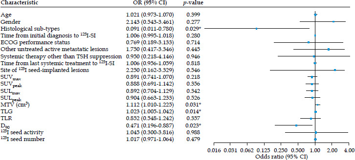
In the univariable logistic regression analysis, histological sub-types, MTV, TLG, and D90 were identified as factors influencing LRR prognosis. Variables with p < 0.05 were entered into a multivariable logistic regression model, and this analysis identified D90 as the significant independent predictor of 6-month efficacy (χ2 = 4.653, p = 0.031). The detailed results are shown in Table 4.
Table 4
Univariable and multivariable logistic regression analysis of prognostic factors for 6-month efficacy (LRR vs. non-LRR) after 125I-SI
[i] LRR – local response rate defined as the percentage of patients achieving complete metabolic response (CMR) or partial metabolic response (PMR) at 6 months (Table 2), non-LRR includes patients with stable metabolic disease (SMD) or progressive metabolic disease (PMD), 125I-SI – iodine-125 seed implantation, OR – odds ratio, CI – confidence interval, ECOG – Eastern Cooperative Oncology Group, TSH – thyroid-stimulating hormone, PET/CT – positron emission tomography/computed tomography, SUVmax – maximum standardized uptake value, SUVpeak – peak standardized uptake value, SULmax – maximum SUV normalized by lean body mass, SULpeak – peak SUV normalized by lean body mass, MTV – metabolic tumor volume, TLG – total lesion glycolysis, TLR – tumor-to-liver ratio, D90 – dose covering 90% of gross tumor volume, 125I – iodine-125
ROC curve analysis for D90 demonstrated moderate predictive value for local response, with an AUC of 0.742 and a cut-off value of 129.85 Gy (Figure 3A). To estimate potential overfitting bias, 1,000 bootstrap re-samples were performed. The optimism-corrected concordance statistics (c-index) was 0.74, suggesting that while some bias existed, the model retained reasonable predictive discrimination (Figure 3B). DCA indicated that clinical decision-making based on this model yielded greater net benefit across a broad range of threshold probabilities, demonstrating strong clinical utility (Figure 3C). The calibration curve of the D90 model showed good agreement between predicted probabilities and observed outcomes (Figure 3D). To visualize the results, a forest plot of significant variables from the univariable logistic regression was constructed, as illustrated in Figure 4.
Fig. 3
A) ROC curves representing the value of D90. B) Bootstrap AUC distribution. C) DCA of D90 models for LRR prediction. D) Calibration curve for LRR prediction
ROC – receiver operating characteristic curve, D90 – 90% of the dose received by gross tumor volume, AUC – area under the curve, DCA – decision curve analysis
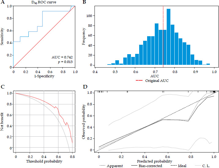
Fig. 4
Forest plot. The round dot represents the odds ratio (OR) for each variable in univariable logistic regression. The horizontal line indicates the 95% confidence interval (CI), while the vertical dashed line represents the line of no effect
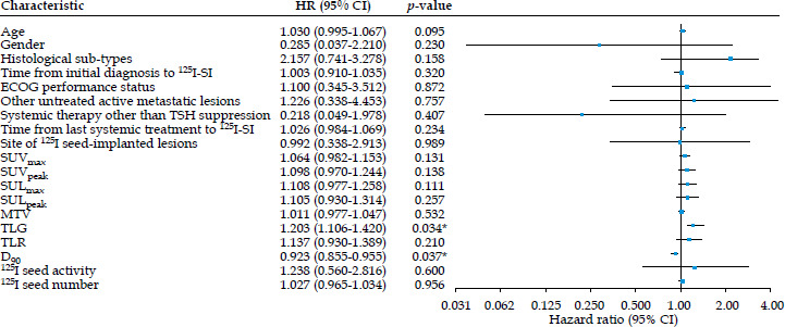
In the univariable Cox regression analysis, TLG and D90 were identified as factors influencing LPFS prognosis. The detailed results are presented in Table 5. These variables were then included in a multivariable Cox regression model, which suggested that TLG was the only metabolic parameter significantly associated with LPFS. A higher TLG was associated with a worse LPFS (HR = 4.106, 95% CI: 2.850-5.620, p = 0.036). To visualize the results, a forest plot of significant variables from the univariable Cox regression was drawn, as depicted in Figure 5.
Table 5
Univariable and multivariable Cox regression analysis of LPFS after 125I-SI
[i] LPFS – local progression-free survival, HR – hazard ratio, 125I-SI – iodine-125 seed implantation, ECOG – Eastern Cooperative Oncology Group, TSH – thyroid-stimulating hormone, PET/CT – positron emission tomography/computed tomography, SUVmax – maximum standardized uptake value, SUVpeak – peak standardized uptake value, SULmax – maximum SUV normalized by lean body mass, SULpeak – peak SUV normalized by lean body mass, MTV – metabolic tumor volume, TLG – total lesion glycolysis, TLR – tumor-to-liver ratio, D90 – dose covering 90% of gross tumor volume, 125I – iodine-125
Fig. 5
Forest plot. The round dot represents the hazard ratio (HR) for each variable in univariable Cox regression. The horizontal line indicates the 95% confidence interval (CI) and the vertical dashed line represents the line of no effect
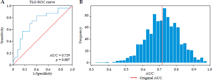
The ROC curve analysis for TLG yielded moderate predictive value of LPFS, with an AUC of 0.729 (Figure 6A). To estimate potential overfitting bias, 1,000 bootstrap re-samples were performed for this model. The optimism-corrected c-index was 0.717, indicating that while some bias existed, the model retained reasonable predictive discrimination (Figure 6B).
Discussion
125I-SI is a feasible and effective treatment option for metastatic RAIR-DTC lesions [4-6]. Given the substantial heterogeneity of RAIR-DTC, a comprehensive assessment of metabolic tumor burden and dosimetric parameters is essential for treatment selection and outcome optimization. Therefore, it is imperative to establish a non-invasive and objective methodology to predict the efficacy of 125I-SI based on pre-treatment PET/CT parameters.
Among all evaluated clinical, metabolic, and dosimetric factors, D90 demonstrated the strongest predictive value for 6-month efficacy, consistent with previous studies on 125I seed implantation [6, 12-16]. This finding was expected, as D90 directly reflects the minimum dose delivered to 90% of the target volume, which is a critical determinant of local tumor control in brachytherapy. The robust performance of D90 in calibration and decision curve analyses further supports its clinical utility in treatment planning, particularly for lesions with complex geometry or proximity to organs at risk.
In contrast, LPFS was more closely associated with TLG. TLG is a composite parameter reflecting both tumor metabolic activity and volume. Thus, it can more comprehensively characterize the tumor’s overall metabolic burden [17]. A high TLG may reflect aggressive biological features, such as hypoxia, high proliferation, or metabolic heterogeneity, which are known contributors to radio-resistance. Even after high-dose local radiation therapy, these tumors retain a substantial risk of local recurrence or progression. This finding aligns with external beam radiotherapy studies, where TLG was inversely correlated with prognosis [18, 19]. These results underscore the importance of pre-operative PET/CT for comprehensive assessment of tumor metabolic burden. Also, they indicate that the predictive role of TLG for efficacy could be extended to the field of 125I seed brachytherapy in future research.
Notably, although a significant predictor of 6-month efficacy, D90 did not show a significant association with LPFS in our analysis. This discrepancy may be explained by the physical half-life of 125I seeds (59.4 days) [20]. By six months post-implantation, the internal radiation dose declines to near-background level [21]. While an initial high dose can eradicate the majority of tumor cells and induce early metabolic response, it may be insufficient to fully eliminate resistant clones in high-metabolic-burden tumors, or to prevent recurrence driven by high proliferative potential. These observations underscore the complex temporal interplay between dose delivery and tumor response [22].
In this study, MTV alone did not emerge as a significant predictor of either LRR or LPFS outcomes. MTV is directly proportional to tumor diameter. As the tumor diameter increases, the proportion of hypoxic cells also increases, potentially leading to elevated radio-resistance of the tumor and, consequently, poorer treatment efficacy [23]. Although other studies indicated that larger tumor volumes often receive implantation of more seeds, this may lead to less uniform dose coverage and sub-optimal dose distribution [14]. Therefore, tumor volume might merely be a surrogate marker for inadequate dose delivery rather than indicating the tumor’s intrinsic radio-resistance. This insight reveals that when using particle implantation therapy for large volume metastatic lesions, caution must be exercised regarding the risk of insufficient dose due to technical limitations.
We acknowledge the inherent heterogeneity of the RAIR-DTC population in this study, including varied histological sub-types (PTC vs. FTC), metastatic sites (bone, lymph node, and lung), and prior treatments (TKI, radiotherapy, or none). This diversity reflects the real-world clinical spectrum of RAIR-DTC, and enhances the generalizability of our findings. Multivariable analyses accounted for these factors and the consistent predictive value of D90 and TLG across sub-groups, suggesting their potential utility across histological and anatomical contexts. Nonetheless, larger, more homogeneous cohorts are needed to validate and refine our findings.
This study has several limitations. First, its retrospective, single-center design introduces selection bias, potentially reducing generalizability and undermine the robustness of statistical analyses. There was no external validation cohort, thus clinical utility requires confirmation in prospective independent cohorts. Second, the relatively small sample size, particularly in sub-group analyses, may impact statistical stability and limit multivariate and survival analyses, potentially affecting the robustness of our findings. Third, incomplete dosimetric data, such as missing post-operative parameters (e.g., V100) and dose-volume data during follow-up, restrict exploration of the “metabolism-volume-dose-efficacy” pathway, and may diminish the predictive power of some parameters. Finally, MTV measurement thresholds depend on factors, such as PET scanner model, tumor type, tumor characteristics, and clinical context. This study used a fixed 40% SUVmax threshold without exploring the predictive value of other thresholds, which may affect measurement consistency.
Conclusions
Iodine-125 seed implantation is an effective treatment for locally metastatic RAIR-DTC lesions. D90 is a reliable predictor of 6-month efficacy, and TLG is significantly associated with LPFS following 125I-SI. Further prospective studies with pre-defined treatment algorithms and external validation are needed to translate these associations into individualized treatment strategies.