
Purpose
Conjunctival melanoma (CM) and conjunctival squamous cell carcinoma (CSCC) are sight- and life-threatening ocular surface malignancies, necessitating aggressive management [1, 2]. Although they represent two different tumor types, they are similar in biological behavior, since both can expand locally and metastasize to regional lymph nodes [1, 2]. Surgical excision with wide margins remains the cornerstone of management, enabling lesion removal, histopathological diagnosis, and margin control [1, 2]. A variety of adjuvant treatment modalities have been proposed, including alcohol de-epithelialization, cryopexy, and topical cytotoxic, antiviral, or immunotherapeutic agents [3, 4]. These agents include mitomycin C, 5-fluorouracil, interferon α, cidofovir, and immune checkpoint inhibitors, such as anti-PD1 (nivolumab, pembrolizumab, and cemiplimab), anti-PDL1 (avelumab and atezolizumab), and anti-CTLA4 inhibitors (ipilimumab) [3, 4]. In addition, sentinel lymph node biopsy using radio-labeled tracers has been proposed, especially in conjunctival melanoma for early detection of regional metastatic disease [5].
In incompletely resected or unresponsive cases, adjuvant radiotherapy using proton beam, external electron beam, or brachytherapy with isotopes, such as iodine-125 (125I), ruthenium-106 (106Ru), or strontium-90 (90Sr), has been employed, showing high local control rates [6-8]. Here, we presented a series of 8 patients with CM (4 cases) and invasive CSCC (4 cases) located at the bulbar conjunctiva, treated during the previous 7 years at the Department of Ophthalmology of the University Hospital of Heraklion using adjunctive 106Ru. In addition, we discussed options, dilemmas, and limitations of the management of such patients.
Case presentations
Operative methodology
All patients had incompletely excised either CSCC or CM, without metastatic disease, located at the bulbar conjunctival area, treated at the Department of Ophthalmology of the University Hospital of Heraklion from 2018 till 2025. Incompletely excised cases with primary location at the conjunctival fornices, palpebral conjunctiva, or lacrimal puncti areas were not included. For such patients, different treatment protocol is applied at our Department, which generally includes stereotactic radiosurgery (Gamma Knife). Treatment planning was performed by radiation physicists with experience in the field (KP and GS), with advisory use of plaque simulator software (Eye Physics, LLC, Los Alamitos, CA, USA) and magnetic resonance imaging (MRI) scanning, as described in the 2nd edition of GEC-ESTRO handbook (chapter 31, uveal melanoma). After epithelial healing process from the initial surgical excision under general anesthesia, adjunctive brachytherapy was performed in all cases using the same 106Ru plaque (CCB, BEBIG Medical GmbH, Berlin, Germany) with a scheduled radiation dose of 100 Gy at a depth of 1 mm, resulting in a mean outer and inner scleral radiation dose of 280.35 Gy and 240.28 Gy, respectively. No adjunctive treatments were applied to the primary tumor bed treated with 106Ru brachytherapy in our patients. A corneal dose was calculated based on isodose curves provided by the plaque simulator software and added for 3 points, i.e., the proximal cornea (the area of limbal cornea closest to the tumor), central cornea (corneal apex), and distal cornea (the area of limbal cornea most distal to the tumor). Mean values of received corneal radiation doses for the proximal, central, and distal cornea were 16.46 Gy, 0.354 Gy, and 0.060 Gy, respectively. Corresponding mean lens center dose provided by the plaque simulator software was 4.76 Gy.
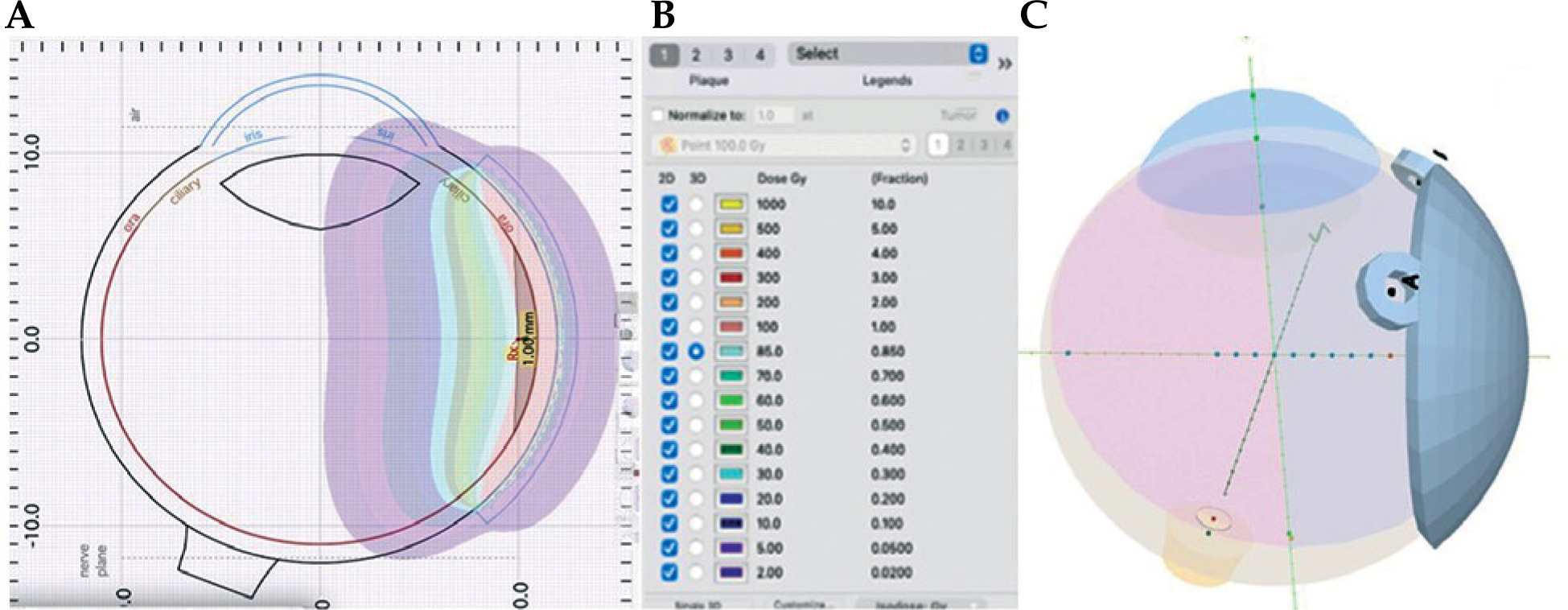
In both CM and CSCC patients, lateral margins were determined by dimensions of the plaque used (CCB, diameter of 20.2 mm). Since the tumor has been already removed, the exact area of potential residual microscopic disease could only be presumed based on previous photographs, if available (cases A, C, D, E, and H). In all patients, footprint of the plaque on the ocular surface (20.2 mm) was centered on the center of lesion, based on pre-operative photos, if available. In all cases, the large size of CCB plaque (with a diameter of 20.2 mm) was sufficient to include both presumed tumor bed and 2 mm safety margin. Due to rapid beta dose fall-off beyond the plaque edge, effective treated area corresponded to the geometric footprint of the plaque. Figure 1 illustrates the pre-operative (A, C-E, H) and pre-brachytherapy (B, F, G) clinical images of lesions included. An example of treatment plan with calculated doses for the outer and inner sclera, lens, optic nerve, and macula, is shown in Figure 2. In addition, the patient characteristics, treatment parameters, and outcomes, are presented in Table 1.
Table 1
Patient characteristics, treatment parameters, and outcomes
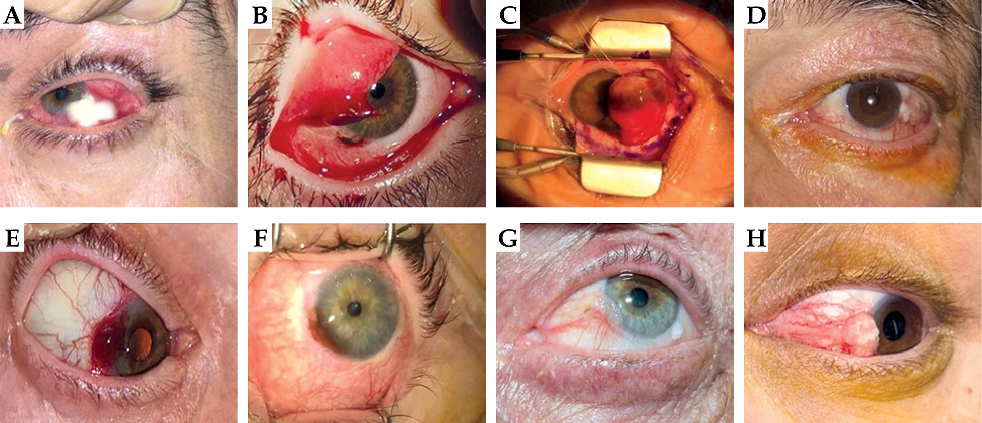
Fig. 2
Treatment plan for patient No. 3 with A) isodose curves, B) calculated doses for each isodose curve, C) and position of the plaque
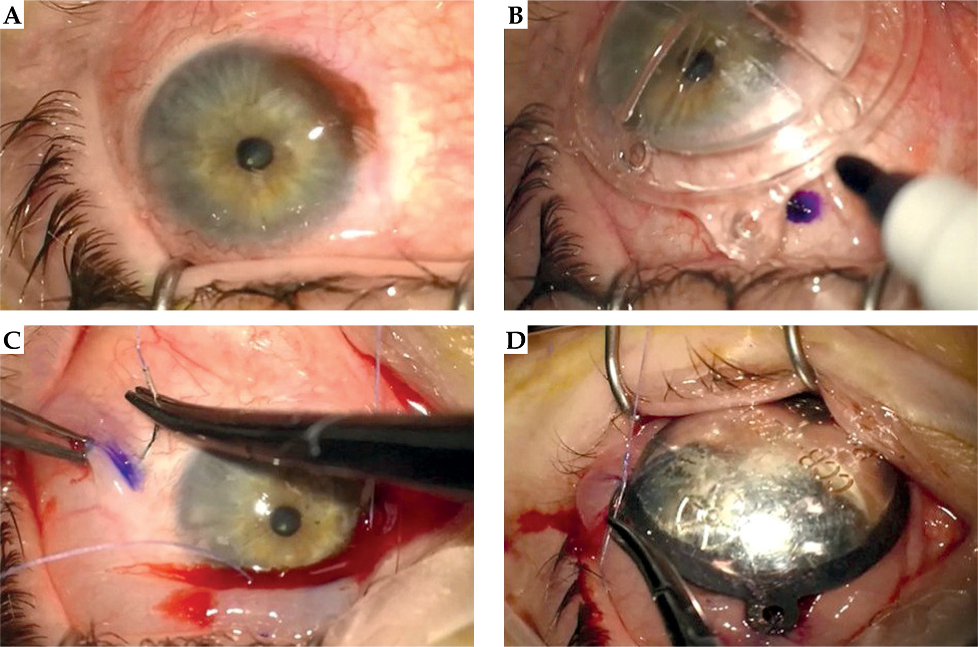
The procedure of transepithelial brachytherapy was performed under local anesthesia in all patients, and included: 1. Marking of the suture insertion sites using a transparent dummy plaque; 2. Insertion of 6.0 Vicryl sutures to engage the superficial sclera at pre-marked sites; 3. Securing the CCB plaque on the ocular surface with pre-placed sutures; and 4. Temporary tarsorrhaphy to hold the eyelids in close apposition to the plaque (Figure 3). On the next day, the tarsorrhaphy was opened and the plaque removed. The exact timing of plaque removal was determined by dosimetric planning, considering the date of plaque construction and target depth (1 mm); but it did not exceed 24 hours in any case (Table 1). Since the plaque covered partly or completely the corneal surface, all patients developed corneal epithelial defects managed by a bandage soft contact lens with topical antibiotics. Toxicity assessment included slit-lamp evaluation for corneal epithelial defects, limbal stem cell deficiency, cataract formation, and lacrimal drainage obstruction. No formal standardized toxicity grading scale was applied. During follow-up (range, 18-75 months; median, 46 months), no adverse effects from the anterior ocular segment, such as corneal epitheliopathy, dry eye disease, lacrimal outflow obstructions, or cataract development, were observed. Local control was defined as the absence of clinically detectable tumor recurrence at the treated site during follow-up. Recurrence was determined based on slit-lamp biomicroscopic examination and, when clinically indicated, imaging studies (MRI). Follow-up assessments were performed at regular intervals according to departmental protocol.
Fig. 3
Surgical technique of ruthenium-106 (106Ru) surface brachytherapy (patient No. 6). A) Initial appearance of the lesion on the ocular surface. B) Application of dummy plaque and marking of suture positions. C) Pre-placement of sutures engaging the superficial sclera. D) 106Ru in place with suture tightening
Case 1
A 51-year-old male presented with a large elevated painless and whitish mass on the infero-temporal surface of the left eye (Figure 1A). The mass has been slowly growing over the past several months, while previous history revealed prolonged significant exposure to sunlight (the patient was a sailor). On examination, the lesion had macroscopic features suggesting of a CSCC. A systemic work-up was done, including MRI scans of the head and neck, to rule out metastatic disease (non-contributory), and the patient underwent excision of the lesion under general anesthesia; no adjunctive treatment was administered. Histopathological examination confirmed the presence of a CSCC with infiltrated deeper margins (along the scleral surface). An MRI scan was performed, excluding the presence of intra-ocular or intra-orbital disease. Subsequently, the patient underwent transepithelial Ru-106 brachytherapy, with a treatment plan covering the entire clinically visible lesion with a large, at least 2 mm, safety margin beyond the apparent lesion borders, considering the 20.2 mm large diameter of CCB plaque used. The position of the plaque was determined by direct observation of the tumor bed on the ocular surface, using pre-operative photographs of the lesion. However, 3 months later, the patient presented with a macroscopic local recurrence. A new MRI scan revealed orbital and intra-ocular extension, and a decision was taken to exenterate the orbit. Histopathological examination of the orbital specimen confirmed the presence of a squamous cell carcinoma, infiltrative to both eyeball and orbital tissues, with clear excision margins along the periorbita. The patient remains disease-free for 75 months post-treatment.
Case 2
A 17-year-old female was referred from another hospital for an incompletely excised CM located at the corneo-scleral limbus of the right eye with topical recurrence. No adjunctive treatment has been applied during the initial excision due to low clinical suspicion of melanoma. On examination, the patient presented with bloody tears, while on eversion of the upper eyelid, a secondary focus inoculated from the primary site in the area of contact with the upper eyelid was identified. Moreover, another secondary focus in the area of lower punctum, probably due to lacrimal dissemination, was found (Figure 1B). A decision was taken to treat the recurrent primary focus with transepithelial 106Ru brachytherapy, whereas the secondary foci were treated with wide excision and adjunctive cryotherapy. This was done since the concave configuration of CCB plaque did not permit anatomical apposition to eyelid conjunctiva. Despite an initial tumor control, the upper eyelid lesion recurred aggressively 3 months later with orbital invasion, without metastatic disease. To avoid orbital exenteration, the recurrent lesion was treated with Gamma Knife and adjunctive systemic immunotherapy using systemic checkpoint inhibitors (ipilimumab plus nivolumab). The patient responded very well and is disease-free for 44 months. With cumulative radiation-induced toxicity by different modalities used in the same patient, dosimetric data from brachytherapy was sent to radiotherapists to plan Gamma Knife treatment without exceeding safety limits. It should be noted that additional treatments (cryotherapy, stereotactic radiosurgery, and systemic immunotherapy) were applied to secondary lesions or subsequent recurrences, but not to the primary tumor bed treated with 106Ru brachytherapy.
Case 3
A 35-year-old female presented with a large elevated melanotic lesion of the left eye (the patient was psychotic and refused treatment until the time of presentation), occupying almost all the area of nasal conjunctiva and overhanging on the corneal surface (Figure 1C). Metastatic and orbital disease were excluded with systemic work-up, and the lesion was removed without adjunctive treatment. Histopathological examination confirmed the presence of a CM with infiltrated surgical margins. Accordingly, following the completion of healing process of the excision area, transepithelial 106Ru brachytherapy was performed with initial tumor control. Despite the large size of the lesion and technical difficulties associated with anatomical constraints in the nasal conjunctiva, the presumed area of tumor bed with a diameter of 12 mm was completely covered by the plaque and with associated 4 mm safety margin. Treatment depth planning remained 1 mm for this patient since MRI scan did not reveal scleral infiltration by the tumor. Unfortunately, the tumor recurred aggressively on one wound margin and invaded the orbit. Apart from a possible anatomical off-set of the plaque resulting in geographic miss, a recurrence may also reflect pre-existing microscopic disease beyond clinically detectable margins. Therefore, considering the orbital invasion and difficulty for the patient to maintain a strict immunotherapy protocol, a decision to exenterate the orbit was taken. Presently, the patient is disease-free at 62 months post-treatment.
Case 4
A 58-year-old male presented with a conjunctival elevated painless lesion at the area of the infero-lateral corneo-scleral limbus of the right eye (Figure 1D), which was surgically removed without adjunctive treatment. Histopathological examination revealed an invasive CSCC with infiltrated margins. Subsequently, the patient underwent systemic work-up that excluded metastatic disease. After the completion of epithelial healing, he underwent transepithelial 106Ru brachytherapy treatment, achieving local tumor control. The patient is disease-free for 60 months.
Case 5
A 54-year-old female presented with an elevated indolent melanotic epibulbar mass of the right eye, which was there for a few months, with progressive enlargement reported (Figure 1E). The lesion was surgically removed without adjunctive treatments applied, and histopathologically proven to be a CM with infiltrated deeper margin. Following the completion of epithelial healing, and exclusion of systemic and orbital disease by imaging and laboratory studies, she underwent adjunctive surface 106Ru brachytherapy, achieving local tumor control. The patient remains disease-free at 24 months.
Case 6
A 50-year-old female was referred to the Department of Ophthalmology of the University Hospital of Heraklion due to a CM of the left eye (Figure 1F), which has been excised elsewhere, with infiltrated margins but recurred topically. No adjunctive treatment was administered. A systemic work-up for metastatic disease was non-contributory. The patient underwent surface 106Ru brachytherapy, and is disease-free for 26 months.
Case 7
A 60-year-old male was referred to the Department of Ophthalmology of the University Hospital of Heraklion due to a CSCC of the left eye, which has been excised elsewhere (without adjunctive treatment) but recurred topically (Figure 1G). The patient has been receiving systemic immunosuppressive medications continually due to heart transplantation. Although previous photographic documentation was not available, we proposed adjuvant surface brachytherapy, with severity of the initial histopathological diagnosis (incompletely excised CSCC) considered. However, the risk of surface brachytherapy failure associated with poor documentation of the initial borders of the lesion was fully explained to the patient along with other options, including observation or proton beam treatment of the entire ocular surface. A comprehensive medical investigation for metastatic disease was non-contributory, and the patient underwent surface 106Ru brachytherapy. He remains disease-free for 48 months.
Case 8
A 43-year-old male presented with an elevated painless conjunctival lesion on the ocular surface of the left eye, partially overhanging on the adjacent corneal surface. He underwent excision of the lesion without adjunctive treatments, and histopathological examination confirmed an incompletely excised CSCC. Following the completion of healing process, he underwent surface 106Ru brachytherapy and remains disease-free at 18 months.
Discussion
This is a presentation of 8 consecutive cases of incompletely excised invasive CSCCs (4 cases) and CMs (4 cases), treated at the Department of Ophthalmology of the University Hospital of Heraklion. Ruthenium-106 transepithelial brachytherapy was administered to the patients, delivered by the same plaque model (BEBIG CCB) with a target dose of 100 Gy at a depth of 1 mm. The results indicate an overall success rate of 75% (6/8 cases), achieving local tumor control in both CSCC and CM cases in a follow-up period of 18-75 months.
Several previous studies have reported satisfactory treatment outcomes of surface brachytherapy in conjunctival malignancies, mostly with 90Sr and 125I plaques. Cohen et al. reported 90% local tumor control using 90Sr beta radiotherapy following excision of CM [9], while Berkowitz et al. showed successful treatment of CM using annulus-shaped 125I plaque brachytherapy [10]. Arepalli et al. employed 125I plaque radiotherapy for scleral-invasive CSCC and reported local tumor control in all treated eyes [11]. Due to its greater penetration depth, 125I may provide advantages for deeper invasion, but possibly at the expense of increased radiation dose to non-target tissues [12-17]. In terms of dosimetry, 125I plaques for ocular surface brachytherapy are typically prescribed to deliver around 100 Gy to 1.5-3 mm compared with beta emitters (i.e., 90Sr) targeting depths of 1 mm [9]. It is because 125I plaques achieve deeper penetration due to their gamma radiation [10, 11]. Ruthenium-106 surface brachytherapy has also been employed for recurrent or incompletely excised conjunctival tumors, though evidence remains largely limited to case series. Rao et al. reported 95% local tumor control in incompletely excised CSCC treated with 106Ru plaque brachytherapy [18]. Also, Grajewski et al. used adjuvant 106Ru plaque brachytherapy following excision, and achieved an 84% local control rate [19]. Interestingly, the case series presented by Grajewski et al. is not limited to incomplete excision, but directly supports 106Ru plaque brachytherapy as a post-excision adjuvant ocular-surface approach.
In the current case series, only incompletely excised lesions were included, and the two cases requiring orbital exenteration likely harbored microscopic orbital disease beyond the plaque footprint at presentation, as described in advanced conjunctival malignancies [20]. Therefore, the results from this case series suggest that transepithelial ocular surface brachytherapy may represent an effective adjunctive modality in selected cases. Local control was achieved in 3/4 of conjunctival melanoma cases (75%) and 3/4 of conjunctival squamous cell carcinomas (75%). Tumor recurrence in cases No. 1 and 3 may possibly be associated with incomplete evaluation of tumor boundaries, but also with inherent technical limitations of the method associated with anatomical position of the lesion, or alternatively, the presence of topical micro-metastatic disease, which could not be predicted in advance. Interestingly, in case No. 2, there was no tumor recurrence on the initial tumor site, which was the only tumor location treated with surface 106Ru brachytherapy, a finding that together with the successful outcomes of cases No. 4-7 supports the feasibility of the methodology employed.
Importantly, no adjunctive treatments, such as topical cryotherapy, immunotherapy, or chemotherapy was administered to patients in this series prior to the application of 106Ru brachytherapy. This shows that by using ocular surface 106Ru brachytherapy as the only adjunctive treatment modality in incompletely excised CMs or CSCCs, local tumor control can be achieved without exposing the eye to cumulative toxicity of other adjunctive treatment modalities, such as mitomycin C, which should not be regarded as a substitute for brachytherapy but as a therapeutic modality with a distinct mechanism of action. In selected cases, particularly in the presence of extensive or multifocal lesions, mitomycin C may act synergistically with local radiotherapy, supporting disease control when used as part of a multimodal treatment strategy rather than as an alternative approach. Concerning radiation dosimetry, the dose of 100 Gy at 1 mm depth with corresponding outer and inner scleral doses of under 300 Gy in all cases, is consistent with previously published 106Ru protocols prescribing 80-110 Gy at depths of 1-2.2 mm [18, 19].
The retrospective design and small number of included cases are the drawbacks of this study. The relatively small sample size implies that findings should be interpreted with caution and considered preliminary. Another limitation is that brachytherapy was delivered following surgical excision, and target delineation was based on the presumed tumor bed location. In several cases, plaque positioning relied on pre-operative clinical photographs, which were not consistently available. Moreover, histopathological margin orientation was not systematically used to guide target coverage. These factors introduce uncertainty in target delineation and may increase the risk of geographic miss, potentially affecting local control. Importantly, patient selection targeted only incompletely excised cases and current 106Ru sources are designed for epibulbar use to treat uveal neoplasms. Therefore, the extrapolated use of the same sources on the ocular surface is limited for epibulbar locations, where geometric conformation with the tumor bed is feasible. Since the protocol employed at our Department for both surface brachytherapy and subsequent follow-up is the same for CMs and CSCCs, we combined both conditions into a single cohort. However, this represents a source of biological heterogeneity, and should also be considered a limitation of the study. While there are 106Ru plaque designs for epibulbar applications in the BEBIG plaques menu, such as CIA, CIB, CIB-2, and COE (BEBIG Medical GmbH, Berlin, Germany), only CCB and COB are available at the Department of Ophthalmology of the University Hospital of Heraklion, whereas 106Ru treatments are mainly intended for intra-ocular lesions. The fact that safety and efficacy outcomes are comparable with previous studies indicates that plaque designs for intra-ocular lesions may alternatively be successfully used on the ocular surface without inducing corneal or stem cell toxicity. Actually, round plaque designs, such as the CCB used in this series, may offer adequate corneo-scleral or corneal radio-therapeutic action, which could be important, since several of the lesions presented in this series, i.e., cases No. 5-8, displayed corneal overhanging or frank corneal expansion. The minimally invasive nature as well as enhanced efficacy and safety profile of 106Ru ocular surface brachytherapy described in this report is a reasonable alternative even in centers with limited plaque resources. However, 106Ru ocular surface brachytherapy is effective only when preceded by adequate pre-treatment planning, and based on a careful assessment of lesion size, extent, and margins. Accurate initial staging is therefore a critical factor in maximizing therapeutic success and minimizing the risk of treatment failure.











