Introduction
IgG4-related disease is an inflammatory-fibrotic, immune-mediated disease which can occur in any organ. Due to its multi-organ involvement, it is often difficult to diagnose as the disease typically resembles malignancy, infection, or other autoimmune diseases [1]. Most often, the affected organs include the pancreas (autoimmune inflammation of the pancreas – AIP), the hepatobiliary tract (sclerosing cholangitis), the salivary glands (chronic sclerosing sialadenitis, Mikulicz’s disease, Küttner’s tumour), the orbital soft tissues (orbital pseudotumor), and the lymph nodes [2].
IgG4-related disease was recognised as a distinct disease relatively recently, i.e., in 2003 [3, 4]. It seems that recognition is still insufficient, which makes it difficult to accurately determine its incidence [3]. According to some sources, the disease is estimated to occur in 2.8–10.8 cases per million people [5]. Most commonly, men are more prone to getting affected (5 : 1) than women in the middle age [5], and it is possible for children to also get the disease [2, 3]. However, in cases regarding head and neck symptoms, the disease frequency is similar in both sexes.
The classifying criteria for IgG4-related disease, made in 2019 by the American College of Rheumatology (ACR) and the European League Against Rheumatism (EULAR), are helpful in diagnostics but should not exclude starting treatment if all the criteria are not met [1]. Treatment includes oral corticosteroids, immunosuppressive drugs and biological agents [5].
The description below shows multidisciplinary approaches to a patient with chronic sinusitis and ocular symptoms, which led to the diagnosis of IgG4-related disease.
Case report
A 61-year-old female patient, with underlying medical conditions (hypertension, diabetes, hypothyroidism), was admitted to the Clinic of Otolaryngology and Oncological Laryngology in Poznan due to changes in inflammatory changes in the paranasal sinuses with accompanying ocular symptoms. The patient’s history includes 30 years of eyelid swelling and itching, divergent strabismus of the left eye, and over the last 6 months, worsening of these symptoms with an additional left-eye vision loss, periodic purulent discharge coming from the right eye, and retrobulbar pain. In the past, the patient underwent functional endoscopic sinus surgery (FESS) three times due to chronic sinusitis.
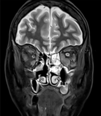
Due to the inability to see from her left eye, the patient was first hospitalized in the Neurology Department. Based on the magnetic resonance imaging (MRI) (Figure 1) of the head, the following was found: a large infiltration measuring 29 × 14 mm and a maximum height of 27 mm, showing heterogeneous contrast enhancement, located outside and inside the cone in the posterior part of the left orbit, involving thickened extraocular muscles (mainly the inferior and medial rectus muscles), surrounding the optic nerve, and reaching the pterygopalatine fossa; a smaller thickening of the extraocular muscles in the right orbit with additional slight infraorbital swelling; The swelling of both eyelids; severe inflammatory changes in the paranasal sinuses, predominantly in the left sphenoid sinus and the left side of the ethmoid sinus.
FIGURE 1
Magnetic resonance imaging scan revealing paranasal sinus changes and extraocular muscle thickening predominantly on the left side
Due to the thickening of the extraocular muscles, Graves thyroid orbitopathy was suspected. Thus, the patient was referred to the Department of Endocrinology for further diagnostic evaluation. Lab results showed an increase in TSH (52.6 uU/ml), low FT4 (8.35 pmol/l), low FT3 (2.2 pmol/l) and positive anti-thyroglobulin and anti-thyroid peroxidase antibodies. These findings, in conjunction with the MRI scan of the orbits, excluded the possibility of thyroid orbitopathy due to Graves’ disease. Simultaneously, positive IgG4 antibody levels of 6.610 g/l and IgG levels of 20.2 g/l were detected. This led to the raised suspicion of IgG4-related disease. Due to the typical systemic involvement of many organs in IgG4-RD, a full body positron emission tomography (PET) scan was performed which showed no abnormalities anywhere other than an increased tracer uptake in the region of the nasal bone and paranasal sinuses (Table 1).
TABLE 1
Comparative table between Graves’ disease and IgG4-related disease
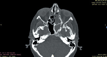
A computed tomography (CT) scan of the paranasal sinuses was performed which revealed: airless and mucus mass filled the left sphenoid sinus, connecting to the orbit due to bilateral disruption of its lateral wall; an airless left ethmoid sinus; both the ethmoid and maxillary sinus were open wide to the nasal cavity; besides the parietal mucosal swelling in the maxillary sinuses, the other paranasal sinuses were normally air-filled (Figure 2).
During the hospitalization, the patient was consulted by an ophthalmologist. Ophthalmic examination revealed conjunctivitis in the right eye, preserved light perception in the left eye, divergent strabismus of the left eye, pronounced eyelid swelling, and limited mobility of both eyeballs.
The patient was referred to the Laryngology Department for a biopsy of the paranasal sinus lesions mentioned before. The patient was qualified for surgery under general anaesthesia in which a biopsy was taken from the pathological mass filling the lumen of the left sphenoid sinus for histopathological examination. In the postoperative period, both oral and topical corticosteroids were recommended. Microscopic examination revealed chronic inflammatory infiltration in the stroma, with large presence of plasma cells (CD138+) – around 100-120/1 HPF – as well as marked fibrosis. Immunohistochemical analysis of plasma cells showed IgG expression, with the majority expressing IgG4 (> 40% plasma cells IgG4+). The histological findings, in addition to the elevated serum IgG4 and IgG, with full correlation of the patient’s clinical findings confirmed the diagnosis of IgG4-related disease. The patient was referred to the Rheumatology Department to start targeted therapy. Treatment included a 40 mg daily dose of prednisone for 2–4 weeks, followed by a taper of 5 mg every 1–2 weeks, reaching 5–10 mg/day by 3–6 months. The patient responded well to the glucocorticoid therapy. Due to the high risk of relapse in IgG4-RD, monitoring and long-term maintenance is crucial in treatment with a low-dose of prednisone recommended for up to 3 years in some patients.
Discussion
According to EPOS 2020, chronic rhinosinusitis (CRS) is classified into primary CRS and secondary CRS. Each of these categories is further divided into localized (unilateral) and diffuse (bilateral) forms, which, in turn, present as two dominant endotypes: type 2 and non-type 2 [6].
In the case of the localized primary chronic rhinosinusitis (CRS), two main forms are distinguished: allergic fungal rhinosinusitis (AFRS, type 2) and isolated rhinosinusitis (non-type 2). Secondary unilateral conditions include odontogenic sinusitis, fungal ball, and tumours. Unilateral sinonasal lesions account for approximately 23% of all nose and sinus-related cases [7]. Retrospective studies indicate that the majority of these cases are caused by chronic inflammation, followed by fungal infections, inverted papilloma, and malignant lesions [7–11]. According to a meta-analysis of 17 studies (including a total of 1,133 patients) by Moss et al. [12], the aetiology of isolated sphenoid sinusitis is associated with chronic rhinosinusitis without nasal polyps (CRSsNP) (28.3%), mucocele (20.3%), fungal sinusitis (12.5%), malignant tumours (7.7%), intracranial lesions (7.0%), benign tumours (5.7%), and chronic rhinosinusitis with nasal polyps (CRSwNP) (3.4%).
The sinus manifestation of IgG4-related disease is a relatively rare phenomenon. The first cases were reported only in 2009 by Ishida et al. [13]. The lack of definitive diagnostic criteria for IgG4-related sinusitis further complicates accurate diagnosis. The described case of a 61-year-old female patient with unilateral paranasal sinusitis inflammation and severe ocular symptoms further represents an example of an atypical presentation of this disease within the sphenoid sinus.
IgG-related disease often presents as a condition that mimics malignancy, severe infection, or another autoimmune disease. In this patient, despite an initial suspicion of Graves’ disease, imaging and laboratory results did not confirm the diagnosis. Instead, an elevated level of IgG4 antibodies guided further investigation toward IgG4-related disease.
An important factor in establishing the diagnosis was the histopathological examination, which revealed a dense inflammatory infiltrate with numerous IgG4-positive plasma cells, ultimately confirming IgG4-related disease. Treatment for IgG4-RD in the discussed case involved the use of glucocorticoids, which are considered first-line therapy. The rapid response to treatment emphasises their effectiveness in controlling inflammation and preventing complications. Additionally, the patient was referred for further specialised care in the Rheumatology Department, in line with recommendations for a multidisciplinary approach to IgG4-RD patients.
Documenting this atypical case aims to raise awareness among healthcare professionals, particularly otolaryngologists, to enable prompt diagnosis and appropriate management.