
Purpose
Brachytherapy is an essential component in the definitive management of cervical cancer [1, 2], enabling highly conformal dose delivery with rapid dose fall-off that limits exposure to surrounding organs at risk [3]. Applicator type and placement are pivotal determinants of dose distribution, target coverage, and ultimately, disease control [4, 5]. In clinical practice, intra-operative pelvic examination plays a key role in guiding applicator selection, with assessment of vaginal canal (VC) dimensions and tumor extent at the time of insertion. Both mechanical capacity and tumor geometry, including stage, are critical factors in determining the appropriate applicator [6, 7]. However, evaluation of these parameters may not be feasible even before the patient has entered the procedure suite and anesthesia initiated, hence information from pre-brachytherapy MRI (pre-MRI) might help supplement the clinical exam.
Image-guided adaptive brachytherapy has improved soft-tissue visualization, target definition, and dose optimization [8-10], which is particularly important given the association between poor implant geometry or applicator displacement and increased local recurrence [4]. Cervical cancer patients frequently have an MRI at the end of external beam radiotherapy (EBRT) to assess the response and aid in planning brachytherapy. From our clinical experience, we observed substantial variability in VC dimensions on these pre-MRIs, quantifiable by MRI-based contouring, and sought to leverage this information to supplement the clinical examination and applicator selection even prior to entering the brachytherapy suite. While standard of care in the treatment of cervical cancer with brachytherapy, this pre-MRI might yield additional information to support procedural planning, including selection of brachytherapy applicators, and thus warrants further study. Advanced knowledge provided by this anatomy-guided approach could reduce procedure and anesthesia time (with associated adverse effects), streamline equipment preparation, improve patient comfort, and optimize applicator positioning, and thereby dosimetry.
This study investigated the relationship between pre-MRI-derived VC volumes and applicator selection. We hypothesized that the pre-MRI upper VC volume is a reliable predictor of applicator selection prior to the procedure, when clinical examination has not yet been performed under anesthesia.
Material and methods
We conducted a single-institution retrospective cohort study approved by the institutional review board. One hundred and twenty-three consecutive patients with cervical cancers treated by two radiation oncologists with brachytherapy for up to four fractions at a single-institution from January 2022 to November 2024, were screened. Applicator selection was determined by a treating physician at the time of examination under anesthesia during first procedure. Eligibility criteria included age ≥ 18 years, completion of pre-MRI with vaginal gel, and treatment incorporating a tandem and ovoid (TO) applicator (Varian Medical Systems, Palo Alto, CA, USA) plus interstitial needles [11] or tandem and cylinder (TC) with interstitial needles [12]. All patients were treated using a hybrid intracavity-interstitial approach.
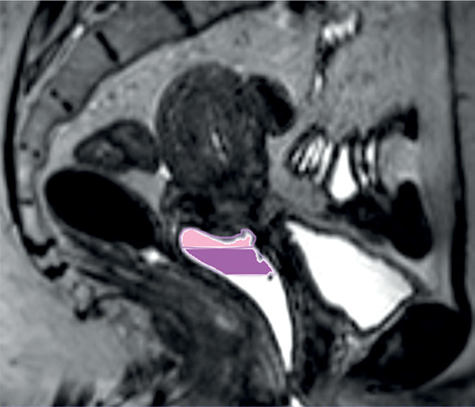
To objectively quantify canal size, the upper VC was contoured on axial T2-weighted images of pre-MRI completed with vaginal gel. This was done according to a standardized protocol of ARIA Eclipse by a personnel trained under the supervision of attending radiation oncologist (Figure 1), and corresponding VC volumes were computed. Vaginal gel during pelvic MRI has been found to improve delineation of the cervix and vaginal walls, enhancing anatomic visualization [13]. This is standardly performed at our institution for gynecologic patients undergoing pelvic MRI using a 200 cc syringe inserted until patient-reported capacity is reached. Without vaginal gel, collapse of vaginal walls precludes accurate volume contouring; thus, patients who completed pre-MRI without vaginal gel (e.g., at another institution) were excluded from the current study. As it was uncertain whether the proximal 1 cm or 2 cm of the canal would be more predictive of applicator utilization, both measurements were assessed.
Fig. 1
Delineation of the upper 1 cm (pink) and 2 cm (magenta) volumes of the vaginal canal measured on pre-brachytherapy MRI
Group comparisons were performed using Wilcoxon rank-sum or Kruskal-Wallis tests, as appropriate. A two-sided p-value < 0.05 was considered statistically significant. All analyses were performed using R version 4.4.3 software (R Foundation for Statistical Computing, Vienna, Austria).
Results
Of the 123 screened patients, 114 met eligibility criteria and were included in the final analysis. The baseline patient demographics and clinical characteristics are summarized in Table 1. Across the cohort, TC applicators were applied in 34% of all fractions, and in the rest, TO applicators with various combinations of ovoids were used. The volume of the upper 1 cm of VC was found to be significantly associated with applicator type utilized in some fractions. The volume of the upper 2 cm of VC was significantly associated with applicator type in all fractions (p < 0.01).
Table 1
Clinical characteristics of the patient cohort with gynecologic cancers treated with intracavitary/ interstitial brachytherapy
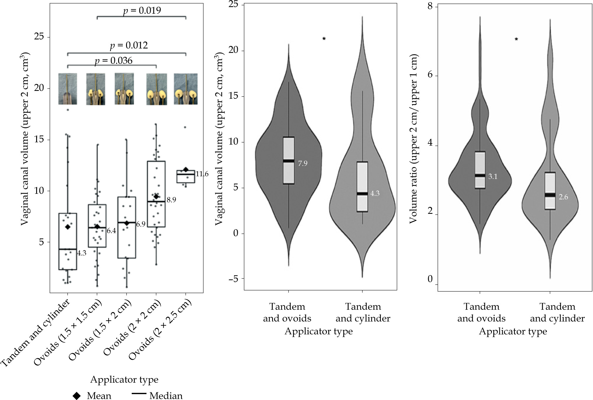
Figure 2 demonstrates a significant trend between increasing upper 2 cm VC volumes and applicator types with increasing ovoid sizes used in the first fraction. The median volumes were 4.3 cm3 (TC), 6.4 cm3 (dual mini ovoids), 6.9 cm3 (mini × 2 cm ovoids), 8.9 cm3 (dual 2 cm ovoids), and 11.6 cm3 (2 × 2.5 cm ovoids). Patients treated with TC applicators had smaller upper 2 cm VC volumes than those treated with TO applicators (mean ±SD: 6.48 ±5.14 cm3 vs. 8.06 ±3.88 cm3; p = 0.045). The volumetric ratio of the upper 2 cm to the upper 1 cm of VC was also lower among patients treated with TC applicators. Larger upper 2 cm VC volumes were associated with lower T stage classification (T1-2: mean 8.4 cm3 ±4.2 vs. T3-4: mean 6.5 cm3 ±4.0; p = 0.032). In contrast, VC volume did not differ significantly by FIGO stage (p = 0.45), N classification (p = 0.35), or M classification (p = 0.42). In exploratory multivariable analysis including both upper 2 cm VC volume and T stage, the associations of both VC volume and T stage with TC applicator use were attenuated.
Fig. 2
Sagittal and coronal T2-weighted MRIs demonstrating A, B) a tandem and cylinder applicator in situ, with central tandem and surrounding cylindrical component visible within the vaginal canal, and C, D) a tandem and ovoid applicator in situ, with bilateral ovoids positioned laterally to the tandem within vaginal fornices
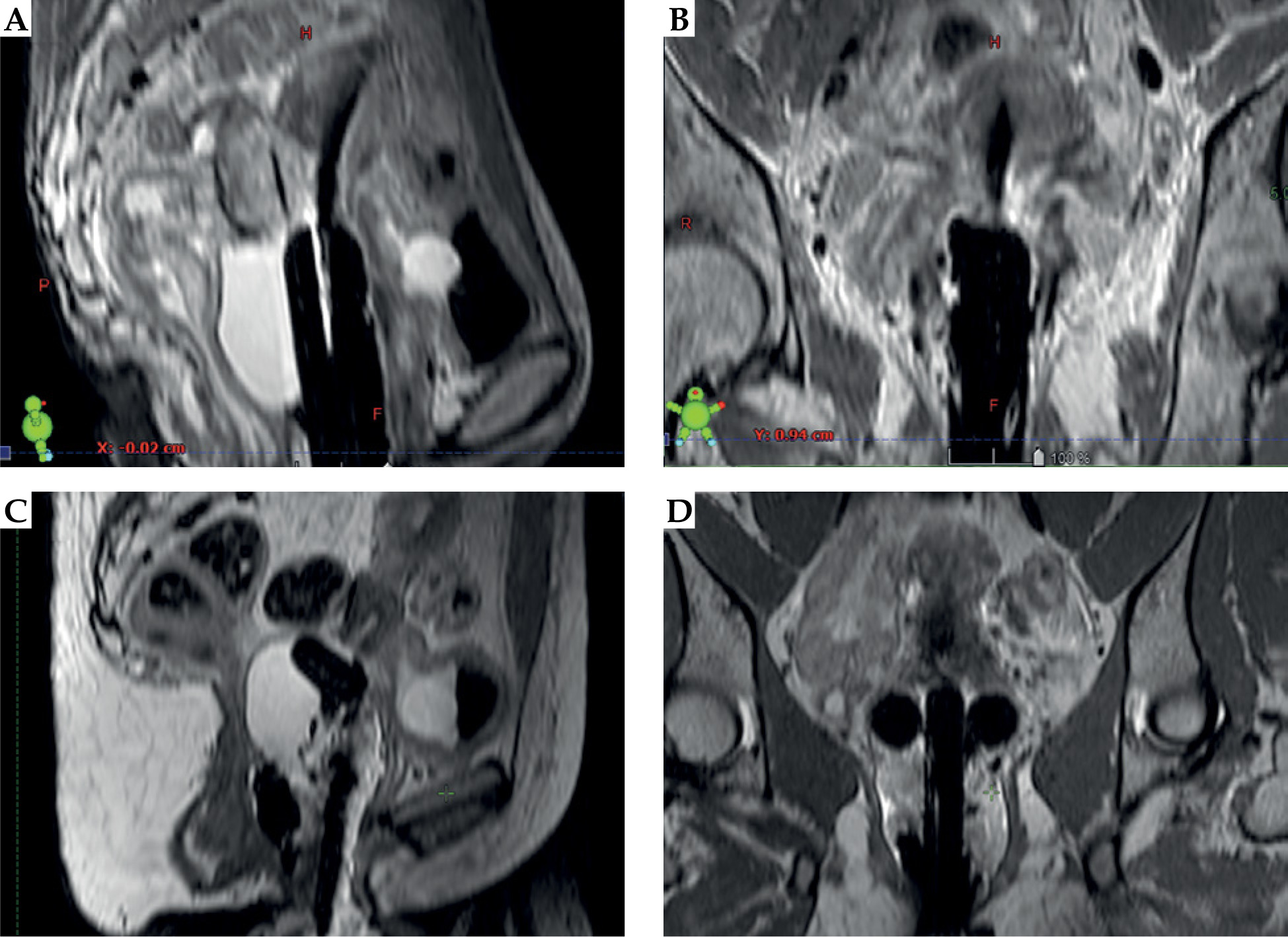
Fig. 3
Distribution of vaginal canal volumes on pre-brachytherapy MRI stratified by applicator sub-type or category. Box and violin plots depict median values, data distribution, and kernel density estimation, with whiskers extending to minimum and maximum values. Statistical significance of differences between groups is denoted by asterisks (*p < 0.05)
Amongst “uniform applicator” patients (for whom one applicator was used in all treatment fractions), the TC applicator was applied for most patients (22%), followed by dual 2 cm (20%) and dual mini ovoids (16%). Nearly all patients (92%) initially treated with a TC applicator continued with this applicator for subsequent fractions. The remaining patients did not complete four total fractions. In contrast, of the 89 patients who were treated with a TO applicator at the first fraction, 14 (15.7%) were treated with a TC applicator for at least one subsequent fraction of their treatment course.
Discussion
Precise applicator placement is critical for achieving optimal target coverage, dosimetry, and procedural efficiency, as sub-optimal placement has been associated with increased local recurrence [4]. Although existing guidance on applicator selection primarily emphasizes tumor-related factors, such as high-risk clinical target volume (HR-CTV) size, and extent [3], our findings suggest that pre-brachy MRI of VC anatomy represents a complementary consideration. Importantly, this information is available prior to the procedure, and may help narrow the range of applicators prepared for and utilized during the treatment, potentially reducing unnecessary equipment use and subsequent re-sterilization. In this study, the upper VC volume measured on pre-MRI with vaginal gel, emerged as a simple anatomic factor associated with applicator choice, with smaller upper VC volumes favoring TC applicators and larger VC volumes favoring TO applicators. Notably, the unidirectional pattern of applicator choice, where 92% of patients treated with TC continued using TC for the remaining fractions, suggests that anticipating TC use upfront may serve as an early triage metric and a practical time-saving strategy by reducing intraprocedural uncertainty and minimizing interfraction applicator changes. Together, these findings highlight a simple and widely accessible approach to support early applicator planning and optimize dosimetry as early as possible in the treatment course.
These conclusions should also be interpreted in the context of local tumor extent. Because upper VC anatomy is influenced by tumor geometry, the VC volume may not represent a purely independent anatomic feature, but may partly reflect the local effects of disease extension. Consistent with this, larger upper 2 cm VC volumes were associated with lower T stage in our cohort, whereas no significant association was seen with FIGO stage. In exploratory multivariable analysis, the associations of both VC volume and T stage with TC applicator use were attenuated, likely owing to limited statistical power and expected collinearity between these related variables. Taken together, these results suggest that the upper VC volume may function as a practical imaging-based metric that captures clinically relevant anatomy for applicator selection, while also reflecting, in part, local burden of disease.
Current guidelines recommend application selection based on tumor anatomy, organ at risk proximity, and institutional applicator availability [3, 14]. Specifically, bulky or laterally extending tumors, inadequate tumor regression following EBRT, unfavorable normal tissue anatomy, and more advanced disease, are recognized indications for incorporating interstitial needles. Stenhouse and colleagues prospectively validated a machine learning model combining tumor geometry and organ at risk features, demonstrating utility in guiding selection between intracavitary and hybrid interstitial applicators [15].
There are some limitations in this study to acknowledge, such as being retrospective in nature and from a single-institution. However, the study provides a novel demonstration of how pre-MRI anatomy can be utilized to inform applicator selection. While the relationship between VC volumes and applicator size may be inferred intra-operatively, this study demonstrates that MRI-derived measurements obtained before brachytherapy can provide objective, pre-procedural information to guide applicator selection prior to clinical exam under anesthesia. By integrating simple VC contours and volume measurements into pre-procedural planning, clinicians may improve workflow efficiency and reduce uncertainty in advance of the procedure. Future studies may explore potential correlations between VC dimensions and the number and positioning of interstitial needles required for optimal implants as well as their potential influence on trajectories of curved needle systems, such as those used with 3D-printed templates, which allow for a narrow vaginal applicator to cover a wider tumor dimension [16].
Conclusions
In conclusion, pre-MRI upper VC volume is a useful predictor of applicator selection in brachytherapy, with volumes < 4 cm3 favoring alternative applicators, such as a TC applicator. These findings suggest that pre-procedural imaging may improve applicator selection efficiency, and the particular size thresholds could be further validated prospectively. Thus, pre-MRI upper VC volume evaluation provides a simple and accessible method for applicator selection.











