Purpose
Cervical cancer remains the fourth most common malignancy among women worldwide, and continues to pose a major barrier to the World Health Organization’s elimination strategy [1]. In many low- and middle-income countries, limited screening coverage and constrained radiotherapy capacity translate into higher rates of locally advanced disease and inferior survival [2]. Dose-escalated image-guided brachytherapy is pivotal for improving local control while respecting organ at risk (OAR) constraints, yet it is technically demanding in patients with bulky tumors, distorted uterine canals, prior surgery, or extensive vaginal and parametrial involvement [3]. These clinical realities have driven interest in three-dimensional (3D)-printed applicators as a way to extend image-guided brachytherapy to anatomically challenging cases without sacrificing geometric precision or workflow robustness [4, 5]. In this context, 3D-printed templates have moved from experimental prototypes to practical tools that stabilize complex intracavitary/interstitial (IC/IS) implants.
Early clinical reports have highlighted that free-hand interstitial needles are difficult to position reproducibly and may diverge asymmetrically beyond the cervical os, particularly in large or eccentric tumors. Even in experienced hands, the geometry of free-hand needles can vary across fractions and operators, yielding inconsistent target coverage and OAR doses [6-8]. These limitations, together with the restricted adaptability of rigid commercial applicators, have motivated the development of patient-tailored 3D-printed templates, which constrain needle trajectories and allow paths to be engineered while avoiding major vessels and bowel loops [9-11].
In parallel, several groups have designed standardized yet highly individualized 3D-printed templates for locally advanced cervical cancer (LACC). The most widely described platforms include non-coplanar needle templates (3D-PNCT) and other customized tumor-conformal molds that guide IC/IS implants under computed tomography (CT) or magnetic resonance imaging (MRI) guidance [4, 5]. These designs aim to preserve the dosimetric advantages of hybrid IC/IS brachytherapy while maintaining reproducible geometry across fractions [12].
More recently, 3D-printed vaginal individualized applicators have been developed to manage complex vaginal vaults and cuff recurrences by embedding channels or sleeves for interstitial needles. These mould-based techniques conform more closely to irregular post-hysterectomy anatomy, improving coverage of high-risk clinical target volume (HR-CTV), and reducing hot spots in the vaginal wall and lower gastrointestinal tract. At the same time, they offer more predictable insertion depth and angulation, and may simplify training for both residents and experienced brachytherapists [13].
Historical European experience with MRI-guided individual vaginal molds for cervix and vault brachytherapy developed before clinical 3D printing, became widely available, showing that fully individualized applicators can achieve excellent local control with acceptable OAR doses in both intact and post-operative settings [14]. These series also emphasized the need for close collaboration between radiation oncologists, medical physicists, and radiologists when designing and commissioning patient-specific applicators, which can be integrated into image-guided adaptive brachytherapy workflows [14].
What began as single-center innovation has now been adopted across diverse healthcare systems and case mixes. Chinese cohorts have shown that 3D-printed non-coplanar templates and customized IC/IS guides can reliably reproduce pre-planned needle geometry and achieve EMBRACE-II-conformant HR-CTV coverage while maintaining comparable OAR sparing, even in large or irregular tumors [5, 9, 15].
A Danish series has incorporated 3D-printed templates into routine MRI-guided adaptive brachytherapy for LACC, using templates selectively in patients with poor response or challenging anatomy after chemoradiation [4, 9, 14]. This experience suggests that 3D printing can be aligned with established guideline-based workflows, and implemented without prolonging procedures or compromising the overall treatment time.
In Southeast Asia, Siriraj Hospital has reported the feasibility of 3D-printed applicators for both primary and recurrent gynecologic malignancies, revealing that these approaches can be adapted beyond high-income settings [16].
Several prior articles have addressed three-dimensional printing in gynecologic brachytherapy more broadly, often combining cervical and non-cervical indications, and including phantom or pre-clinical work without a specific focus on template-assisted IC/IS workflows in cervical cancer [4, 17]. Other reviews summarize image-guided adaptive brachytherapy for locally advanced cervical cancer, but do not systematically analyze 3D-printed devices or template-based implantation strategies [3, 18]. In contrast, the present narrative review used a primary structured search window from January 2020 to April 2026, and concentrated on contemporary human studies of 3D-printed template-assisted cervical brachytherapy, with structured tables detailing applicator design, dosimetry, and clinical outcomes. Earlier landmark reports on individualized MRI-based molds and hybrid IC/IS applicators were incorporated through backward citation of key series rather than exhaustive coverage of all pre-3D printing prototypes [14, 19].
Against this background, this review summarized contemporary 3D-printed template-assisted brachytherapy for cervical cancer, with a focus on technology, dosimetry, clinical outcomes, and practical implementation within modern radiotherapy programs. In this context, 3D-printed template-assisted brachytherapy may also be viewed as a practical form of personalized interventional radiotherapy, translating patient-specific imaging information into reproducible IC/IS needle trajectories.
Material and methods
We conducted a narrative literature review with structured literature search and evidence mapping elements, focusing on 3D-printed templates and applicators used for cervical brachytherapy. The aim was not to perform a formal systematic review or meta-analysis, but to map the clinical, dosimetric, technical, and implementation literature relevant to 3D-printed templates, applicators, molds, and needle guides used in image-guided cervical and gynecologic brachytherapy. PubMed and PubMed Central were searched for English-written articles published between January 2020 and April 2026. The final search was updated on April 28, 2026. The primary search window was selected to capture contemporary clinical implementation of 3D-printed template-assisted brachytherapy after wider integration of image-guided adaptive brachytherapy, additive manufacturing, and template-specific quality assurance into clinical practice. Earlier landmark studies on individualized molds, hybrid IC/IS applicators, and image-guided adaptive brachytherapy were included through backward citation when they provided essential conceptual, dosimetric, or workflow context [14, 19].
Studies were eligible if they described a 3D-printed template, applicator, mould, vaginal cylinder, interstitial cap, needle guide, or related patient-specific device used for cervical or gynecologic brachytherapy in human subjects, or if they provided directly applicable technical, dosimetric, quality-assurance, automation, modelling, or training evidence. Appropriate reports included clinical cohort studies, feasibility studies, technical reports, dosimetric comparisons, planning studies, phantom or training studies, quality assurance analyses, consensus documents, and relevant reviews. Both primary cervical cancer and recurrent pelvic or vaginal disease were deemed eligible, because template-assisted brachytherapy is frequently used in anatomically complex primary disease, post-operative vault recurrence, salvage treatment, and re-irradiation settings. Articles limited to external beam radiotherapy, proton therapy, or heavy ion therapy without a brachytherapy component were not considered significant to the review. Conference abstracts, editorials, and letters without sufficient extractable information were not used as primary evidence.
For each relevant report, we extracted study-level information regarding country or region, clinical setting, patient population, disease status, device type, printing technology, and materials when available, imaging modality, brachytherapy technique, dose and fractionation, target-volume parameters, organ at risk parameters, implantation accuracy, quality-assurance procedures, and reported clinical outcomes. Particular attention was given to commonly used image-guided adaptive brachytherapy metrics, including HR-CTV D90, D98, V100, and V150 as well as bladder, rectum, sigmoid, and bowel D2cc or related high-dose volume parameters. Practical and implementation-related information, including procedure time, number of verification scans, anesthesia considerations, channel labeling, template fixation, sterilization, source transit verification, failure mode and effects analysis, and staff training, were also extracted if reported. Since the included literature was heterogeneous in design, indication, device type, comparator, dose prescription, imaging workflow, follow-up duration, and outcome reporting, no pooled quantitative synthesis was attempted. Instead, findings were organized thematically into sections on technology, dosimetry, clinical outcomes, practical implementation, quality assurance, training, and future directions [14, 19].
Titles, abstracts, and full texts were screened by two authors using the eligibility criteria, with emphasis on studies reporting clinical implementation, dosimetric outcomes, workflow design, quality assurance, or implementation issues. Potentially relevant or uncertain records were discussed with senior authors to determine thematic relevance and inclusion. The overall search and selection process is summarized in Figure 1, and the full list of included studies, along with key guideline and consensus documents informing this review, is provided in Supplementary Table 1. In total, 1,213 records were identified through the core and supplementary searches. After deduplication, 861 records were screened by title and abstract. Of these, 811 were not considered, and 50 publications were retained for evidence mapping, including 23 representative clinical, dosimetric, and technical studies summarized in Tables 1 and 2, 11 supporting guideline, quality-assurance, automation, modelling, and training reports summarized in Supplementary Table 1, and 16 contextual background reports cited in the Introduction and Discussion. Two pre-2020 landmark reports were retained through backward citation for historical and workflow context. In Figure 1, the evidence mapping workflow of this narrative review is documented, and should not be interpreted as a PRISMA flow diagram for a formal systematic review. Since this article was designed as a narrative review rather than a formal systematic review or meta-analysis, protocol registration was not undertaken.
Fig. 1
Literature identification and evidence mapping workflow for the narrative review. The diagram summarizes the structured literature search and evidence mapping process used to identify contemporary studies on 3D-printed templates, applicators, molds, and needle guides for cervical and gynecologic brachytherapy. PubMed and PubMed Central were searched for English-written articles published between January 2020 and April 2026, with the final search updated on April 28, 2026. The core and supplementary searches identified 1,213 records before deduplication. After removal of duplicates and overlapping records, 861 records were screened, 811 were not retained for evidence mapping, and 50 publications were retained. These included representative clinical, dosimetric and technical studies summarized in Tables 1 and 2, supporting guideline, quality assurance, automation, modelling, and training literature summarized in Supplementary Table 1, and contextual background reports cited in the Introduction and Discussion. This figure is intended to document the evidence mapping workflow of a narrative review, and should not be interpreted as a PRISMA flow diagram for a formal systematic review
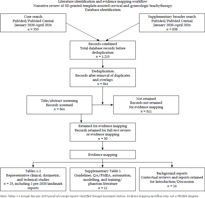
Table 1
Clinical studies of 3D-printed templates and applicators for cervical and gynecologic brachytherapy
[i] The primary structured search covered English-written publications from January 2020 to April 2026. Earlier studies published before 2020 were included selectively through backward citation, when they provided landmark proof-of-concept evidence or essential technical background for individualized 3D-printed applicators and template-assisted image-guided brachytherapy workflows
3D – three-dimensional, BT – brachytherapy, LACC – locally advanced cervical cancer, EBRT – external beam radiotherapy, HDR – high-dose-rate, LDR – low-dose-rate, IC – intracavitary, IS – interstitial, IC/IS – intracavitary/interstitial, IGABT – image-guided adaptive brachytherapy, CTV – clinical target volume, HR-CTV – high-risk clinical target volume, OAR – organ at risk, QA – quality assurance, TPS – treatment planning system, 3D-PNCT – three-dimensional-printed non-coplanar template, 3D-NG – three-dimensional-printed needle guide, GU – genitourinary, GI – gastrointestinal, FMEA – failure mode and effects analysis, TNT – tandem and needle template, VIA – vaginal individualized applicator, DMBT – digital monarch butterfly tandem
Technology
Three-dimensional-printed applicators for cervical brachytherapy now span a spectrum from simple add-on guides to fully individualized molds. Early work focused on patient-specific templates that reproduced a pre-planned needle geometry for single complex cases, but the approach has since evolved into a set of more standardized solutions, which can be integrated into routine workflows. Across centers, the common goal is to reduce geometric uncertainty, extend IC/IS techniques to challenging anatomy, and do so in a way that remains practical even where hardware resources are limited [20].
The main clinical and technical characteristics of the published 3D-printed systems for cervical brachytherapy are summarized in Table 1. Most series describe templates designed from pre-treatment CT or MRI, with channels or sleeves that guide intracavitary tandems and interstitial needles according to a virtual plan. These devices are typically used in conjunction with established image-guided adaptive brachytherapy (IGABT) workflows and adapted to local resources, including in-house printing capacity, access to advanced imaging, and institutional policies on sterilization and re-use.
In parallel, modular re-usable templates have been developed to retain the familiarity and robustness of commercial cylinders while adding the flexibility of customized channel angulation [21]. Systems, such as re-usable vaginal cylinders with detachable 3D-printed caps or sleeves, allow centers to introduce template-guided IC/IS techniques without abandoning their existing applicator inventory [10, 13, 22]. These designs favor standardized diameters and connector interfaces, so that a single-printed component can serve multiple patients after appropriate cleaning and sterilization.
Hybrid IC/IS designs have also matured through closer integration with three-dimensional imaging and treatment planning. Tumor-adaptive needle templates (TNT) and similar platforms use MRI- or CT-based planning to define a set of needle trajectories that can be reproduced across fractions, often with pre-defined depth markers and labelling, which simplify communication between physician, physicist, and radiation therapists. When combined with intraoperative imaging, these templates support reproducible IC/IS implants that follow the planned geometry rather than relying on real-time free-hand adjustments [12, 23]. Chinese groups have further advanced patient-specific non-coplanar templates (3D-PNCT) for CT-guided interstitial brachytherapy, designing needle paths in Mimics software to respect vascular and bony constraints, and achieving sub-millimeter placement accuracy with minimal need for re-imaging [24, 25]. Altogether, these hybrid systems demonstrate that when MRI and CT pre-planning are employed to define safe channels before printing, procedural quality assurance (QA) can be supported by numbered pathways, locking screws, and intraoperative imaging to deliver reproducible IC/IS implants.
A further development is the incorporation of curved flexible needles into patient-specific 3D-printed template systems. Emerging clinical implementation studies suggest that MRI-based anatomical modelling can be used to design individualized templates, which guide both straight and curved needle trajectories in bulky or parametrial invasive cervical cancer [26]. This approach may extend template guidance beyond fixed straight trajectories, and may be particularly useful for lateral parametrial extension, posterior bulky disease, or anatomy where rigid needles cannot adequately cover HR-CTV without increasing organ at risk exposure.
Material and manufacturing choices now reflect a deliberate balance between biocompatibility, mechanical strength, dimensional stability, and sterilization compatibility. Earlier reports have often used polylactic acid or similar low-temperature plastics suitable for low-cost printers, but these materials can deform during steam sterilization and may not meet regulatory requirements for repeated clinical use [27, 28]. Desktop 3D printers have also been used to produce intracavitary applicators for high-dose-rate (HDR) cervical brachytherapy with acceptable mechanical stability and CT visibility [29]. More recent workflows favor medical- or dental-grade photopolymers and high-temperature plastics, which tolerate steam or low-temperature plasma sterilization without significant warping. However, these materials and printers are more expensive, and outsourcing fabrication to certified vendors can introduce delays and additional costs that may limit adoption in resource-constrained settings.
Quality assurance and workflow integration have evolved alongside the technology. Most programs now incorporate routine checks of template dimensions against digital design, verification of channel patency and angulation, and end-to-end testing of the imaging and planning workflow before first clinical use [15, 30]. Several groups have applied failure mode and effects analysis (FMEA) to identify vulnerabilities, such as mislabeling of channels, incorrect template orientation, and collision between needles and bony structures, and have responded by introducing template-specific checklists, labelling conventions, and staff training. These measures create a feedback loop between additive manufacturing and safe clinical delivery [31].
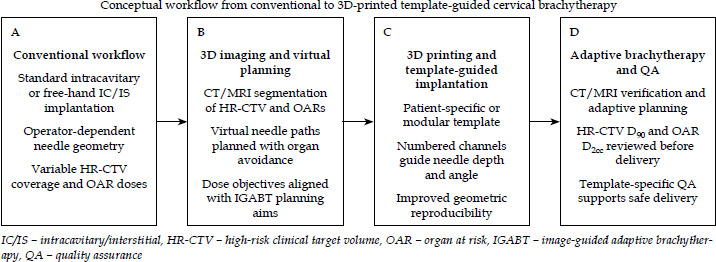
Overall, international experience points toward convergent design principles, i.e., patient-specific or semi-standardized templates, which encode the planned geometry, modular re-usable components that fit existing applicators, and material choices that support safe sterilization and repeated use. Within this framework, and as schematically illustrated in Figure 2, centers can select simpler or more sophisticated solutions according to their caseload and resources while still achieving more predictable needle placement and high-dose conformity in challenging pelvic anatomies.
Fig. 2
Conceptual workflow from conventional to 3D-printed template–guided cervical brachytherapy. The diagram illustrates the transition from standard intracavitary or free-hand intracavitary/interstitial implantation to 3D imaging, virtual needle planning, template-guided implantation, and adaptive brachytherapy with template-specific quality assurance. Patient-specific or modular templates can translate imaging-based plans into more reproducible needle geometry while preserving image-guided adaptive brachytherapy principles
Dosimetry
Across the emerging 3D-printed template literature, reported dosimetric studies generally suggest improved target coverage and organ at risk sparing compared with conventional cylinders, standard applicators, or free-hand IC/IS techniques, although reporting formats and comparators vary across studies. In a CT-based series comparing single-channel cylinders with customized multichannel sleeves, the 3D-printed sleeve elevated HR-CTV D98 from 370 to 446 cGy per fraction, while keeping bladder and rectal D2cc within tolerance limit [11, 15]. A separate dosimetric trial confirmed these trends: a patient-specific applicator increased HR-CTV D90 to 633.8 cGy vs. 601.8 cGy and D100 to 465.6 cGy vs. 415.3 cGy, improved high-dose conformity indices, and reduced rectal D2cc from 402.0 to 374.3 cGy as well as bladder D2cc from 458.9 to 414.5 cGy, all with high statistical confidence.
Head-to-head comparisons of free-hand hybrid implants reinforce the value of template guidance for both deep target coverage and organ protection. In one institutional series, template-assisted fractions achieved HR-CTV D90 of 629.4 ±19.3 cGy compared with 613.0 ±15.9 cGy for free-hand insertions, accompanied by reductions in bladder D2cc from 458.1 to 431.1 cGy, and parallel decreases across D1cc and D0.1cc constraints [20]. In another study using a pre-plan based guidance template, tandem depth was maintained within ±0.3 cm of the virtual trajectory across fractions, and HR-CTV D90 increased over the first four fractions while rectal and bladder D2cc tended to decline relative to the initial free-hand benchmark [23]. These per-fraction gains translated into cumulative HR-CTV D90 of approximately 92 ±7 Gy EQD2, while rectum, bladder, sigmoid, and bowel D2cc remained below 63 ±6.5, 79 ±10, 66 ±7, and 61 ±10 Gy3, respectively – values closely associated with EMBRACE-II planning aims.
Across these reports, the quantitative impact of 3D-printed templates on HR-CTV coverage, V100, and organ at risk D2cc compared with cylinders, conventional tandem-ovoid applicators, or free-hand IC/IS implants is summarized in Table 2. As shown there, most template-based regimens achieve HR-CTV D90 values in the 90-95 Gy EQD2 range, and consistently lower bladder and rectal D2cc than non-templated techniques, with the largest relative gains seen in large or irregular targets. To improve interpretability across heterogeneous studies, dosimetric findings are described according to the reporting format used in the original publications. Some studies reported per-fraction physical doses, whereas others reported cumulative EQD2 values after external beam radiotherapy and brachytherapy; the reporting context is therefore specified when these metrics are discussed.
Table 2
Dosimetric and clinical impact of 3D-printed template-assisted brachytherapy compared with conventional techniques
[i] Studies published before 2020 are included as foundational or proof-of-concept reports identified through backward citation, and are not intended to represent an exhaustive pre-2020 literature search
3D – three-dimensional, BT – brachytherapy, EBRT – external beam radiotherapy, HDR – high-dose-rate, LDR – low-dose-rate, LACC – locally advanced cervical cancer, IC – intracavitary, IS – interstitial, IC/IS – intracavitary/interstitial, IGABT – image-guided adaptive brachytherapy, CTV – clinical target volume, OAR – organ at risk, LC – local control, PFS – progression-free survival, OS – overall survival, CSS – cancer-specific survival, LRFS – loco-regional relapse-free survival, D90/D95/D98/D100 – minimum dose received by 90%/95%/98%/100% of the target volume, V100/V150 – percentage of volume receiving 100%/150% of the prescribed dose, D2cc/D1cc/D0.1cc – minimum dose to the most exposed 2/1/0.1 cm3 of an organ at risk, DMBT – digital monarch butterfly tandem, TNT – tandem and needle template, CI – conformity index, HI – homogeneity index, EI – external index, GU – genitourinary, GI – gastrointestinal, CRT – chemoradiotherapy
Emerging curved needle and AI-assisted template systems may further expand the dosimetric flexibility of template-assisted brachytherapy. Early reports suggest improved procedural reproducibility, acceptable HR-CTV coverage, and feasible organ at risk sparing in anatomically complex or parametrial invasive disease [26, 32]. These data remain preliminary, however, and require prospective validation before they can be interpreted as evidence of superiority over established template-guided or free-hand approaches.
Tumor size appears to modulate the magnitude of benefit. In one analysis comparing 3D-PNCT-assisted CT-based IC + ISBT plans with conventional applicators or free-hand arrays, lesions ≥ 30 mm showed modest improvements in CTV D95/D98/D100 (53.0/46.7/33.4 Gy vs. 52.0/45.4/32.2 Gy; p ≤ 0.043), but marked reductions in bladder and rectal D2cc (69.7 vs. 83.4 Gy and 67.4 vs. 76.7 Gy; both p < 0.001) [5]. Smaller tumors experienced both robust target coverage (CTV D95 53.4 Gy) and workflow efficiency gains, suggesting that templates can support dose escalation and organ preservation even as needle trajectories become less forgiving. A separate multichannel vaginal applicator study demonstrated similar patterns, with mean HR-CTV D90 increasing from 83.4 to 88.6 Gy and D98 from 75.4 to 79.7 Gy, compared with non-printed techniques, and a larger proportion of patients meeting EMBRACE-II criteria. The increased benefit in D90 was disproportionately greater when HR-CTV exceeded 30 cm3.
Post-operative cases impose unique geometric constraints that templates can help standardize. In a cohort of non-central pelvic recurrences after hysterectomy, a 3D-printed template-assisted iodine-125 (125I) implant series identified a survival-linked D90 threshold of 108.5 Gy EQD2; exceeding this benchmark was more readily achievable in tumors ≤ 3 cm than in larger lesions [33]. In intact cervical cancer, CT-guided 3D-printed non-coplanar templates enabled needle dense reconstructions, which improved conformity and heterogeneity indices while keeping bladder and rectal D2cm3 at approximately 114 and 267 cGy per fraction, respectively [24]. A larger multi-institutional analysis stratifying 377 implants by HR-CTV volume showed that 3D IC + ISBT configurations usually delivered higher HR-CTV D90/D98 and lower rectal D2cc than tandem-ovoid insertions, with the 50-60 cm3 sub-group gaining around 0.35 Gy in D90 and 0.30 Gy in D98 [9].
Commercial intrauterine tandems remain an important comparator. In a planning study contrasting a nylon PA12 applicator with a Fletcher-Williamson metal system, HR-CTV D90 remained close to 100% of the prescription for both, but the PA12 tandem roughly halved bladder D2cc (65-81% vs. 87.3% of the prescription) at the cost of slightly higher rectal exposure, underscoring the need for material-specific attenuation studies before widespread adoption [29]. Template-guided needle selection can also rationalize catheter numbers without sacrificing coverage. An automated catheter reduction approach preserved HR-CTV D98 within 0.31% and limited bladder, rectum, and sigmoid D2cc shifts to ≤ 0.10 Gy; although removal of a single catheter from the optimized plan reduced D90 by 1.5 Gy, highlighting the narrow dosimetric margin within which 3D-planned implants operate [21].
Taken together, these studies outline a coherent dosimetric pattern. Template-assisted workflows tend to stabilize HR-CTV D90/D98 within the 90-95 Gy EQD2 range, improve volumetric coverage in both intact and post-operative settings, and repeatedly lower bladder and rectal D2cc compared with cylinders, conventional applicators, or free-hand IC/IS implants. The magnitude of benefit increases with tumor complexity, supporting template-guided interstitial supplementation as a practical strategy for achieving EMBRACE-level targets without exceeding organ tolerances.
Clinical outcomes
Chinese multi-institutional experience with 3D-printed template guidance covers both HDR and 125I low-dose-rate (LDR) salvage settings, providing an important benchmark for local control after re-irradiation. The national expert consensus on seed implantation for recurrent cervical cancer summarized multicenter data, and demonstrated that CT-guided, 3D-printed non-coplanar templates (3D-PNCT) enable 125I implants to deliver prescription doses around 120 Gy, achieving 3-year local control of approximately 75% and a median overall survival (OS) of 17 months, with only isolated grade 3 proctitis and rare fistulas reported [30]. Within the same consensus, a randomized trial comparing template-assisted 125I seed implantation with concurrent chemoradiation, revealed superior short-term local control (1-12 months) and improved 1-5-year OS in the template arm, underscoring the durability of LDR salvage when implants conform to pre-planned 3D navigation. Complementary Chinese salvage cohorts extend these findings to HDR brachytherapy. In patients with recurrent cervical cancer, patient-specific 3D-PNCT-guided HDR interstitial brachytherapy (ISBT) achieved an objective response rate of 66.7%, 2- and 5-year local progression-free survival (LPFS) of 30.0% and 25.7%, and a median OS of 23.2 months, with 66.7% pain relief among symptomatic patients despite 71% having extra-pelvic disease [25]. Even in this heavily pre-treated population, grade 3-4 toxicity occurred in only 20%, mainly fistulas and severe dermatitis or mucositis, supporting the safety of repeated template-guided implants when needle trajectories are constrained.
Prospective European data confirm that 3D-printed templates can achieve EMBRACE-II dose-volume aims in bulky primary tumors without increasing morbidity. In an Aarhus series of 101 women treated with a 3D-printed tandem-needle template (TNT), mean HR-CTV D90 reached 93 Gy EQD2 and 3-year local control was 85% overall – 95% for parallel-only and 75% for parallel-plus-oblique configurations, while 3-year cancer-specific survival was 68% [34]. Moreover, grade ≥ 3 late complications were limited to about 10%, indicating that even anatomically challenging cases can retain contemporary local control and toxicity profiles when needle geometry is individualized [34]. North American experience is consistent. After implementation of customized 3D-printed needle guides for complex cervical and vaginal targets, the 2-year cumulative local failure rate fell from 22.0% in non-3D workflows to 8.2% (adjusted hazard ratio, 0.31), without significant changes in bladder or rectal D2cc, indicating that template-based drill guides can enhance local control without compromising OAR safety [21].
Smaller feasibility studies provide higher resolution insight into progression-free survival (PFS) and toxicity. A Thai pilot study on in-house 3D-printed applicators reported a 92.9% complete response rate at 3 months; among recurrent cases, 2-year local relapse-free survival, distant metastasis-free survival, PFS, and OS were 74.1%, 88.9%, 64.8%, and 88.9%, respectively [16]. Genitourinary and gastrointestinal grade ≥ 2 toxicities remained low (7.1% acute and ≤ 21.4% late), with only a single persistent grade 3 vaginal stenosis despite cumulative EQD2 to mucosa approaching 170 Gy, indicating that individualized applicators can maintain pelvic control while containing mucosal sequelae [16].
Early clinical reports on curved needle template systems are encouraging but remain immature. Available data suggest feasibility and acceptable short-term toxicity in selected bulky or parametrial invasive tumors, while re-irradiation cases continue to require careful patient selection, conservative dose constraints, and long-term monitoring for late toxicity [26]. A recent individualized curved needle re-irradiation case report also illustrates the feasibility of treating difficult recurrent anatomy, but the occurrence of grade 3 proctitis and cystitis underscores the need for careful patient selection, conservative dose constraints, and long-term late toxicity monitoring in salvage settings [35]. These reports are best interpreted as early technical and clinical signs rather than definitive outcome evidence.
Modern 125I salvage programs that integrate intraoperative 3D-printed guides further highlight the interplay between target size, D90, and outcome. In a 32-patient cohort with non-central pelvic recurrence, 125I seed brachytherapy guided by CT and 3D-printed templates, yielded 6-, 12-, and 24-month local control rates of 88.0%, 63.2%, and 42.1%, respectively, with a median OS of 13.3 months and no major procedure-related complications [33]. Tumor diameter < 5.3 cm and immediate post-operative D90 > 108.5 Gy were independently associated with improved survival, reinforcing the importance of both anatomical selection and dosimetric quality in template-assisted salvage [33]. Altogether, HDR and LDR experiences from Chinese multicenter cohorts, European prospective programs, and early-phase feasibility trials converge on a common indication: 3D-printed templates raise expectations for local control and PFS in both primary and recurrent cervical cancer brachytherapy, while keeping severe toxicity uncommon when strict image-guided planning and QA protocols are observed. These outcome signals are consistent with the dosimetric gains summarized in Table 2, supporting 3D-printed templates as a means of safely achieving dose-escalated IC/IS brachytherapy in both primary and recurrent settings.
Practical issues
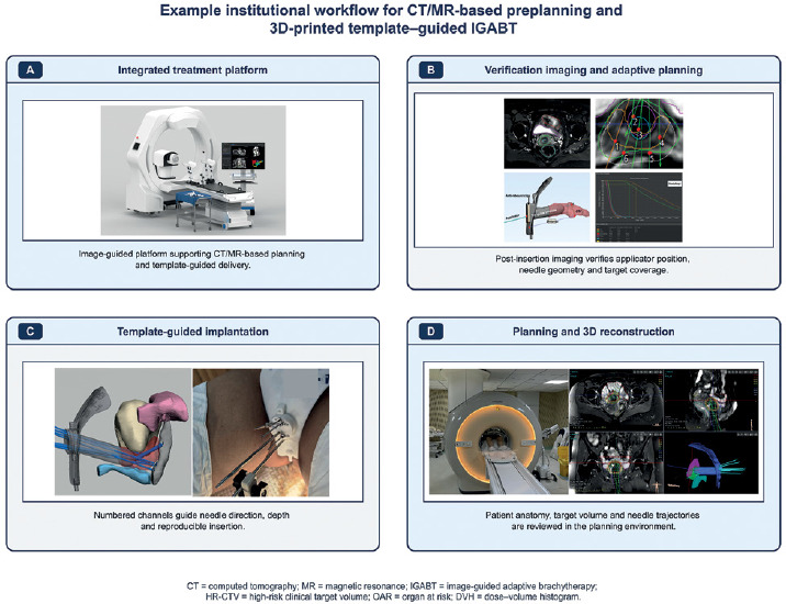
Template-assisted implants depend on disciplined, imaging-rich workflows, so that individualized hardware reliably translates into reproducible dosimetry. In CT-based programs, pre-planning typically occurs about one week before HDR brachytherapy: HR-CTV and OARs are contoured, virtual needles are reconstructed with ≥ 5 mm spacing, and collision-free oblique trajectories are finalized before printing the guide. The schematic overview of the transition from conventional free-hand implants to 3D-printed template-guided image-guided adaptive brachytherapy (IGABT) is presented in Figure 2, while Figure 3 illustrates a practical CT/MRI-based pre-planning and treatment workflow from a representative institutional program. In a Chinese CT-guided series, this approach halved insertion time (≈10 vs. 19 min), reduced the number of CT checks per application (2 vs. 4), and stabilized geometry in difficult parametrial or pelvic wall disease compared with free-hand IC/IS techniques [5]. MRI-based planning before each fraction remains crucial in pulsed-dose-rate (PDR) or HDR/PDR schedules, where the number, position, and angulation of needles must be adapted to evolving tumor regression while maintaining EMBRACE-conformant coverage. Hybrid workflows increasingly combine intraprocedural transrectal ultrasound to set needle depths with post-implant MRI and CT for HR-CTV/OARs contouring. This indicates that template adoption does not lessen the need for GEC-ESTRO-compliant segmentation on 2.5-3.0 mm CT or MRI datasets [15, 21]. Patient-specific systems, such as TNT tandem-needle template at Aarhus or the Stanford VIA and 3D-NG platforms, provide additional well-labelled channels in narrowed vaginal canals while preserving dosimetry, but their print-to-patient pipelines still require rigorous contour propagation, needle verification, and post-implant quality checks [13, 21, 34].
Fig. 3
Example institutional workflow for CT/MRI-based pre-planning and 3D-printed template-guided image-guided adaptive brachytherapy. The figure shows representative institutional components of template-assisted cervical brachytherapy, including an integrated treatment platform, verification imaging, and adaptive planning, template-guided implantation, and planning with 3D reconstruction. This workflow illustrates how image-based planning, individualized template geometry, implantation guidance, and dose-volume evaluation can be integrated into clinical practice
Procedure logistics generally benefit from these guides. In most programs, CT- or MRI-planned templates shorten implantation time to roughly 10-20 minutes, minimize repeat verification scans, and give anesthesia teams greater confidence when extensive parametrial coverage is required, even for very large or asymmetric targets [5, 9]. The TARGIT and TARGIT-FX tandem-anchored systems further reduced the overall procedure time by roughly 30% in large-tumor cohorts, and improved the proportion of fractions meeting EMBRACE-II goals, while all surveyed residents reported easier needle placement and interest in using the technique in future practice [10, 12]. Some institutions still rely on spinal or epidural anesthesia for complex interstitial insertions, highlighting ongoing demands for anesthetic resources [18]. Phantom-based training with multimaterial pelvic models, originally developed as educational tools, now also supports ultrasound-guided caudal blocks and simulation of difficult tandem or needle paths, potentially improving patient comfort and broadening the adoption of hybrid IC/IS approaches [8, 36, 37].
Manufacturing and sterilization choices are tightly coupled with resource constraints. BioMed Clear resin and selective laser sintered PA12 nylon are biocompatible, thermally stable, and compatible with steam, plasma, or gas sterilization, enabling repeated use when budgets preclude single-patient builds [13, 22]. In contrast, workflows that print a new applicator or template for each patient using validated SLA resins simplify infection control, but demand higher printer throughput and robust quality systems [21]. Economic considerations extend to training; a multimaterial pelvic phantom described by Campelo and colleagues provides transparent QA of needle paths and costs on the order of a few hundred US dollars, substantially less than many commercial phantoms [8]. Low-cost 3D-printed obturators have also been proposed as simpler options for less invasive gynecologic brachytherapy in constrained settings [38]. Nevertheless, several clinical series note that comprehensive 3D workflows require CT- or MRI-based modelling, additional planning scans, and engineering support, which may limit implementation in centers without dedicated imaging physicists or in-house printing capacity [4, 38].
Quality assurance (QA) lessons closely mirror those of external beam radiotherapy. A repeat failure mode and effects analysis of a 3D-printed IC/IS program identified 54 potential failure modes, with needle-track design, channel labelling, and template commissioning carrying the highest risk priority numbers, prompting standardized checklists, and revised design rules [31]. Emerging knowledge-based and anatomy-aware planning frameworks can serve as dose prediction and plan checking tools, flagging sub-optimal DVHs and accelerating approval when combined with reproducible template geometry [39, 40]. Additional safeguards include applying direction-specific displacement tolerances derived from phantom and in vivo source tracking studies as well as using transparent or modular prints, which permit immediate visual inspection of channel patency before imaging and loading [8, 41]. Statistical process control methods have further been used to monitor HDR cervical brachytherapy plan quality over time and detect drifts in D90 or OAR doses in routine practice [42]. Collectively, successful deployment of 3D-printed templates depends on tightly integrating imaging-based pre-planning, multidisciplinary training in anesthesia and device handling, scalable sterilization strategies, and pro-active QA frameworks, so that the dosimetric advantages demonstrated in early series can be realized safely in routine clinical practice.
Template-specific quality assurance (QA) becomes more important as device geometry becomes more complex. In curved needle or AI-assisted workflows, QA should verify not only template dimensions and channel patency, but also source transit through non-linear pathways, needle compatibility, fixation stability, and correspondence between the printed template and the approved digital design. Such workflows shift brachytherapy QA from applicator availability alone toward an integrated verification chain involving imaging, digital design, manufacturing, sterilization, needle insertion, and source delivery [26].
Future directions
Evidence gaps and trial design
Future work needs to move beyond the predominantly retrospective, single-center series that currently define the field in order to generate more generalizable evidence of benefit. Embedding 3D-printed templates within standardized workflows illustrated in Figures 2 and 3, will be essential if they are to move from innovative local projects to broadly reproducible clinical practice. Existing reports showing that 3D-printed templates improve EMBRACE-II goal attainment, organ sparing, and local control, even in challenging anatomy, are encouraging but remain limited by modest sample size, short follow-up, or the absence of robust control arms, which constrains reliable estimates of recurrence, toxicity, and quality of life gain [16, 23, 34]. Real-world analyses based on EMBRACE-II treatment principles further highlight the importance of patient selection, standardized image-guided adaptive brachytherapy workflows, and prospective outcome benchmarking when translating protocol-based planning aims into routine clinical practice [43]. Prospective, multi-institutional trials comparing clearly defined template-assisted workflows with the best available free-hand or commercial applicators, should therefore be prioritized, ideally incorporating translational endpoints that link needle placement fidelity and HR-CTV coverage to biological response and patient-reported outcomes. A similar level of rigor is required in salvage settings, where early success of 3D-printed non-coplanar HDR or 125I seed implants for non-central pelvic relapse has been demonstrated without formal comparator arms or granular normal tissue profiling [18, 33].
Automation and adaptive workflows
Automation and quality management warrant parallel maturation. Failure mode and effects analyses have already emphasized unreasonable needle track design, labelling errors, and intraoperative misplacements as high-risk steps that currently lack systematic mitigation, underscoring the need for template-specific checklists, digital design validation, and formal commissioning pathways [31, 42]. Knowledge-based planning, machine learning for dose prediction, and auto-segmentation have shown that they can compress contouring and optimization timelines, and flag sub-optimal OAR doses in conventional cervical brachytherapy [39, 40, 44-46]. Recent AI-assisted template and integrated optimization studies suggest that the next generation of 3D-printed brachytherapy may be defined less by printing alone than by the coupling of anatomical modelling, needle path selection, dwell-time optimization, and real-time verification [32, 47]. However, these approaches remain in early phase and should be prospectively evaluated before routine clinical adoption.
True adaptive planning also requires coordinated data capture throughout implantation, not faster optimization alone. Studies that combine insertion metrics with dosimetry, explore dose-based pre-planning to minimize catheter number, or evaluate automated optimization of curved channels, all point towards richer decision support layers, but remain retrospective and focused on technical surrogates rather than patient outcomes [9, 23, 44, 48, 49]. Future protocols should prospectively embed real-time imaging, predictive analytics, and explicit re-planning triggers, for example, when applicator displacement or D2cc thresholds are exceeded, and then test whether such adaptive intent improves local control, toxicity, and overall treatment time at scale.
Equity and implementation
Finally, equitable dissemination needs to be built into research agendas from the outset. Low-cost, re-usable templates, and interstitial caps could extend hybrid IC/IS implants to centers that lack commercial applicators, but they require pragmatic implementation studies to quantify sterilization logistics, printer requirements, engineering support, and achievable dosimetry in resource-constrained settings [27, 38, 42]. Analgesia and skill-training innovations, such as caudal block phantoms, multimaterial pelvic models, and hybrid training courses that use 3D-printed IC/IS applicators on mannequins, should be evaluated as part of capacity-building packages, which target pain control, procedural safety, and resident competency [8, 50]. Without deliberate investment in scalable manufacturing standards, QA pathways, and supportive education, the advantages of 3D-printed templates risk remaining confined to high-resource centers rather than re-shaping cervical cancer brachytherapy practice globally. Ultimately, prospective multi-institutional comparisons, integration of automation into routine workflows, and deliberate capacity building in low- and middle-income settings, will determine the future of this technology. These efforts will decide whether 3D-printed templates become a standard component of cervical brachytherapy or remain confined to high-resource centers.
Conclusions
Three-dimensional-printed templates and applicators represent a clinically relevant strategy for improving individualized image-guided brachytherapy in cervical cancer. By translating patient-specific imaging information into reproducible needle geometry, these devices can support IC/IS implantation in bulky tumors, parametrial extension, distorted anatomy, and recurrent pelvic disease. Contemporary evidence suggests dosimetric advantages, including improved HR-CTV coverage, better conformity, and similar or reduced organ at risk doses, compared with standard applicators or free-hand approaches. Early clinical series also suggest feasible local control and acceptable toxicity in selected primary and salvage settings, including emerging AI-assisted and curved needle template approaches, although these findings remain preliminary and require longer follow-up.
Mainstream adoption, however, will depend on addressing several persistent challenges. The current evidence base is dominated by single-center, short-term series, whereas randomized or multi-institutional trials confirming durable gains in tumor control, toxicity, and quality of life are still lacking [4, 5]. Quality assurance pipelines also require further standardization. Failure mode analyses have highlighted vulnerabilities in template design, labelling, and intraoperative placement, which must be mitigated through rigorous commissioning, template-specific checklists, and workflow-focused training [31, 42]. In parallel, manufacturing consistency, regulatory oversight, and integration with adaptive planning tools or source tracking technologies, are still evolving, creating additional barriers for centers with limited engineering or imaging support. Bridging these gaps through harmonized validation studies, shared design, QA standards, and formal credentialing, will determine whether 3D-printed template-assisted brachytherapy becomes a more widely adopted component of cervical cancer brachytherapy, or remains a specialized solution for highly selected cases.