Introduction
Congenital heart disease (CHD) occurs in approximately 9/1,000 newborns and requires surgical treatment in one-third of these newborns during the first years of life [1, 2]. Due to its size and location, the thymus gland obstructs access to the heart during surgery, making thymectomy a routine procedure in early surgical interventions for CHD. Removing the thymus also eliminates the risk of the swollen gland compressing the heart and major blood vessels in the early postoperative period [3]. In developed countries, more than 90% of children with CHD survive into adulthood [4]. For this reason, the effect of early thymectomy has been a subject of research.
In 1987, Brearley et al. first reported lower T-cell levels in thymectomized children than in controls [5]. Subsequent studies, taking into account other components of the immune system, the age at which thymectomy was performed, the time elapsed after thymectomy, and whether the thymus was partially or entirely removed, analyzed the effects of thymectomy [6–9]. Although observational studies suggest that early thymectomy increases the incidence of infection, there is still no clear consensus on this issue [10].
This article discusses a case of T-cell deficiency and increased lower respiratory tract infection rate following thymectomy performed in the first year of life due to CHD.
Case report
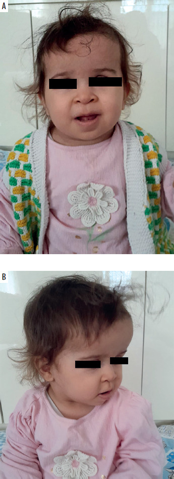
A 13-month-old female patient with a history of premature birth was followed up at the pediatric cardiology outpatient clinic due to a ventricular septal defect, patent ductus arteriosus, and tricuspid insufficiency. At 9 months of age, the patient underwent cardiac corrective surgery with partial thymectomy. Postoperatively, the patient had a history of multiple hospitalizations and antibiotic use due to recurrent lung infections. Physical examination revealed macrocephaly (+2.2 SDS), frontal bossing, bitemporal narrowing, hypertrichosis on the forehead, depressed nasal root, anteverted nostrils, long philtrum, and mildly low ears (Figures 1 A, B). The parents were not related, and the family had no known immunodeficiency.
FIGURE 1
A, B – Macrocephaly, frontal bossing, bitemporal narrowing, depressed nasal root, anteverted nostrils, long philtrum, and mildly low ear findings are notable in the patient’s phenotype
During outpatient follow-up, the patient was admitted to the ward for further investigation and treatment due to high fever resistant to antibiotic therapy, vomiting, poor oral intake, and respiratory distress. The respiratory virus panel detected influenza A and respiratory syncytial virus (RSV). Due to the patient’s persistent fever despite treatment, delayed clinical improvement, and laboratory findings suggestive of immunodeficiency, intravenous immunoglobulin (IVIG) therapy was initiated at 0.5 g/kg. The patient was discharged after achieving clinical improvement without complications.
No abnormalities were detected in the patient’s routine hematological and biochemical tests (Table 1). Immunological tests showed IgG: 535 (n: 748–851) mg/dl, IgA: 21 (n: 42.9–52.6) mg/dl, IgM: 66 (n: 96–117) mg/dl, anti-HBs: negative, anti-Rubella IgG: negative, anti-CMV IgG: positive (Table 2). The patient’s childhood vaccinations were administered according to the Ministry of Health vaccination schedule, including the 6-month vaccination; the 1-year vaccination was postponed due to infections. Flow cytometric lymphocyte analysis showed CD3+ T cells: 47% (n: 51–81.8), CD4+ T cells: 12% (n: 27.6–55.6), CD8+ T cells: 34% (n: 12.7–30.9), CD19+ B cells: 7.5 (n: 11–34.4), CD16+56+ NK cells: 28.4% (n: 2–26.3) and recent thymic emigrant (RTE) -T cells %18.5 (n: 42.5–82.2) (Table 3).
TABLE 1
Hematological and biochemical parameters of the patient
TABLE 2
Immunological evaluation of the patient
TABLE 3
Percentages of lymphocyte subpopulations before and after IVIG treatment
Chromosome analysis, microarray analysis, and fluorescence in situ hybridization (FISH) analysis for DiGeorge syndrome were performed for the patient regarding genetic syndromic disorders (dysmorphia). The chromosome analysis of the patient, who was found to have 46, XX, was reported as usual in the microarray and FISH analysis results. The clinical exome sequencing of the patient revealed a heterozygous (start loss, c.1 deletion, p.Met1?, rs922092315) in the SHH (Sonic Hedgehog signaling molecule) gene, which is a potential pathogenic mutation. No genetic changes associated with immunodeficiency were identified. The patient’s genetic results explain the developmental delay, cardiac defects, and syndromic appearance observed in the patient, but do not explain the immunological picture. The patient, who is under clinical follow-up, showed a decrease in the frequency of infections and hospital admissions due to infections after IVIG treatment. The patient continued immunoglobulin replacement therapy for approximately 1 year (receiving intravenous treatment for 6 months and then switching to subcutaneous treatment due to vascular access issues). At the end of the 1-year treatment, CD4+-T and CD8+-T cell levels and CD19 and IgG levels were within the normal range (Table 3). At the end of 1 year, the patient’s cellular and humoral immune deficiency improved, and the frequency of infections decreased, leading to the discontinuation of IVIG therapy. Although the patient’s abnormal values improved, due to the risk of infection during the winter months and the fact that the RTE values did not return to normal completely, we could not discontinue immunoglobulin replacement therapy. However, we decided to continue with weekly subcutaneous injections and to discontinue/pause the treatment as we approach summer. (The patient’s family has provided written informed consent for the publication of this case report.)
Discussion
The routine removal of the entire thymus or part of it during surgical treatment of congenital heart defects significantly affects the formation of the immune cell pool and the subsequent preservation of the phenotype and functionality of pure T cells in the patient’s body.
Various publications investigating the long-term outcomes of early thymectomy are available in the literature. In a study conducted by Broek et al., 26 patients who underwent thymectomy due to CHD in the first month of life were compared with 31 healthy children. The results showed that thymectomized patients had a decrease in the absolute number of T cells and a reduction in the ratio of naive CD4+-T and CD8+-T cells compared to healthy controls of the same age. Additionally, while there were no significant changes in B cell subsets in thymectomized patients, a decrease in total IgG levels was observed [7]. In another study, 11 patients with early thymectomy (< 6 months) were evaluated intermittently over 18 years; a significant decrease in the number of naive CD4+-T and CD8+-T cells was observed in thymectomized patients compared to the control group [8]. In another study by van der Broek et al., a decrease in the number of naive CD4+-T and CD8+-T cells was detected in thymectomized patients; however, at the end of 10 years, the CD4+-T and CD8+T-cell counts in these patients were found to be similar to those of healthy controls [9]. Similarly, in a study published by van der Gent et al., 39 patients who underwent thymectomy due to CHD at the age of 1.5 years were followed up until the age of 31. In the first 5 years following thymectomy, the levels of naive and total CD4+-T and CD8+-T cells in the blood and TREC levels in CD4+-T cells were significantly lower than in healthy controls of the same age. Five years after thymectomy, the counts of pure CD4+-T and CD8+-T cells and TREC levels were within the normal range [11]. In our case, flow cytometric lymphocyte analysis showed decreased CD3+-T cells: 47% (n: 51–81.8), CD4+-T cells: 12% (n: 27.6–55.6), mildly elevated CD8 T cells: 34% (n: 12.7–30.9), an inverted CD4+/CD8+ ratio, very low recent thymic emigrant T cells 18.5% (n: 42.5–82.2), mildly low CD19+ B cells: 7.5 (n: 11–34.4), and relatively elevated CD16+-56+NK cells: 28.4% (n: 2–26.3). A transient reduction in B cell percentage was also detected despite increased NK cell percentage. We also observed a decrease in naive CD4+-T and naive CD8+-T cell counts in our patient. At the end of 1 year of IVIG therapy, we observed that CD4+-T cells, CD19+-B cells, and naive CD8+-T cells had reached normal levels in our patient. Although the naive CD4+-T cell count remained below the reference range, we observed an increase at the end of 1 year. After 21 months of replacement treatment, we have observed that the percentage and absolute counts of T, B, and NK cells have returned to normal values and that these immune system cell deficiencies were temporary. Given that winter is approaching and RTE percentage values are still low, we plan to continue replacement treatment for a while longer. We would like to pause treatment at least during the summer to see if there is any improvement.
A Swedish study compared 5,664 patients who underwent cardiac repair surgery and thymectomy with 2,276 control patients who underwent cardiac surgery without thymectomy; a significant increase in the incidence of atopy, asthma, and infection was found in the thymectomy group [10]. In our case, the frequency of respiratory infections increased, particularly after thymectomy. Additionally, the treatment processes for respiratory infections in our patient were prolonged. A decrease in infection frequency was observed during IVIG therapy. Notably, while respiratory infections previously required hospitalization and intensive care unit admission, they were managed ambulatory and without complications following treatment.
A study conducted by van der Broek et al. in 2017 demonstrated that neonatal thymectomy did not alter measured B cell subpopulations but caused a decrease in total IgG levels [12]. In a study conducted in 2004, 19 children who underwent thymectomy (partial or complete) during the neonatal period due to CHD were compared with a healthy control group of the same age group. The age of patients included in the study ranged from 5 to 16 years. The study showed that IgA and IgG1 concentrations were significantly lower in the thymectomy group [13]. In a survey by Kurobe et al., patients who underwent complete or partial thymectomy due to CHD were compared; 3 years after surgery, IgG antibody titers against measles and rubella viruses were significantly lower in patients who underwent complete thymectomy. The complete thymectomy group also showed an increase in the frequency of hospital admissions associated with infectious diseases such as bronchitis and pneumonia [14].
On the other hand, studies have reported immunoglobulin levels in thymectomy patients to be at the same level as healthy controls [5, 15]. Our patient had a history of subtotal thymectomy. In our patient with low serum IgG, IgA, and IgM levels, IgG subclass levels were not evaluated due to the patient’s age. Anti-rubella antibody titers could not be assessed because IVIG therapy was initiated before the rubella vaccine.
Our patient had a history of frequent hospital admissions, intravenous antibiotics, and inhaler treatment due to recurrent bronchiolitis attacks. In the literature, some studies have found more extended hospital stays and higher average antibiotic use in patients who underwent thymectomy [11, 14], while others have not found significant differences [6, 7, 15]. These differences in the studies are thought to stem from the inability to standardize the degree of thymectomy (partial, subtotal, complete) across studies and from varying follow-up periods.
In addition to our patient’s clinical history, based on current laboratory findings, the prolonged duration of the most recent bronchiolitis attack, its resistant course to treatment, and the identification of immunodeficiency findings, we initiated IVIG therapy and planned close clinical monitoring. Due to vascular access issues, the patient continued to receive immunoglobulin subcutaneously. During the treatment process, we observed a marked decrease in the frequency of infections and the need for hospitalization, and after 1 year, cellular and humoral immunity returned to normal levels.
Conclusions
Temporary or permanent cellular/humoral changes may occur in the immune system of patients undergoing thymectomy. These changes should be kept in mind when monitoring patients. The immunological values we determined in our patient returned to normal 1.5 years after thymectomy. However, despite numerous studies, the long-term results of thymectomy and its course in different patients are still poorly understood. Therefore, longer-term and more detailed studies are needed in these patients to determine thymectomy’s delayed/long-term follow-up results.