


Introduction
Spinal cord infarction (SCI), also referred to as ischemic myelopathy or spinal cord injury, is a relatively rare disease characterized by high death rates. The available statistical data show that it accounts for merely 1% of all classified strokes [1]. The consequences of SCI are usually extensive and permanent [2]. The wide spectrum of possible neurological aftermaths of ischemia to spinal cord structures usually leads to the patient’s considerable disability, significantly lowering their quality of life. The most commonly reported consequences of SCI primarily include paraplegia and urinary incontinence [1]. Despite a well-explored etiopathogenesis of this condition, literature contains virtually no reports on physiotherapeutic management of such patients [3]. Rehabilitation of patients post SCI is multi-faceted, takes a long time, and is targeted at minimization of the existent motor and functional deficits. Therefore, the conducted therapeutic actions should focus on the areas of existent limitations and difficulties in the patient’s activities of daily living, leading to eliminate them or compensate for them [2].
This paper is an example of a conceptual plan for late-stage rehabilitation of a patient post SCI. The case described is unique in that it presents a long-term (4-year) outpatient rehabilitation process, focused mainly on soft tissue techniques, assessed using objective tools (the Fugl-Meyer Assessment (FMA) scale, dolorimeter, inclinometer) and the CARE protocol for clinical reports. The chosen concept is not widely described in literature. Therefore, the case study discussed here allows us to present the possible benefits and limitations of the adopted rehabilitation model, as well as the requirements related to monitoring its effects.
Condition etiopathogenesis and diagnosis
Transport of blood to the spinal cord is an immensely complex process. The reason behind this is to deliver proper blood supply to the structure in its entirety. Moreover, vascular variability is not uncommon. It has been observed that blood reaches some areas of the spinal cord on a preference basis [4]. As a result, the spinal cord is much less prone to atherosclerosis and its consequences compared to the brain [5]. Atherosclerotic plaque disease in neighboring arteries is not a prognostic factor for spinal ischemia [6]. SCI or ischemia is rare and is sometimes misdiagnosed as inflammatory myelopathy in acute conditions [7].
Spinal cord injuries related to blood vessels account for 5.8% of acute myelopathy cases and approximately 1.2% of stroke cases, while spinal cord ischemia is diagnosed 100 times less frequently than stroke [8].
In studies conducted by Zalewski et al. on groups of 100 people, it was found that 76% had one or more vascular risk factors in their medical history: hypertension – 61 (46%), smoking – 61 (46%), hyperlipidemia – 57 (43%), and diabetes – 21 (16%) [9]. Aortic diseases [10] and musculoskeletal overload due to strenuous physical exercise (weightlifting) are also indicated as direct causes of SCI [11].
The spinal cord is supplied by the anterior spinal artery (ASA) and a pair of posterior spinal arteries (PSA) [12]. Spinal cord ischemia is classified as:
– infarction of the root artery area (bilateral infarction of the anterior or posterior spinal artery and unilateral infarction),
– extensive hypoperfusion of the spinal cord (central and transverse infarction) [13].
Depending on the extent and location of SCI, the following syndromes are distinguished:
– anterior spinal cord syndrome – results in blockage of the anterior vertebral artery, causing ischemia of two-thirds of the spinal cord surface supplied by this artery. It is the most common cause of SCI. The most common location is the mid-thoracic level. It manifests as incomplete motor paralysis below the site of injury. Damage to the neurological pathways responsible for pain and temperature sensation, known as anterior cord syndrome, is also evident [14];
– posterior spinal cord syndrome – develops as a result of ischemia of the posterior vertebral artery.
It manifests itself as a disturbance of proprioception and vibration sensation, hypotonia, ataxic gait, positive Romberg’s sign, and absence of deep tendon reflexes [15];
– central cord syndrome – is a non-vascular injury to the spinal cord, particularly evident after extensor hyperreflexic injuries in the cervical spine. It is the most common injury among incomplete spinal cord injuries. It manifests itself as a significant impairment of sensory and motor functions in the upper limbs compared to the lower limbs [16].
The onset is generally sudden, and the symptoms peak within 30-45 minutes. Diagnosis is made based on magnetic resonance imaging (MRI). The supporting sign on MRI is a local hyperintense spinal cord signal, so-called “owl eyes”, on axial T2-weighted images [14], although it is worth pointing out that MRI may seem completely unremarkable in up to 24% of patients with acute symptoms [17]. The role of computed tomography (CT) in assessment of suspected spinal cord ischemia is limited because of low sensitivity, but this modality may serve for a quick assessment of curable causes, such as epidural hematoma or displaced fracture compromising blood supply [18].
Case description
In May 2018, a woman was admitted to the hospital’s neurology ward with a stroke unit.
General information: 56 years old, married, resident of Poland, not professionally active. Higher education.
Main symptoms on admission: pain in the lumbosacral region (L-S) radiating to the lower limbs, worsening for about 4 hours. Progressive weakness of the lower limbs, urinary disorders (anuria).
Medical history: long-standing hypertension, multi-level discopathy of the thoracic and lumbosacral spine. No chronic diseases in the immediate family. No neurological pathologies in the immediate family.
Psychosocial history: smoking and alcohol – denies, physically active (on average 3 times a week physical activity such as jogging or Pilates), high financial stability, no stress factors.
Neurological examination at the neurology ward showed:
–no meningeal signs,
–no upper limb paresis,
–superficial sensory disturbances from Th11 level, no control of trunk muscles at the L-S level – MRC 0/1 (Medical Research Council), functional assessment
–1/7 FIM (Functional Independence Measure) in terms of balance in a sitting position,
–paralysis of the lower limbs – AIS A (ASIA Impairment Scale),
–bilateral positive Babinski reflex,
–lack of deep and tendon reflexes in the lower limbs, neurogenic urinary retention.
An MRI scan revealed a high signal and increased thickness of the spinal cord in the Th8-L1 segment of an ischemic nature, probably of arterial etiology. The edematous changes affected the entire gray matter and central white matter along the entire length of the described segment.
After approximately two weeks of pharmacotherapy with anticoagulants and thrombolytics, a follow-up MRI scan showed a gradual improvement in the radiological image.
The patient underwent a neurological assessment in order to plan the physiotherapy process. The examination revealed:
– profound paresis of the lower limbs,
– superficial and deep sensory disturbances,
– significant deficit in active mobility of the knee and ankle joints,
– sphincter dysfunction.
Two functional scales were used to assess the patient’s independence in basic activities of daily living: Lawton Instrumental Activities of Daily Living (IADL) and the Barthel scale.
The results obtained were:
Based on the above results, the patient was classified as partially independent, requiring support in some activities of daily living.
All of the above information was obtained from the medical records provided by the patient and presented in a comprehensive form.
The patient began outpatient rehabilitation at a rehabilitation clinic approximately 16 months after diagnosis. The course and results of rehabilitation are presented as a case study in this paper.
History taking and physical examination
Patient history
– Severe pain as the dominant symptom, localized in the lower spine and lower limbs, radiating along the hip bones and sacroiliac joints toward the hips and knees.
– Described a feeling of discomfort in movement, characterized by a “pulling” sensation in the lower limbs and lack of control over foot movement while walking.
– Using a dolorimeter in the area of the hip bones, pain at a level of 0.8 KGS.
– Secondary pain in the upper spine, without neurological symptoms.
Physical examination
Information on the assessment of active and passive ranges of motion is presented in the table below (Table 1).
Table 1.
Table of lower limb joint mobility according to SFTR (Shoulder, Flexion, Thumb/Total Range, Rotation), with assessment according to the Modified Ashworth Scale (MAS)
Assessment of posture and gait
– In a standing position: forward tilt of the pelvis, deepening of the lumbar lordosis, compensatory slight forward tilt of the torso, protraction of the shoulders and head.
– Inclinometer measurements: 11° flexion and 6° internal rotation in the hip joints. No full active extension in the knee joints – 8° flexion.
– Gait: paretic with compensatory dominance of adductors and internal rotators of the lower limbs. Short, irregular, slightly asymmetrical stride. Lack of fluidity in transferring weight from heel to toes.
– Extended double-support time and shortened single-support phase – qualitative observation made during walking in clinical conditions. Both feet remain in contact with the ground for most of the gait cycle, indicating instability and a need for increased safety.
– When fatigued, there is a noticeable reduction in the ability to actively lift the foot, resulting in it touching the ground during the transfer phase.
– Patient walking with an orthopedic cane.
Case analysis
The rehabilitation was divided into 3 stages to analyze the effects of regional and global therapy on the nature of musculoskeletal disorders. In addition to implementation of specific therapeutic concepts, each stage involved FMA of lower extremity for the neurologic patient, measurements of pain levels with a dolorimeter, and measurements of angular disorders in hip and knee joints in a standing position.
The following criteria for therapy termination were set for bioethical reasons:
– full or temporary resignation by the patient,
– inflammation involving fever directly post therapy,
– evident subcutaneous extravasation (Bruise Visibility Scale > 4),
– evident deterioration in posture or gait patterns directly post therapy, lasting for more than 24 hours,
– exacerbation of pain compared to baseline from before therapy, persistent for more than 48 hours.
Rehabilitation in subsequent stages was carried out at different locations, selected on the basis of the severity of the patient’s subjective feelings during palpation. At each stage, the frequency and duration of therapy were strictly defined in accordance with the internal regulations of the outpatient clinic where the rehabilitation process was carried out. A detailed description of the methods used and their effects is presented in Table 2.
Table 2.
Summary of treatment stages according to the CARE guidelines
The following types of therapeutic interventions were selected:
Stage I
– Mobilization of soft tissues using the myofascial release technique – areas sensitive to pain indicated by the patient during the palpation assessment were treated as local muscle/fascia dysfunction (continuum distortion). These points were treated by applying constant local pressure with the thumb at high intensity (up to 10 kg/cm2) for a maximum of 2 minutes or until the pain subsided.
Stage II
– Soft tissue mobilization using myofascial release techniques – techniques as in stage I, supplemented by release within the trigger band, i.e. relaxation of painful areas during palpation by applying constant pressure with the thumb with identical load and moving in the direction of pain and based on the fascial tape pattern according to Thomas Meyers’ concept until the pain subsides or the pain zone is left.
– Supplementing and supporting soft tissue techniques and relieving the thumb by using Small Bar used in Instrument Assisted Soft Tissue Mobilization (IASTM) tools and the M-Stick tool for point fascial therapy based on the same working methodology.
– Kinesitherapy involving locomotion training on a treadmill to re-educate gait, performed after myofascial release to consolidate the effects of therapy in the patient’s daily functioning.
Stage III
– Mobilization of soft tissues using myofascial release techniques in selected areas throughout the body according to the same methodology.
– Kinesitherapy including balance exercises performed with the use of:
sensory integration cushions – lunge exercises with one leg forward and to the side, with or without lower limb support (2 sets of 15 repetitions); free standing and half-squats with sensory cushions under both lower limbs (2 sets of 15 repetitions).
BOSU balance platform – free standing and half-squat exercises with and without support, involving the upper limbs in transferring 1 kg balls from hand to hand for about 10 minutes or until fatigue prevents further safe performance of the exercise.
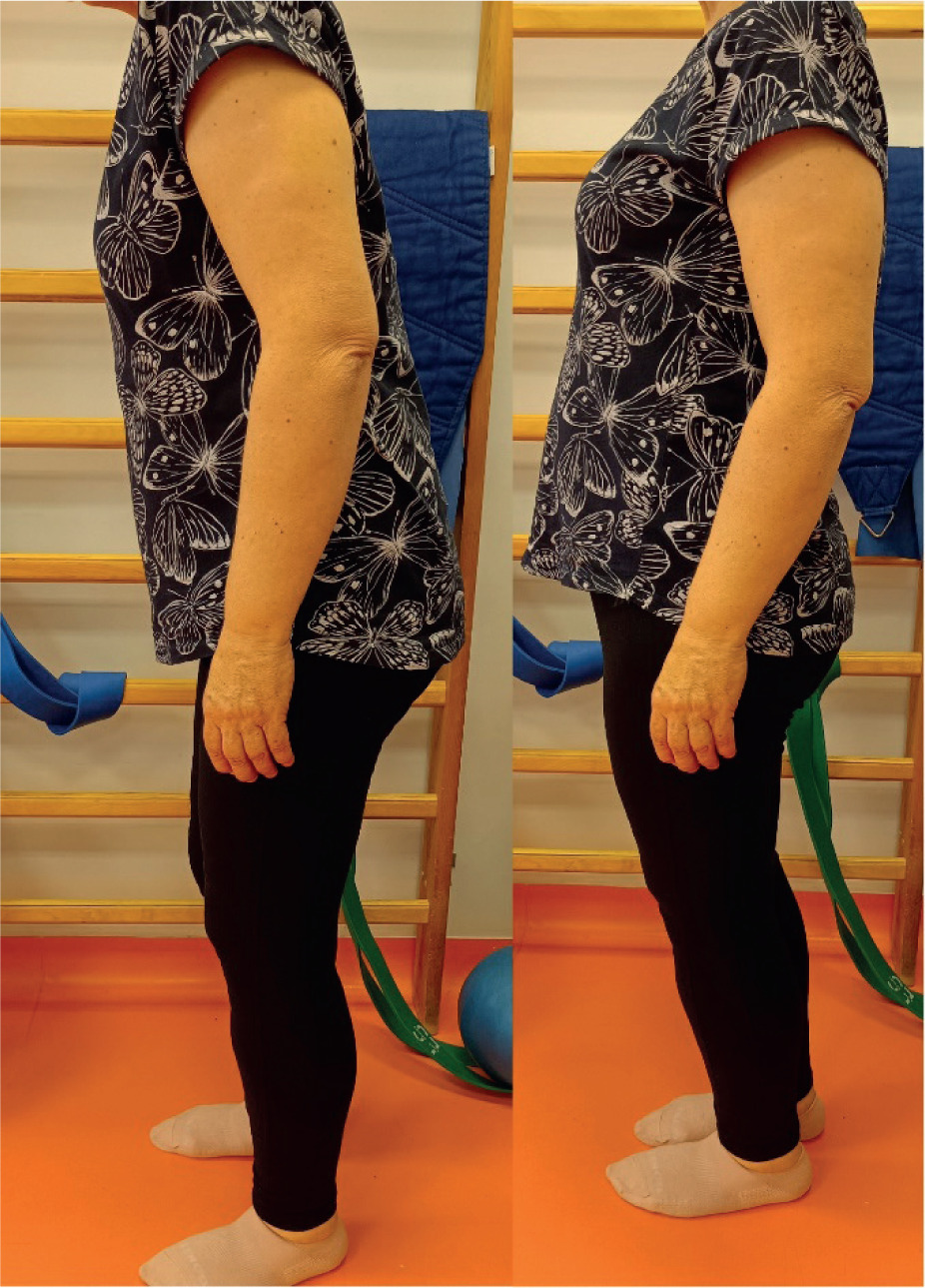
The final examination showed progression in the patient’s gait pattern during the weight-bearing phase of the lower limb in terms of foot rollover and propulsion (Figure 1).
No physical therapy treatments were used at any stage. In the final interview, the patient reported episodic pain in the pelvis and thoracolumbar spine, especially in situations of increased stress. There was a quantitative improvement in gait function, measured by the distance covered without feeling pain or fatigue, as well as a qualitative improvement towards normalization of the gait pattern. In order to prolong the effects achieved during physiotherapy, the patient was recommended physical activity in the form of walks lasting no longer than 60 minutes at a time. No imaging or laboratory tests were performed during the intervention. The patient participated in all therapy sessions without any absences. Physiotherapy treatment, apart from pain in response to fascial techniques, did not result in any adverse events that would warrant its discontinuation. The intervention produced a number of quantitative end results (Table 3).
Table 3.
Comparison of information from the preliminary and final examination
Discussion
Therapy with a neurologic patient requires determination of stage goals suitable to the current condition of the patient. In view of the paramount, controlling function of the nervous system, it is characterized by the complexity and multi-faceted nature of treatments. As a rule, it involves therapies targeting restoration and/or compensation of lost fine and gross motor skills, which – by design – aims to improve the patient’s independence in activities of daily living. The reported case partially matches the analysis developed on the basis of larger groups. The study by Nedeltchev et al. [19] on a group of 54 SCI patients has shown that after a follow-up of a mean 4.5 years, 41% of patients were able to walk independently, and 31% could walk with outpatient support devices. Despite this relatively high rate of patients retrieving their walking function post SCI, the long-term death rate reported for that cohort was at 9% [19]. In turn, in a study conducted on 41 patients in 1999-2020, Ros Castelló et al. observed motor deficits in 39 patients (95.1%), pain in 20 patients (48.8%), sensory deficits in 33 patients (80.4%), and autonomic dysfunction in 24 patients (58.5%), which confirms the numerous and rough nature of SCI symptoms [20]. Other studies involving long-term biopsychosocial follow-up (mean > 7 years) compared the functional results of patients with cerebrovascular accident and SCI, showing that patients in that group had higher employment rates and lower death rates, but at the same time, higher chronic pain rates and reduced functional independence (Modified Rankin Scale).
In the presented case study, soft tissue therapy was the dominant form of physiotherapy treatment. The strengths of this approach were its safety and the possibility of individual adjustment of intensity, as well as its direct impact on pain reduction and improved tissue mobility. The limitation was the lack of a control group and the fact that the therapy was conducted on an outpatient basis at a facility contracted by the National Health Fund, which limited its availability and continuity.
It is claimed that loss of motor and sensory function, including paresthesia and paralysis, may – despite specialist interventions – persist in some cases, increasing the risk of cardiovascular and pulmonary disease. Some SCI patients still suffered from chronic pain [21]. This can be a difficult obstacle to overcome and hinder the patient’s progress toward achieving an acceptable level of independence in their daily life.
Even though over 400 randomized controlled trials into SCI have been conducted, the vast majority were assessed as having poor methodology and focused not on commonly used treatments but rather on modern technologies such as robotics, epidural spinal cord stimulation, and transcranial magnetic stimulation [22]. There is still a lack of high-quality research confirming the effectiveness of classic manual methods, although from the researchers’ point of view, this topic may be extremely attractive.
Concluding remarks
In literature, rehabilitation outcomes are affected by numerous determinants, for instance the extent of SCI, patient’s age, the patient’s general health, intensity of physiotherapy provided, its quality, time elapsed from the incident, and the patient’s motivation.
This case study achieved its main goal, which was to present the effects of regional and global therapy on the type of musculoskeletal disorders occurring secondary to SCI. The authors’ observations are an inspiration for performance of a study with a larger study group to increase the merit of the results and confirm the theory of effectiveness of soft tissue therapy as a method applicable to physiotherapeutic management involving rehabilitation of SCI. Despite the numerous unpleasant painful sensations accompanying the therapy, the patient did not decide to stop. The intensity of the suggested manual techniques was increased gradually, which gave the patient space to accept the therapist’s pressure strength. A factor that facilitated the process was, without doubt, providing education to the patient and raising her awareness as to the therapeutic mechanisms of the stimuli applied. In addition, the patient was motivated to further rehabilitation by the quick and subjectively visible effects of the therapy.
Conclusions
SCI is a condition which may cause permanent disability in affected patients. In physiotherapeutic management, it is vital that the therapeutic goals be targeted at reduction in the patient’s motor deficits. The authors’ experiences, presented as a case study of an SCI patient, showed that soft tissue therapy may be one method effective in rehabilitation of such patients. It is necessary to conduct a study on a larger patient group with objective measurement methods, which will legitimize the determination of effectiveness of myofascial release therapies in interventions on patients post SCI.