


Giant condyloma acuminatum termed as a Buschke-Löwenstein tumour (BLT) is an invasively slow-growing wart-like skin lesion of the anogenital region [1]. Although the disorder is histologically benign, its management is challenging due to its large size, local tissue destruction, and high recurrence rate [2]. We report a case of genital BLT associated with a low-risk human papillomavirus (HPV) infection which was treated successfully with radical excision and reconstructive surgery.
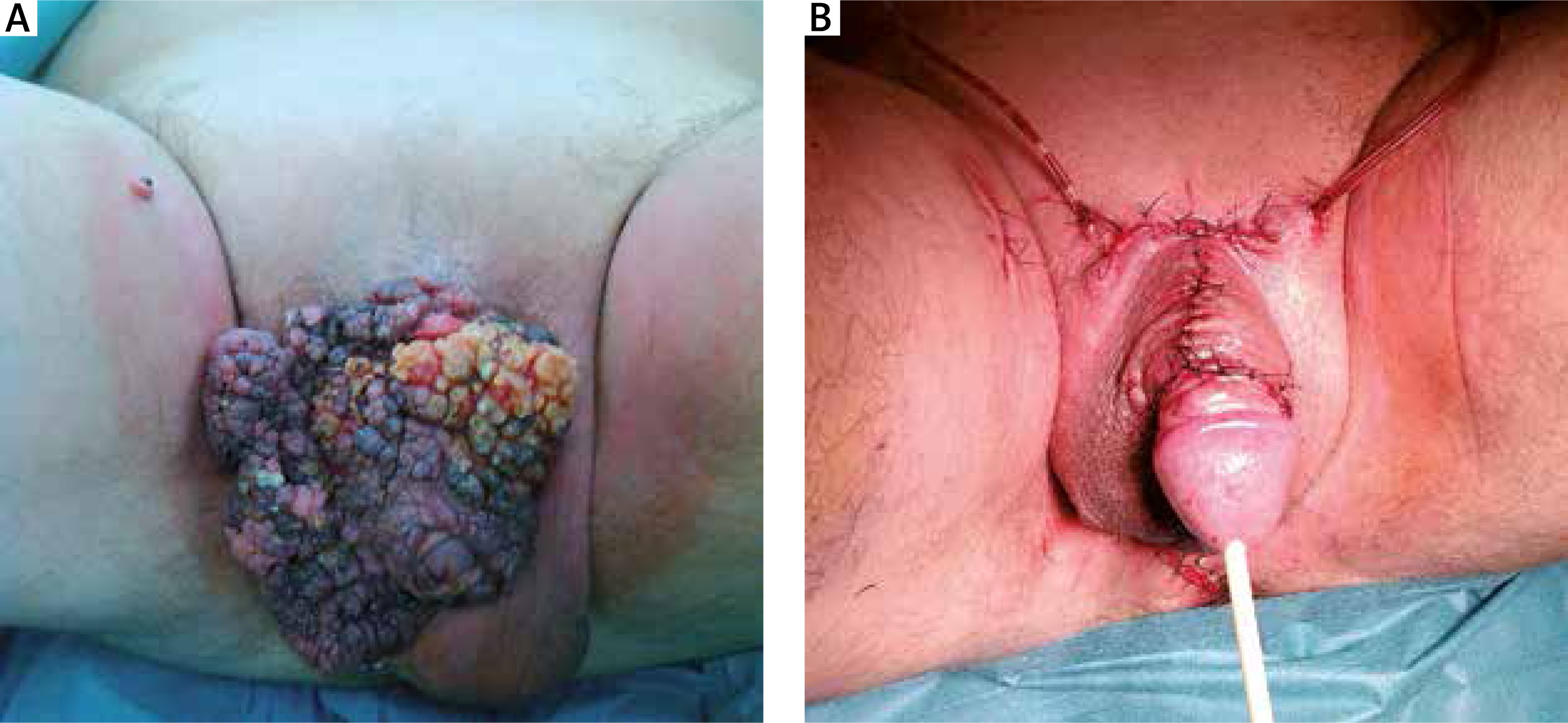
A 66-year-old man was admitted to the dermatological outpatient clinic due to a large polypoid lesion in the genital area (Figure 1 A). The single brownish small wart had first appeared on the penis 19 years before and had grown gradually expanding also the perineal region. The lesion was painless, but caused disfigurement of the penis and made sexual intercourses impossible. Nevertheless, the patient had not sought any therapy. He denied any sexual risk behaviours. The patient was suffering from hypertension, benign prostatic hyperplasia and degenerative joint disease. The family history of skin disorders was negative.
Figure 1
A – Cauliflower-like, large, yellowish-grey papillary tumour covering almost the entire surface of the penis skin and a part of the scrotum; a small “satellite” of the tumour seen on the right thigh; B – the affected region after excisional and reconstructive surgery
In the conducted digital rectal examination, the wall of the anus and anal canal were unaffected. The prostate gland was enlarged, smooth, symmetrical, without palpable changes. Urination was effortless. The mucosa of the glans penis was normal. Lymph nodes were not palpable. The diagnostic tests towards other sexually transmitted diseases was negative. The skin biopsies were taken and the patient was referred to the department of urology for further diagnostics and treatment. Histopathological examination of the genital skin lesion confirmed the clinical suspicion of giant condyloma acuminatum. Moreover, HPV detection by polymerase chain reaction method (PCR) from the biopsy specimens was performed and revealed the presence of HPV 6 and 11 genotypes (Figure 2 B).
Figure 2
A – The biopsy of the surface of the lesion with arborescent papillae showing prominent fibrovascular cores, surface koilocytosis, and no significant atypia; B – HPV 6 and 11 detection (arrow) with PCR method
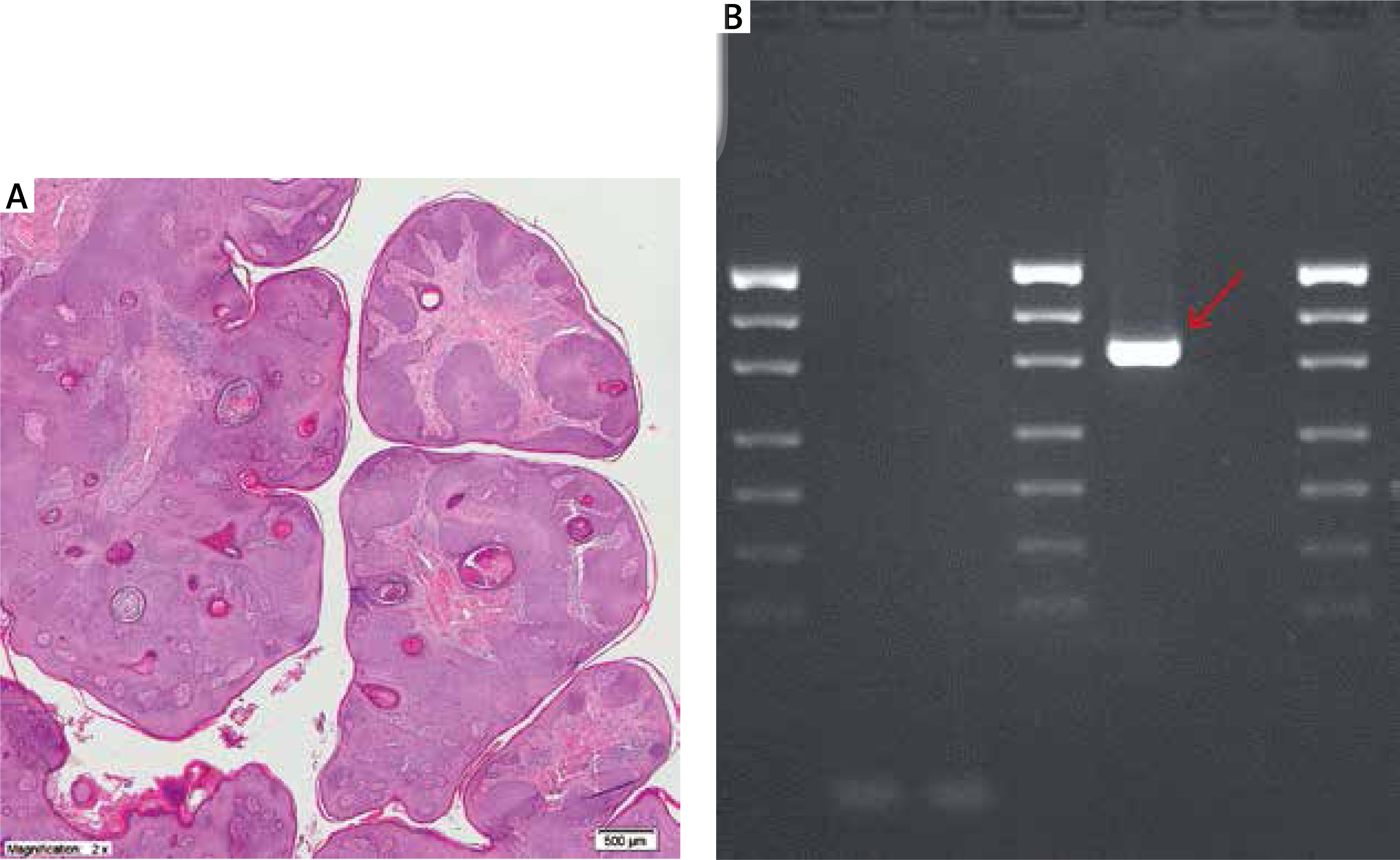
The therapy consisted of radical excision with the follow-up reconstruction by rotation flap surgery (Figure 1 B). The patient was discharged from hospital in good condition with recommendation of antibiotic treatment and daily wound dressings. All the surgical material was forwarded for histopathological examination which confirmed the previous diagnosis of BLT (Figure 2 A). No recurrence was observed during 5 months of follow-up.
The giant condyloma acuminatum was originally described by Buschke in 1986 [3] and Löwenstein in 1925 [4] as a penile lesion. The disorder belongs to a group of ‘semi-malignant’ verrucous carcinomas due to their destructive local evolution [5], however its terminology is still controversial [6]. Some authors consider BLT to be a regional variant of verrucous carcinoma while others classify it as a separate entity [7–9]. Chu et al. analysed 42 known cases of BLT in the English literature and reviewed their behaviour and management [7]. The disease was reported to have a significant rate of recurrence and a risk of malignant transformation to squamous cell carcinoma (SCC) with no distant metastases [7]. Trombetta et al. noticed the presence of neoplastic foci in as many as 50% of the 52 reports of patients undergoing surgery for BLT [10].
The BLT is considered a relatively rare sexually transmitted disease with an estimated incidence of 0.1% in the general population [11]. The ratio between women and men is 1 : 3, and the mean age of occurrence is about 50 years [11].
The etiopathogenesis of BLT also remains unclear, however, its association with HPV infection has been highlighted [1]. Low-risk HPV DNA (types 6 and 11) has been most commonly identified in skin samples of BLT, strongly suggesting their pathogenic role in tumour development. The presence of these two HPV types was also detected in our case. The risk factors for BLT also include an HIV infection, homosexuality and immunosuppression [11, 12].
The diagnosis of BLT is usually based on the patient’s history, clinical features and histopathological examination. In certain cases, due to the risk of deep local invasion especially in the anorectal region, imaging studies are also required. Before the beginning of therapy, it is important to obtain a skin biopsy. The BLT is a histologically benign disorder, nevertheless careful histopathological examination is necessary to exclude the presence of SCC transformation. The histopathological presentation with condyloma features (koilocytosis and fibrovascular cores) but with more prominent bulbous expansion into the underlying tissue and endophytic pattern of growth without presence of atypia is crucial to confirm BLT and exclude other benign or malignant lesions. The BLT of the anogenital region can be misdiagnosed mainly because of the chronic friction and risk of maceration. There are some rarely occurring lesions in the anogenital area clinically similar to BLT such as giant seborrheic keratosis [13].
Early surgical resection of condyloma acuminatum prevents the BLT development [8]. The BLT management is challenging due to its size and degree of local invasion [12]. The tumour should be treated radically as topical chemotherapy, laser resection or cryosurgery are not sufficient in terms of disease control and recurrence. Wide radical excision with the follow-up reconstruction seems to be the treatment of choice, and it was also implemented in our case. Neo-adjuvant chemo-radiation therapy with local excision may be considered in the treatment of BLT with SCC transformation, but also in unresectable or recurrent disease [14]. The effectiveness of adjuvant immunotherapy is still uncertain. Due to a risk of recurrences, follow-up visits are strongly required. In our case, we excluded presence of malignant transformation and so a wide excision and reconstructive surgery exclusively seemed to be the best choice of treatment. Unfortunately, the follow-up period was only 5 months because the patient did not keep his further appointments with the doctor.
The presence of genital BLT certainly causes discomfort, adversely affects sexual life and, therefore, it decreases the quality of patient’s life [15]. An early diagnosis, appropriately aggressive therapy and vigilant surveillance are crucial to improve patient outcome. A long-term follow-up period can decrease the risk of recurrence.
In conclusion, we present a case of BLT associated with HPV 6 and 11 genotypes successfully managed surgically. We point out that although several treatment modalities have been proposed, a wide surgical excision with reconstructive surgery seems to be the optimal therapeutic strategy in such disfiguring tumours of the genital region with a risk of relapses and even malignant transformation.