
Introduction
According to current classifications, both en coup de sabre morphea (ECDS) and progressive hemifacial atrophy (PHA) are considered a subtype of linear morphea [1–6].
ECDS is a rare acquired disease of the connective tissue located in the fronto-parietal region, leading to excessive collagen accumulation, sclerosis of the skin and deeper structures with subsequent atrophy. Patients struggle not only with dermatological problems. They are also described also ipsilateral abnormalities in the stomatognathic system. Moreover, this problem seems to be significantly underestimated [7–9].
PHA is an extremely rare, acquired, progressive disease leading to unilateral atrophy of the skin, subcutaneous, muscle and bone tissue in the face, usually with relative sparing of forehead tissue. It involves dermatomes associated with one or more branches of the trigeminal nerve. It leads to irreversible deformations and asymmetry, dysfunction of facial structures [3]. Morphological disorders may be accompanied by neurological and ophthalmological disorders as well as impaired functioning of the stomatognathic system. An increase in the degree of facial asymmetry and its changed proportions may lead to stigmatization, affecting the patient’s psychosocial functioning [10].
In this paper, we summarize the disturbances of the stomatognathic system in patients with craniofacial morphea, together with an analysis of current treatment options.
Etiopathogenesis
The aetiology of the above-mentioned diseases is still unknown. Some researchers indicate that there is a close spatial relationship between the location of skin, dental and bone lesions in the course of morphea ECDS and PHA and the corresponding zones inhabited by individual groups of neural crest cells. The occurrence of ECDS and PHA is supposed to be a proof of the presence of factors in the embryonic life that trigger dysmorphogenesis processes related to the migration of neural crest cells [11–13]. The segmental structure of the hindbrain in the initial period of embryonic life determines the migration of cells in the form of bands. Most of the structures that build the face originate in the 2nd and 4th rhombomeres of the hindbrain (NCC – Neural Crest Cells – from the 4th rhombomere are controlled by the HOX B1 and B2 genes, whose expression depends on the level of BMP and TGF-β proteins), with whose neural crest cells move to the 1st and 2nd gill arches, giving rise to skin, muscle, cartilage and bone tissue of the face, also taking part in the morphogenesis of dentine, pulp, cement and periodontal tissues. These cells also build sensory ganglia of the trigeminal nerve and the facial nerve have the ability to influence angiogenesis by building the vascular endothelium. The intercellular interaction, the relationship between cells and the intercellular matrix, and the chemotactic interaction between the neural crest cells and the adjacent plaques guarantee the proper shaping of the facial structures. Due to the extensive role of NCC in the differentiation of facial structures, they are perceived as a presumed causative factor of Parry-Romberg syndrome and other abnormalities associated with facial malformations [14–16]. It is said that the inducing factor that initiates the occurrence of the linear form of morphea and PHA may be an injury [1, 17].
Classification
Guerrerosantos classified 4 types of PHA, determining the possible selection of reconstructive therapy [18] (Table 1).
Table 1
PHA subtypes according to Guerrerosantos
The Inigo classification, based on the degree of atrophy of the bony structures and branches of the trigeminal nerve, divides the PHA syndrome into three categories [16, 19, 20] (Table 2).
Table 2
PHA subtypes according to Inigo
PHA patients suffer from complex abnormalities of the stomatognathic system. The lesions appear ipsilaterally to the skin foci in the area of the cheek, mouth, tongue, mandible and maxilla. Tissue atrophy within the cheek area results in its collapse. As a result of the atrophy of the tissues that build the mouth, the corner of the mouth is lifted, the lip is shortened on one side and, in extreme cases, the teeth are exposed involuntarily. The inability to close the lips freely increases the likelihood of diseases of the oral mucosa. The inability to close the edges of the lips also impairs the swallowing process [16, 20–27]. Structural deficits in the course of PHA translate into functional impairments in chewing, swallowing, speech and breathing. Patients develop malocclusion in the form of complete crossbite (due to jaw hypoplasia) and tooth crowding, their rotation and inclination. Incorrect positioning of the teeth in the dental arch contributes to the occurrence of irregularities within the periodontal tissues and tooth loss. Atrophy of the masticatory muscles, shortening of the body and ramus of the mandible then generate dysfunction in the functioning of the temporomandibular joints. Any changes in the position of the dental arches or the mandible generate proprioceptive disorders of muscle tone in the stomatognathic system, which, in the absence of compensation, lead to functional disorders of the masticatory system. Disturbed synchronization of mandibular head movements leads to myoarthropathy. Unilateral atrophy, smoothing of the tongue impair its functions in terms of nutrition and speech. The balance of forces exerted on the teeth through the cheeks, lips and tongue is disturbed. During swallowing, the deformed tongue is unable to rest on the front part of the palate and incisors. The accompanying atrophy of the hard and soft palate tissues exacerbates this problem. At the same time, the correct respiratory system is disturbed [27].
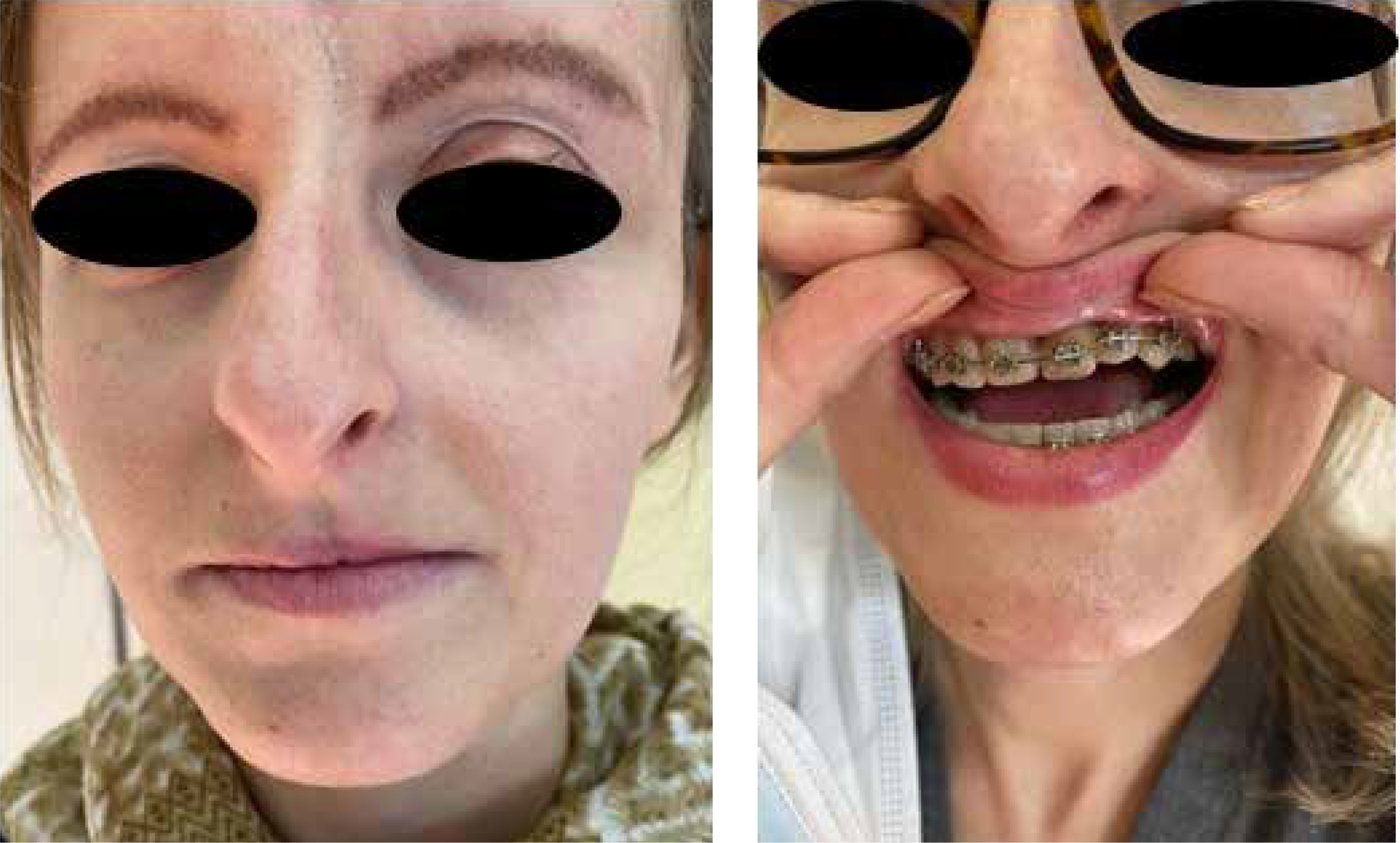
Delayed tooth eruption, intrusions, impacted teeth, stones and pulpal necrosis are described. The structure of tooth crowns usually does not differ from the norm. On the other hand, there are anomalies within the tooth roots, manifested by their distortion and shortening [16, 20, 23–27]. Trigeminal neuralgia manifests itself in the form of acute pain (rarely dull and continuous) lasting from several seconds to several minutes, triggered during chewing, brushing teeth, washing the head [28] (Figure 1).
Treatment
Due to the potential involvement of the disease in tissues other than the skin, the patient requires interdisciplinary care in the field of dermatology, neurology, ophthalmology, laryngology, maxillofacial surgery, endodontics, periodontology and dental prosthetics [22].
The first stage of treatment is the inclusion of systemic therapy. Its purpose is to inhibit inflammation, and thus the progression of fibrosis, and thus, it is possible only with its early implementation.
There is a need for optimization in determining the end of immunosuppressive therapy in morphea. It is usually recommended to continue treatment until remission with subsequent, slow dose reduction [5, 29, 30]. The need for meticulous follow-up also in the period of remission is emphasized. The appearance of further morphea foci should be monitored [31, 32]. Saxton-Daniels et al. report that in nearly 90% of paediatric patients with morphea, despite treatment, new lesions appear or the existing ones expand, therefore systemic treatment does not protect against reactivation of the disease [31]. Hasty decision-making with regard to performing reconstructive procedures that improve the aesthetics of the face may lead to another acceleration of the rate of tissue destruction [5, 8, 29, 33].
However, it should be emphasized that after achieving remission, properly planned therapy in the field of maxillofacial and maxillofacial orthopaedics allows for the correction of the asymmetric pattern of hard tissue growth and thus enables the rehabilitation of the stomatognathic system [16, 20, 34].
A specific therapeutic algorithm for changes in the face, including the stomatognathic system in PHA and ECDS, has not been developed so far [8]. In the case of distortions, the following therapeutic methods are acceptable: collagen, hyaluronic acid, platelet-rich plasma (PRP) [19, 35, 36], autologous adipose tissue [19, 36] – liposuction combined with subsequent lipoinjection, and in the case of more advanced atrophy and deformation – surgical reconstructions using autologous skin, fascial, muscle, fat, bone grafts, pedunculated flaps and alloplastic materials [27, 30, 37–44].
The task of augmentation techniques is the volumetric supplementation of tissue defects resulting from atrophic processes [19, 36]. A thorough examination of the head should be performed not only to assess existing disorders, e.g. on the part of the CNS, but also before the planned reconstructive procedure [13].
The degree of destruction and the extent of changes determine the method of correction. Mild and moderate defects are treated mainly with biomaterials and autologous adipose tissue. Adipose tissue grafting is a common procedure for soft tissue reconstruction, but with a 20–80% probability of reabsorption (to prevent its effects to some extent, the so-called volume overcorrection at the augmentation site is used) [19, 36, 45–47]. Its pioneers were Illouz, Fournier and Otteni. Coleman’s atraumatic procedure of aspiration, decantation, centrifugation and filtration improved the method. Matsumoto et al. described the so-called cell-assisted lipotransfer technique – CAL (Cell Assisted Lipotransfer), consisting in the use of adipose tissue graft enriched with ASC cells (Adipose derived Stem Cells), separated (with the centrifugation-assisted enzyme method) from the layer of cell sediment, the so-called stromal vascular fraction (SVF), devoid of adipocytes, but rich in ASC stem cells and preadipocytes. ASC cells have the ability to differentiate into adipocytes, support angiogenesis, contribute to the regeneration of adipose tissue, some of them can survive as stem cells, which has a positive effect on the transplantation process, have anti-inflammatory and anti-apoptotic effects [45]. It is believed that in the case of a deficit of the necessary volume of adipose tissue, stem cell fractionation is not justified and then its entire resource should be used in the lipotransfer process [45]. Multipotent stem cells derived from adipose tissue are rich in CD13+, CD72+, CD 90+, CD34+, CD31-, CD45 populations and have the ability to differentiate into ecto-, endodermal and mesenchymal cells and secrete growth factors, immunomodulatory factors and cytokines. However, it has not been determined what ASC cell titre is necessary to obtain an optimal augmentation effect. The literature abounds in many questions related to the CAL technique, in particular with regard to the increased risk of carcinogenesis due to increased paracrine mechanisms and the optimal ratio of adipose tissue volume to SVF fraction [48–51]. Laloze et al. reported that the CAL technique in the case of significant atrophy, asymmetry and deformation does not eliminate the need for surgical reconstructive intervention, therefore it is more useful when it is necessary to supplement the volume of adipose tissue to about 100 ml [45]. Balaji documents that in women with PHA, more satisfactory aesthetic results were obtained using adipose tissue obtained from the buttocks area, rather than the abdominal area. It suggests that donor adipocytes from the buttocks region are larger in size, more advanced lipogenic capacity, show higher survival rates and better adaptability [48]. A retrospective analysis of the augmentation methods used by Rodby et al. showed the effectiveness of autologous lipoinjection in patients with various stages of PHA.
It was documented that in the study group, 42% of patients had a moderate form of the disease, 41% – a mild form, and 19% – a severe form. As tissue loss increases, there is a need to use more adipose tissue and perform more procedures. In the case of mild forms of PHA, an average of 38 ml was used, moderate – 81 ml, and heavy – 129 ml of adipose tissue obtained by aspiration [44].
Studies show that the use of fillers containing hyaluronic acid (HA) is an optimal, minimally invasive therapeutic option that effectively corrects volumetric defects and restores the contour of the face in case of its deformation. Cross-linked HA is characterized by biocompatibility, stability of the structure at the site of implantation, low degree of immunogenicity, reduced rate of degradation and high water absorption. Its undoubted advantage is the identical structure within each living creature, which minimizes the risk of immunogenicity. HA is a scaffold for migrating and proliferating fibroblasts, stimulates angiogenesis and proliferation of adipocytes, has an antioxidant effect, indirectly stimulates neocollagenogenesis (collagen type I, II) and the production of elastin and extracellular matrix proteins [52]. The application of hyaluronic acid in the study by Owczarczyk-Saczonek et al. took place 6 months after the stabilization of the disease, with a post-treatment observation period lasting from 6 months to 3 years. In Sharad’ analyses, a 3-year stabilization period was followed by augmentation, the effects of which were assessed for 12 months. According to the authors, this method is safe and does not lead to exacerbations or reactivation of the disease [52, 53].
In the literature, we also find a single study describing the use of injections with the use of autologous concentrate of growth factors obtained from peripheral blood (CGF) in a patient suffering from morphea ECDS, who refused systemic treatment and in whom 6 months of topical treatment with Halometasone failed [54]. CGF is a carrier of growth factors (including VEGF – vascular endothelial growth factor, TGF-β – transforming growth factor, PDGF – platelet-derived growth factor, IGF – insulin-like growth factor) regulating proliferation, migration, remodelling of the intercellular matrix, differentiation and angiogenesis. CD 34 stem cells present in the concentrate show the ability to lead cells from the resting phase (G0) into the cell cycle, affecting the durability of blood vessels, neovascularization and angiogenesis. CGF was applied once a month for 3 months. After 3 months, improvement of skin elasticity, reduction of hyperpigmentation, reduction of telangiectasia, reduction of the degree of tissue atrophy, induction of hair growth within the eyebrows were observed. During 24 months of observation, there was no deterioration of the clinical situation, no side effects of the therapy were found. It should be noted, however, that all observations made were only subjective assessments made by the patient and the physician [54].
The therapy may be supplemented by the use of a fractional ablative laser (FAL). Laser therapy implemented before HA augmentation aims to improve tension and eliminate skin hyperpigmentation. The advantage of using FAL is the short healing period (24–48 h), the possibility of absorbing therapeutic substances through the temporarily “open” epidermal barrier. Together with the repair processes, a cascade of inflammatory reactions is launched. Homogeneous collagen, melanin and degraded keratinocytes are removed followed by their replacement with newly synthesized cells. Implementation of therapy consisting of FAL and HA implantation is expected to lead to satisfying aesthetic effects, confirmed by subjective assessment of patients and clinical assessment (LoSDI reduction from 5 to 1) [52, 53, 55].
While the reconstructive therapy of mild and moderate forms often gives satisfactory aesthetic effects (structural, aspiration and combined methods of obtaining adipose tissue), the severe course of PHA and the associated significant tissue atrophy necessitate the search for more complex treatment methods [56, 57]. A serious challenge in reconstructive surgery in the case of PHA is the atrophy of the skin and subcutaneous tissue, combined with the loss of its elasticity. This results in difficulties in creating an appropriate space adapted to receive the implant [58]. Extensive volume deficiencies are of choice supplemented with microvascular, free soft-tissue grafts (dermal, fascial-dermal, myocutaneous or muscular), taken from various donor sites. In particular, these are the antero-medial surface of the thigh, the lateral surface of the shoulder, the rectus abdominis muscle. Obtaining a satisfactory aesthetic effect using a flap taken from the area of the scapula has also been described [58–60]. Ortega and Sastaque describe the IMECS (Inframammary Extended Circumflex Scapular flap) technique using adipose tissue grafting, Platelet Rich Plasma (PRP) and the Intergra template, which is a scaffold for the augmentation material used [19].
Bone grafts are also taken to reconstruct extensive defects. The most common donor sites are the iliac crest and ribs. A less frequently described reconstruction technique is the MOC (Mandibular Outer Cortex) method [61, 62]. It is fundamental to optimally plan the operation, which today is based on a thorough photographic and computer analysis of previously recorded images. Digital pre-operative analysis allows for bone condition assessment, graft mapping, and the design and printing of surgical templates [58, 61].
The literature reports only a few patients whose missing teeth were successfully replaced with osseointegrated implants. It remains impossible to clearly define the prognosis for this type of prosthetic solution. Removable full dentures, removable partial dentures, implant-supported partial dentures, and implant-supported overdentures are alternative methods to the above solution [22].
Despite the use of various therapeutic methods that slow down or stop the course of the disease and reconstructive methods, in the case of a significant degree of atrophy of the facial structures, the effects of these interventions tend to be unsatisfactory, and they are also associated with a higher risk of complications. The use of augmentation techniques using autologous adipose tissue is still gaining popularity. Nevertheless, the short observation time results in uncertainty regarding long-term therapeutic effects. There is a need for randomisation, meta-analyses and prospective studies to help optimize aesthetically and functionally satisfactory intervention techniques [34, 45, 63–65].
Summary
ECDS and PHA lead to irreversible morphological and functional defects of a dermatological, neurological, ophthalmological, otolaryngological, dental and aesthetic nature, which may potentially even have a destabilizing effect on basic aspects of life, such as breathing, eating or speaking. The occurrence of these abnormalities may result in stigmatization and social exclusion of the patient. Therapeutic management should include interdisciplinary care, which should include treatment aimed at stopping the disease process, but also procedures aimed at restoring the resulting defects [43, 65–70]. The need to conduct randomized clinical trials, verify the effectiveness of selected treatment methods, systematize and standardize diagnostic and treatment guidelines as well as measurement methods, enabling an objective assessment of tissue volume in places affected by the disease, the need to establish standards for the time that should elapse from remission to starting augmentation therapy [71].


