
Pathological processes involving internal organs can manifest themselves through a variety of skin lesions. In the case of neoplasms, these include typical paraneoplastic syndromes, which themselves do not constitute neoplastic lesions [1, 2]. More rarely, in the case of 1–12% of neoplasms, metastatic lesions from primary foci located in internal organs appear on the skin [2–6]. The involvement of the skin in the course of neoplastic diseases means that the neoplasm has gained access to the systemic circulation, which confirms its late stage and significantly worsens the prognosis. The survival time of patients with metastases to the skin is usually less than 1 year [3, 7]. An interesting phenomenon is the occurrence of a metastasis in the skin already involved in a neoplastic process, usually benign, which is referred to as a “tumour-to-tumour metastasis” [4].
Cutaneous paraneoplastic syndromes can manifest under different forms, and usually include a variety of hyperkeratotic and sclerotic lesions. In the case of lung cancer, these are predominantly hyperkeratoses, including Bazex syndrome (cornification, nail eczema), ichthyosis acquisita, dermatomyositis and acanthosis nigricans [1]. Metastatic cutaneous lesions originating from lung cancer can take the form of hard, indolent, mobile, erythroid nodules covered with normal or inflamed skin. Typically, there is one or several nodules, but some cases include several hundred.
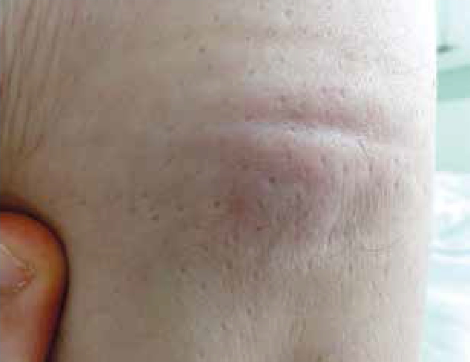
We report a case of primary skin presentation of lung cancer in a 66-year-old male patient, an ex-smoker, who was admitted to the hospital in medium-severe condition due to general weakness, exercise-induced dyspnoea and dry cough. On admission to the department, in the subcutaneous tissue and on the skin of the patient, palpable and macroscopically visible nodules were observed, 2–3 cm in diameter, tender on palpation, of a rosy-bluish colour (Figure 1). These lesions were located on the front of the chest near a post-sternotomy scar, and in the dorsal area of the neck, as well as in the left supraclavicular, left-groin and left-thigh areas. The exanthems were of a polymorphic nature, and varied in terms of their size, colour and tenderness. As indicated by the patient, they appeared a month before admission to the department. During the patient’s hospitalisation, new cutaneous lesions appeared and the old ones evolved. The lesion in the left groin area became spontaneously painful and showed signs of “fluctuation”. The semi-liquid content was collected and sent for histopathological and bacteriological examination. A nodular lesion, 3 cm in diameter, appeared on the left side of the neck. The skin on the surface of the lesion was normal. Neck ultrasound revealed a mixed echogenic focal lesion and single neighbouring lymph nodes with preserved fatty hila. The submandibular and parotid glands were normal. Single nodules were also revealed in the thyroid area. Computed tomography of the chest performed 2 weeks before the hospitalisation revealed a tumour 8 cm in diameter in the right lung hilum. Three nodules, up to 10 mm in diameter, probably metastatic, were identified in the subcutaneous tissue, and a focal lesion was observed in the right adrenal gland.
During hospitalization, the patient underwent bronchoscopy with endobronchial ultrasonography (EBUS). Specimens of the mucous membrane of the bronchi as well as the mediastinal nodes were collected. A non-small-cell lung cancer not otherwise specified (NSCLC NOS) was identified in the specimens of the bronchial mucosa and lymph nodes. Chemotherapy was planned and pending the start of the treatment and the result of the histopathological examination of the skin specimen, the patient was discharged home in stable condition. Several days after the discharge, the patient’s family informed the treating physician of the patient’s death. Three weeks after the collection of the skin specimen, the poorly differentiated carcinoma was identified in the skin and subcutaneous tissue. The immuno-histochemical test results were ambiguous; the pathologist suggested the probable points of origin of the neoplasm – the lung, thyroid gland and salivary glands.
Malignant lung tumours include predominantly carcinomas. Lung carcinomas can be divided into small- and non-small-cell cancer, with the latter including squamous carcinoma, adenocarcinoma, and large-cell carcinoma [8, 9]. Non-small-cell carcinomas constitute 85% of diagnosed lung carcinomas [10]. It should be noted that every type of lung cancer can metastasize to the skin [6], and in 25% of patients with lung cancer the skin is the first place of metastases [8]. Some studies show the predominance of lung large-cell carcinoma as a primary tumour developing metastases in the skin [2, 10]. According to other researchers, adenocarcinoma has the highest incidence of skin metastasis [7, 11–13].
Non-small-cell lung cancer not otherwise specified constitutes a maximum of 10% of all diagnosed cases of lung cancer. Due to its poor differentiation, it is characterised by significant clinical malignancy [9].
From the beginning of our patient’s hospitalisation, we suspected a cause-and-effect relationship between the nodular lesion in the lungs and the subsequently appearing skin exanthems. Nevertheless, the early death of the patient made it impossible to complete planned diagnostic tests, which would include the biopsy of the thyroid, salivary glands, and unidentified nodular masses in the neck area.
In the reported case, the skin lesions appeared to be metastatic lesions with their most probable point of origin located in the lungs. In our clinical practice skin metastases of lung cancer are observed infrequently; however, the case in question reminds us of such a possibility.
By describing this case, we want to emphasize the necessity of a thorough physical examination performed by general practitioners and dermatologists, as they are usually the first doctors to whom patients with skin lesions refer. It is worth noting that among many benign skin changes also those which result from malignancy can be found, and careful diagnostics can prevent a delay in treatment of the primary disease.


